MRI is a highly informative non-invasive diagnostic that allows you to obtain three-plane images of any area of the human body with a slice thickness of 0.1-0.3 cm.
The research does not involve the use of ionizing radiation and is based on the principle of magnetic resonance. Under the influence of the field induced by the apparatus, hydrogen protons in water molecules move with the release of a certain amount of energy. Changes are captured by detectors and transmitted to the central computer.
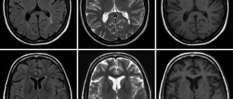
Photos of cerebral structures on magnetic resonance scans
MRI demonstrates which areas of the brain are responsible for speech or body movement, shows painful changes in the pathways (functional diagnostics), features of blood flow (angiography), which is important for neurosurgeons and neurologists to choose treatment. The study can be performed without harm to the health of pregnant women and newborns who have reached the age of one month.
How is an MRI of the brain done? Magnetic resonance scanning does not require hospitalization, and the patient receives a detailed description on the day of treatment. The most informative images are those taken on high-field equipment with a power of 1.5 Tesla or more.
Contrast is used according to indications. Intravenous administration of a chelated gadolinium enhancer improves imaging capabilities and facilitates more accurate interpretation of pathological processes.
The advantages of MR scanning compared to CT include:
- obtaining more reliable data when assessing the posterior cranial fossa and brainstem due to the absence of bone artifacts;
- the ability to detect small changes (from 1 mm) associated with tissue swelling;
- high contrast;
- possibility of making cuts in three planes.
Benefits of examining the brain using MRI
First of all, it should be noted that MRI is safe, because the patient is not exposed to ionizing radiation. Therefore, MRI can be safely performed even at short time intervals (for example, to monitor the dynamics of treatment). Other advantages of this diagnostic method include the following facts:
- The pictures are as informative and clear as possible: this result cannot be achieved by other methods;
- Magnetic resonance imaging is a non-invasive study that does not involve the use of any medications, etc.;
- Ability to monitor the functional activity of all parts of the brain;
- Quick receipt of study results, the possibility of consulting a doctor immediately after the MRI;
- If necessary, the diagnostic process can be recorded on video.
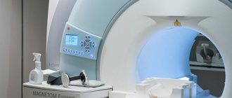
Expert equipment
The Clinical Hospital on Yauza uses the latest generation 1.5 T Philips Ingenia fully digital magnetic resonance imaging scanner, which provides:
- Reduced scanning time (less time to lie in the magnet)
- The maximum possible diameter of the tunnel (73 cm) - the patient’s waist circumference (circumference) is up to 225 cm
- Highest quality images
- Full range of studies due to maximum equipment
- Additional information through advanced software
- Environmental control system (music from the patient’s playlist, choice of lighting, personal climate control)
- Automated quality control system for research and descriptions
- IT platform with triple control of research results - with the support of professors, leading specialists from Russia, Europe and Israel (“second opinion”)
Deciphering and preparing a medical report usually takes about 20 minutes after the diagnostic procedure. In complex cases, to increase the reliability of the research results, the description time can be increased. Images are written to disk or flash card.
Indications for MRI of the brain
Brain diagnostics are carried out according to the doctor’s indications. As a rule, MRI is prescribed in the following cases:
- Suspicion of the presence of benign or malignant neoplasms, as well as metastases in the brain;
- Manifestation of epilepsy of various types;
- Previous head injuries, suspected concussion or functional damage;
- MRI of the brain is performed after operations, for example, after removal of tumors, to track dynamics;
- Suspicion of meningitis and other inflammatory diseases located near the brain;
- Symptoms of cerebrovascular accident.
Also, a diagnostic procedure can be carried out if the patient complains of the following symptoms:
- Unreasonable fainting;
- Absent-mindedness, memory lapses;
- Frequent causeless headaches;
- Disorientation in space;
- Impaired sensitivity of the upper or lower extremities, face;
- A sharp decrease in hearing or vision;
- Dizziness accompanied by nausea and vomiting.
What does the cost depend on?
The main factor determining the cost of MRI of the brain is the power of the tomograph. The higher the induction capabilities of the device’s magnet, the more expensive it is to perform tomography on it. However, the quality of the examination and diagnostic value will also increase.
The price of a brain MRI usually includes preparation, examination and interpretation of the tomograms. Additionally, the patient will need to pay the cost of contrast if the doctor ordered an MRI of the head with contrast. The decision to use contrast enhancement is made either by the attending physician or by the radiologist if, during a non-contrast scan, he detects signs of diseases that are validated during brain MRI with contrast. The cost of administering the drug depends on the patient's weight. On average, with a weight of 70 kg, 10 ml of contrast agent will be required, and the price of the contrast agent will be about 3,000 rubles.
MRI of the brain is the cheapest at night, when the maximum discounts apply. In addition, some medical centers provide additional benefits to people with disabilities, pensioners, and students. You can find out about such discount programs when making an appointment for an MRI.
| Service | Price according to Price | Discount Price at Night | Discount Price During the Day |
| from 23.00 to 8.00 | from 8.00 to 23.00 | ||
| MRI of the brain | 3300 rub. | 2690 rub. | 2990 rub. |
| MRI of cerebral vessels (arteries) / MR angiography of cerebral vessels | 3300 rub. | 2690 rub. | 2990 rub. |
| MRI of the brain and cerebral vessels | 6600 rub. | 5380 rub. | 5980 rub. |
| MRI of the pituitary gland (without contrast) | 3500 rub. | 2690 rub. | 2990 rub. |
| MRI of the pituitary gland with contrast | from 6500 rub. | not implemented | from 6900 rub. |
| MRI of the pituitary gland and brain | 7800 rub. | 5380 rub. | 5980 rub. |
| MRI of the central nervous system (MRI of the brain, MRI of the cervical, thoracic and lumbosacral region) | 13200 rub. | 9590 rub. | 10890 rub. |
| Comprehensive head diagnostics (MRI of the brain, MRI of cerebral vessels, ultrasound of neck vessels, consultation with a neurologist) | 10900 rub. | 7500 rub. | |
| Contrast administration (based on patient weight) | from 4000 to 6000 rub. | from 4000 to 6000 rub. |
What determines the cost of tomography?
Tomograph power
Applying Contrast
Personnel qualifications
Promotions and discounts
How to properly prepare for manipulation
Magnetic resonance imaging of the brain does not require special preparation. There is no need to follow special diets, stop taking medications, etc. The only rule is to abstain from alcohol before the study. It is also not recommended to smoke at least 2-3 hours before the test. Experts recommend avoiding increased physical activity. Before the study, you need to relax as much as possible, avoid stressful situations, etc.
If you are worried or even afraid of your reaction to the confined space in the tomograph, your doctor may recommend taking mild sedatives. It is better to avoid taking strong tranquilizers, because they can cause distortion of the clinical picture. Before the examination, you need to remove all metal objects and jewelry. Before an MRI, it is better not to drink a lot of water so that during the examination you will not want to go to the toilet - you cannot move during the examination.
It is recommended to take your passport, money (if MRI is performed for a fee), and a personal outpatient card with you for the study. If a study has already been carried out previously, take the results of previous diagnostics with you, this will help the doctor determine the dynamics of changes in the brain.
Diagnostic algorithm
Let's look at how magnetic resonance imaging of the brain is performed:
- First, the patient must remove all jewelry, as well as elastic bands and hairpins, if they have a metal insert. If your clothes have metal buttons, hooks, etc., you need to remove your clothes; if necessary, you will be given a robe.
- The patient lies on his back on a special retractable couch. A radio-receiving RF coil is placed above the head, receiving the signal, which, when scanned, is converted into an image.
- The specialist secures the legs and arms with special straps. This is necessary to ensure that the person does not move during the examination, as this may affect the quality of the images.
- The couch moves into the tomograph, where the scan is performed.
- During the manipulation, noise is heard - do not be alarmed, this is the norm. To reduce the intensity of sounds, patients may be offered earplugs.
- There is nothing to worry about - even if you feel ill or very scared from a confined space, you can always press the panic button or contact a doctor through the microphone.
- In general, diagnosis usually takes no more than half an hour. At the end of the manipulation, the couch moves out again, and the doctor releases the patient from the straps.
- The patient receives diagnostic results after 30-40 minutes. If necessary, you can immediately consult a doctor.
All that is required of the patient is to lie as still as possible, preferably not to yawn or talk (although if you really need to contact the doctor, you can do this). Experts recommend wearing loose, comfortable clothes, lying down comfortably and relaxing. Only if this rule is followed will the diagnostic results be as informative as possible.
After the diagnosis, the patient does not feel any changes, the state of health does not change, since the body is exposed to harmless magnetic radiation that does not cause changes in the functioning of organs and the condition of tissues. For this reason, various health consequences and complications are excluded.
Contraindications
Head tomography is not prescribed if the patient has absolute contraindications, but is allowed at the discretion of the doctor in case of relative contraindications. Diagnostics is prohibited if the following conditions exist:
- the patient has prosthetic heart valves, pacemakers or neurostimulators;
- presence of insulin pumps, inner and middle ear prostheses, cochlear implant;
- the patient has an Ilizarov apparatus;
- the presence of metal implants, ferromagnetic particles, fragments in the body.
Relative indications include:
- tremor, the patient’s inability to hold his breath for a long time during examination;
- presence of dental braces, dentures, stents, vena cava filters;
- heart failure, clip instead of gallbladder;
- pregnancy;
- claustrophobia;
- severe pain while standing still;
- coronary artery bypass grafting.
What pathological processes can be identified?
During the diagnostic procedure, many pathologies can be identified even at an early stage. With the help of research it is possible to discover:
- Inflammatory processes;
- Aneurysms;
- Multiple sclerosis;
- Strokes and micro-strokes;
- Cysts, lipomas and other benign neoplasms;
- Malignant tumors;
- Hydrocephalus;
- Encephalopathy;
- Epilepsy;
- Vascular atherosclerosis;
- Meningitis;
- Hemorrhages in the brain structures.
If you conduct a study with a contrast agent, you can also clearly see various vascular pathologies, for example, narrowing of the lumen, cerebral varicose veins, venous malformations, etc.
Diagnostic results
A highly qualified doctor interprets the images. The photographs and conclusion can be provided in printed form or recorded on electronic media. If magnetic resonance imaging does not reveal any abnormalities, the report contains approximately the following information:
“The sub- and supratentorial structures were visualized in axial, sagittal and frontal projections. The median structures are without features, the localization corresponds to the norm. The cortex and white matter are well developed, with normal MR signal intensity. Convexital grooves of the cerebellum and cerebrum are within normal limits. The ventricles of the brain have the same shape and size. The basal cisterns and subarachnoid spaces are normal. There are no symptoms of impaired outflow of cerebrospinal fluid and increased intracranial pressure. Neoplasms and other pathological processes were not identified.”
Do not try to interpret the diagnostic results yourself - entrust this to specialists. The doctor will competently decipher the conclusion, tell you whether there are pathological processes, whether any treatment is required, what the prognosis is, etc.
If necessary, effective therapy will be prescribed. Be healthy!