What are the meninges?
The human brain is surrounded by three membranes:
- The pia mater is adjacent directly to the brain tissue. Of the four-legged vertebrates, only mammals have it. Consists of loose connective tissue.
- Arachnoid. Located between the dura and pia mater. Below it is the subarachnoid space, which contains cerebrospinal fluid and blood vessels.
- Dura mater. It is located outside, adjacent to the bones of the skull. Consists of dense connective tissue. In some places it splits and forms cavities - sinuses, through which venous blood flows.
Inflammation of the soft and arachnoid meninges is called leptomeningitis, and the dura mater is called pachymeningitis.
Meningitis is a disease that can be life-threatening and lead to severe complications. Only a timely visit to a neurologist can help prevent them.
Meningococcal infection and vaccination
The main group of people subject to vaccination against meningococcal infection are children living in closed communities, as well as those going on vacation to organized summer holiday camps. Polysaccharide meningococcal vaccines, such as Meningo A+C or Mencevax, are widely used in medical practice.
These vaccines are intended for immunization of children from 2 years of age and adults using a single dose of vaccine, followed by revaccination after 2 to 3 years.
Thus, meningococcal infection is certainly one of the diseases against which it is easier and cheaper to vaccinate in order to protect yourself from possible unpleasant complications. Naturally, the feasibility and safety of each vaccination must first be discussed with your attending physician, and, if necessary, with a specialist, an immunologist.
Types of meningitis
There are different classifications of the disease, one of which, depending on which meninges is affected by the inflammatory process, we have already considered. The following types of meningitis are also distinguished:
The inflammation can be serous (most often associated with viral infections) or purulent (bacterial infections).
Depending on the cause of the disease:
- Bacterial. Most often they are caused by meningococcal bacteria. But other bacteria can also be the cause, for example, syphilitic meningitis.
- Viral. Meningitis can be caused by rubella virus, mumps virus, chickenpox virus, etc.
- Fungal. In people with reduced immunity, fungi of the genus Candida cause infection (in this case it is one of the forms of candidiasis).
- Protozoans. Some parasites, such as Toxoplasma, may be the cause.
- Mixed. For example, if a bacterial infection joins a viral infection.
The disease can develop at different rates. Depending on this criterion, fulminant, acute, subacute, and chronic courses are distinguished. The pathology can occur in mild, moderate and severe forms.
Treatment of meningitis
Treatment of meningitis in adults is determined by its causes.
For a bacterial infection, intravenous antibacterial drugs are prescribed as quickly as possible. The choice of antibiotics and their combinations is determined by the sensitivity of microorganisms. Until the doctor receives the results of the bacteriological analysis from the laboratory, he prescribes broad-spectrum drugs.
Glucocorticoids are also used - drugs from the adrenal cortex that suppress inflammation. They help prevent brain swelling and seizures.
For viral infections, antibacterial drugs are ineffective. Usually, in mild cases, treatment is limited to bed rest, drinking plenty of fluids and painkillers, anti-inflammatory, antipyretic drugs. For more severe cases, the doctor prescribes glucocorticoids. For herpesvirus infection, special antiviral drugs are used.
Fungal infections are treated with antifungal medications. For non-infectious meningitis caused by other causes, glucocorticoids are usually used. If inflammation of the meninges is caused by cancer, you need to focus on effective antitumor treatment.
In some cases, the patient's condition can deteriorate significantly within a matter of minutes. If you experience headaches, fever, or strange spots on the skin, you should consult a doctor - timely initiation of treatment at a neurology clinic in Moscow will help prevent serious complications, and perhaps even save a life. Our neurologists are ready to see you. Make an appointment by calling +7 (495) 230-00-01. We know how to help.
Take care of yourself, book a consultation now
Message sent!
expect a call, we will contact you shortly
Inflammation of the meninges can be severe and lead to serious complications. The earlier treatment is started, the greater the chance of recovery without consequences. And in order to receive medical help in a timely manner, you need to know about the main symptoms of the disease, and if they begin to bother you, immediately consult a doctor.
Diagnosis and treatment
Neurologists make the initial diagnosis of meningococcal meningitis during a clinical examination, during which a spinal tap is performed to obtain cerebrospinal fluid.
Then the laboratory technicians perform a bacterioscopy of the cerebrospinal fluid. The diagnosis is confirmed by growing microorganisms found in the cerebrospinal fluid or blood. The laboratory also studies biological fluids using polymerase chain reaction. To make the optimal choice of antibacterial agents, laboratory technicians identify the serotype of the pathogen and determine sensitivity to antibiotics. A patient with meningococcal meningitis is hospitalized at the neurology clinic of the Yusupov Hospital. Antibacterial therapy begins immediately after a spinal puncture. Patients are prescribed antibacterial or antiviral drugs immediately after receiving the results of a cerebrospinal fluid analysis and identification of the infectious agent. If lumbar puncture is contraindicated, antibiotics are selected empirically. The following antibiotics are used for initial therapy:
- Penicillin;
- Ampicillin;
- Chloramphenicol;
- Ceftriaxone.
They are administered intramuscularly and intravenously.
Before the first administration of the antibiotic, an infusion of glucocorticoid hormones is given. To reduce cerebrospinal fluid pressure, dehydration therapy is carried out with furosemide, Lasix or mannitol. Infusion therapy is carried out with solutions that have a detoxifying effect and replenish fluid loss by the body. They are administered intravenously. Timely establishment of an accurate diagnosis and adequate antimicrobial therapy allow doctors at the Yusupov Hospital to quickly stabilize the condition of patients with infectious meningitis and prevent the development of complications. If there is reliable information about a severe allergy to beta-lactams, vancomycin is prescribed for pneumococcal meningitis, and chloramphenicol is prescribed for meningococcal meningitis. Patients with identified risk factors for listeria infectious meningitis are prescribed intravenous amoxicillin in addition to third-generation cephalosporins. Dexamethasone is used simultaneously with antibiotics for pneumococcal and hemophilic infectious meningitis.
If the nature of infectious bacterial meningitis is not established, antibacterial therapy is carried out for 10–14 days. For meningitis caused by meningococcus, the duration of antibiotic use varies from 5 to 7 days. Meningitis caused by Haemophilus influenzae type b requires antibiotic therapy for 7-14 days. The course of antibacterial treatment for listeria meningitis is 21 days, and for meningitis caused by Pseudomonas aeruginosa or gram-negative microorganisms, it can last for 21–28 days.
Symptomatic therapy for infectious meningitis involves the use of diuretics, infusions of fluid replenishers, and the administration of antipyretic drugs and analgesics. For increased intracranial pressure, a 20% mannitol solution is prescribed. It increases plasma pressure, and thereby promotes the transfer of fluid from brain tissue into the bloodstream. Diacarb prevents the formation of cerebrospinal fluid. Furosemide inhibits the reabsorption of sodium in the renal tubules, resulting in increased diuresis.
In the presence of convulsive syndrome, doctors prescribe diazepam, sodium hydroxybutyrate, and aminazine to patients with infectious meningitis. From the very first minutes of a patient’s admission to the neurology clinic, doctors administer oxygen therapy. If indicated, the patient is given tracheal intubation and artificial ventilation.
The first signs of meningitis are a characteristic triad:
- Headache.
- Rigidity (increased tone) of the neck muscles. If you lay the patient on his back and ask him to relax his neck, and in the meantime passively bend his head forward, then the chin will not be able to be brought very close to the chest.
- Increased body temperature.
If these signs are absent, then the likelihood of acute meningitis is low. If all three occur, they are very pronounced and increase over a short time, you should immediately call a doctor.
Other possible symptoms: intolerance to loud sounds and bright lights, nausea and vomiting, increased irritability, drowsiness, confusion, coma. With viral infections, manifestations of the disease are usually preceded by muscle pain, increased fatigue, weakness, and loss of appetite.
The first signs of meningitis are a reason to immediately consult a neurologist for advice. If treatment is not started immediately, the consequences can be very serious.
Symptoms
The clinical picture of infectious meningitis is represented by a group of general infectious, cerebral and meningeal symptoms. General infectious signs of meningococcal infection include the following symptoms:
- General malaise;
- Increase in body temperature to high levels;
- Joint and muscle pain (arthralgia and myalgia);
- Increased heart rate;
- Redness (hyperemia) of the face;
- Inflammatory changes in peripheral blood.
General cerebral symptoms of infectious meningitis are headache, nausea, vomiting, confusion or depression of consciousness, generalized seizures. Headache is caused by irritation of the meninges due to their inflammation and increased intracranial pressure. It is bursting in nature. Vomiting is the result of an acute increase in intracranial pressure. Due to CSF hypertension, patients suffering from infectious meningitis may develop Cushing's triad:
- Rare breathing;
- Increased systolic blood pressure;
- Bradycardia (decreased heart rate).
Convulsions and psychomotor agitation, which are periodically replaced by lethargy or impaired consciousness, occur in cases of severe infectious meningitis.
The patient may develop mental disorders in the form of delusions and hallucinations. Meningeal symptoms themselves, which are caused by irritation of the meninges, include increased sensitivity to various irritants (hyperesthesia) and signs of a reflex increase in the tone of the occipital muscles. If a patient with meningitis is conscious, he cannot tolerate loud conversation and noise. The patient's headache intensifies from strong sounds and bright lights. He prefers to lie with his eyes closed in a darkened room, into which no extraneous sounds penetrate.
Patients suffering from infectious meningitis develop a stiff neck and Kernig's sign. Doctors detect stiff neck muscles when the patient's neck is passively flexed, when due to spasm of the extensor muscles it is not possible to completely bring his chin to the sternum. Kernig's sign is checked in the following way:
- The doctor passively bends the lower limb of the patient, who is lying on his back, at an angle of 90º at the knee and hip joints;
- Then he tries to straighten the patient's leg at the knee joint;
- If a patient has meningeal syndrome due to a reflex increase in the tone of the leg flexor muscles, it is impossible to straighten his leg at the knee joint.
With infectious meningitis, this symptom is positive on both sides.
The upper Brudzinski symptom is defined as follows: when the head of a patient lying on his back is passively adducted to the sternum, his lower limbs bend at the knee and hip joints. To test the average Brudzinski sign, the patient's legs should be flexed while pressing on the symphysis pubis. The lower Brudzinski sign is manifested by flexion of the other leg with passive flexion of one limb.
Meningitis rash
In 10-20% of children suffering from infectious meningitis, hemorrhagic rashes appear at the onset of the disease. They are represented by primary infiltrative elements that do not contain cavities that externally resemble spots. The rash resembles allergy rashes and is maculopapular in nature. Patients may have 1-2 small spots or a widespread massive star-shaped rash that tends to merge.
If a person has symptoms of meningitis: is it contagious?
When there is inflammation of the meninges of a bacterial nature, the infection is transmitted from person to person. Infection can occur through airborne droplets, during coughing, kissing, or prolonged stay in the same room with a sick person. Meningococcus bacteria are especially dangerous. Previously, they caused mass epidemics, but now single cases of the disease are being recorded.
Viral infections are also transmitted from person to person. But, if you become infected from a patient, this does not mean that you will also develop inflammation in the meninges. Viruses cause disease only under certain conditions if a person is predisposed.
Fungal infections are also contagious, but they primarily affect people who have severely weakened immune systems.
What are the dangerous signs of meningitis in adults?
- Sepsis is a systemic inflammatory response in the body, which manifests itself in the form of decreased blood pressure, high or abnormally low temperature, rapid heart rate and breathing, and excessive activation of blood clotting. The work of all organs is disrupted.
- Hemorrhage into the adrenal glands. Often leads to death.
- Brain swelling. In this case, convulsions occur.
- Brainstem herniation. If the swelling is too severe, the brain is pressed into the opening that leads from the skull into the spinal canal. The patient gradually loses consciousness, his pupils stop responding to light, and breathing may stop.
- Impaired outflow of cerebrospinal fluid. As a result, meningitis can lead to hydrocephalus.
- Damage to the brain and cranial nerves. Leads to neurological disorders.
Meningitis is a relatively rare disease, and its symptoms can occur in many other pathologies. If the symptoms are very strong and increase quickly, this should prompt you to immediately consult a doctor. But even if you have even a slight suspicion of this disease, it is better to play it safe and call a doctor.
You can get qualified medical care in our neurological center by calling
We will call you back
Message sent!
expect a call, we will contact you shortly
There are quite a few reasons that can lead to inflammation of the meninges. Some of them are associated with infectious processes, others are caused by non-infectious pathologies.
Meningitis in children
For all meningitis, meningeal syndrome , which includes cerebral, local symptoms and changes in the cerebrospinal fluid. General cerebral symptoms are characterized by manifestations in the form of a general reaction of the brain to irritation. Local symptoms occur due to swelling and damage to the cranial nerves. Sometimes symptoms of loss are observed.
A mandatory sign of meningitis is inflammatory changes in the cerebrospinal fluid with cell-protein dissociation (a significant increase in cells in the cerebrospinal fluid).
The meningeal symptom complex includes:
Increase in body temperature to high levels. With tuberculous meningitis, high fever is usually not observed.
Headache. In its pathogenesis, irritation of the receptors of the meninges is important. Headache is the main constant symptom of the disease. It is usually diffuse, intensified by sudden movements, sound and light stimuli.
Vomiting occurs without nausea and is not associated with food intake. More often it occurs when changing body position. After vomiting, the headache does not decrease.
Muscle contracture - rigidity of the muscles of the neck and back, Kernig and Brudzinski symptoms. Young children may experience the hanging sign (Lesage). Muscle contracture during meningitis occurs due to increased activity of the reflex apparatus and irritation of the roots due to increased pressure of the cerebrospinal fluid.
Autonomic disorders , which include dissociation between body temperature and pulse, pupillary disorders, skin petechiae, mental disorders (disorder of consciousness, asthenia).
Motor disorders , manifested in the form of symptoms of damage to the motor cranial nerves, focal epileptic seizures due to irritation of the motor cortex.
Sensitivity disorders , which occur in the form of general hyperesthesia. It is based on irritation of the dorsal roots and intervertebral nodes.
Pachymeningitis
Pachymeningitis is divided into cerebral (inflammation of the dura mater of the brain) and spinal (inflammation of the dura mater of the spinal cord). Based on the nature of the lesion, cerebral pachymeningitis is divided into serous, hemorrhagic and purulent . Serous pachymeningitis occurs in various infections and is characterized by a low-symptomatic or asymptomatic course (headache, vague meningeal symptoms, as well as signs of compression of the brain by the edematous membrane). Hemorrhagic pachymeningitis is characterized by the presence of hemorrhage (a condition in which hemorrhage or bleeding occurs) in the dura mater, which is clinically manifested by headache, focal and meningeal symptoms, and agitation. Occurs with atherosclerosis and hypertension. Purulent pachymeningitis is usually secondary and is associated with a primary purulent rhinootogenic infection. Purulent pachymeningitis is characterized by headache, vomiting, congestive changes in the fundus and blood changes may be observed.
Spinal pachymeningitis can be serous or purulent . Serous is characterized, as a rule, by a benign course, purulent is associated with the presence of purulent foci in the body (furunculosis, osteomyelitis, etc.).
Among spinal pachymeningitis, the following are distinguished.
Spinal epiduritis (inflammation between the layers of the membrane). In this case, the membranes at the level of the upper thoracic region are affected. Patients experience clinical symptoms of spinal cord compression (radicular pain, motor, sensory and pelvic disorders).
Chronic hyperplastic epiduritis. It is most often caused by spinal injury. The clinic is characterized by limited mobility of the spine, radicular pain in the spine. Remissions are characteristic; the protein content in the cerebrospinal fluid is increased.
Cervical hypertrophic syphilitic pachymeningitis occurs as a type of moderate compression of the spinal cord in the neck. In this case, radicular pain, symptoms of compression in the form of flaccid paresis of the arms, spastic paresis of the lower extremities, conduction-type sensitivity disorders, and dysfunction of the pelvic organs are observed.
Tuberculous spinal pachymeningitis, in which clinical symptoms depend on the degree of damage to the spinal cord.
Arachnoiditis
The following forms of the disease are distinguished: adhesive (formation of adhesions), cystic (presence of cysts), cystic-adhesive, or mixed arachnoiditis. Based on the causes of occurrence, arachnoiditis is divided into rheumatic, post-influenza, tonsillogenic, traumatic and toxic. Depending on the course, acute, subacute and chronic arachnoiditis are considered. convexital arachnoiditis is distinguished (damage to the frontal, parietal, temporal lobes and central gyri), basal and posterior cranial fossa.
Arachnoiditis can also be focal and widespread. Pathological anatomy is characterized by fibrosis and proliferation of connective tissue.
Clinical symptoms are quite wide: headache, nausea, vomiting, epileptic seizures, changes in the fundus. Shell symptoms may be absent or mild. Focal neurological symptoms depend on the location of the lesion. With a pronounced diffuse process, intracranial pressure increases and dropsy develops.
Convexital arachnoiditis is a predominant lesion of the soft membranes of the convexital surface of the cerebral cortex. In this case, the leading clinical symptoms are dysfunction of the frontal, temporal, parietal lobes and the region of the central gyri. The clinical picture is dominated by subjective complaints (headache accompanied by nausea and vomiting, sweating, dizziness, etc.) over objective symptoms (unevenness of tendon reflexes, pathological reflexes on the side opposite to the lesion, convulsive seizures, etc.).
Optochiasmal arachnoiditis is characterized by predominant damage to the soft membranes of the brain in the area of the optic chiasm (the point at the base of the brain where the fibers of the 2 optic nerves cross and diverge) and the intracranial part of the optic nerves. The leading symptom in the clinic is a decrease in visual acuity and changes in visual fields. Individual sections of the visual fields may fall out, and concentric narrowing of the visual fields, hemianopsia and blindness may also occur. Along with this, autonomic disorders (sleep disturbances, changes in carbohydrate and water-salt metabolism, etc.) are common.
Arachnoiditis of the posterior cranial fossa appears due to damage to the soft membranes of the brain in the area of the lateral and large cistern, as well as in the craniospinal region with possible disruption of the circulation of cerebrospinal fluid in the posterior cranial fossa. The course of the disease is severe, with meningeal and cerebral clinical signs, disorders of brain stem function, cerebellar disorders, congestion in the fundus, and damage to the cranial nerves.
Arachnoiditis of the cerebellopontine angle occurs most often as a result of otitis media. The clinical picture is characterized by clearly defined focal symptoms with weak cerebral symptoms. Focal symptoms are manifested by damage to the cranial nerves, cerebellar disorders, and mild pyramidal disorders. Pyramidal disorders occur on the side opposite to the lesion, cerebellar disorders on the same side.
In the cerebellopontine angle there are also tumor processes that are benign. Cranial nerves are often involved in the process, and pyramidal and cerebellar disorders may be observed. Treatment of the tumor is surgical.
Differential diagnosis of arachnoiditis and tumor presents significant difficulties. Arachnoiditis, unlike a tumor, is characterized by a remitting course and less severe focal neurological symptoms. However, cystic arachnoiditis and brain tumor are space-occupying processes that may have a similar clinical picture.
Spinal arachnoiditis can be adhesive, cystic or mixed. According to prevalence – diffuse and focal. Adhesive spinal arachnoiditis is accompanied mainly by radicular symptoms. Cystic are characterized by the clinical appearance of an extramedullary tumor.
Leptominengitis
Meningitis affecting the soft membrane of the brain. This type is divided into two volumetric groups: purulent and serous , depending on the nature of the inflammation process and changes in the cerebrospinal fluid. The development of the disease has an acute, subacute and chronic course. Meningitis is divided into primary and secondary. Primary meningitis can be purulent (meningococcal, pneumococcal, etc.) and serous (lymphocytic choriomeningitis, meningitis caused by ECHO and Coxsackie viruses, etc.). Secondary meningitis manifests itself as a complication of purulent otitis media, furunculosis, lung abscess, open head injury, and general infections (tuberculosis, mumps, syphilis, etc.).
Purulent meningitis
Purulent meningitis is a group of diseases caused by various pathogens (meningococci, streptococci, staphylococci, Haemophilus influenzae Afanasyev-Pfeiffer, Escherichia coli, Pseudomonas aeruginosa, fungi, salmonella, etc.), affecting mainly the soft membranes of the brain and spinal cord. Children of all ages, especially young ones, are susceptible to the disease, this is due to insufficient development of immunity and a weakening of the blood-brain barrier.
Features of the development of purulent meningitis in newborns
The entry points for infection are the umbilical vessels, the infected placenta when the mother has pyelitis or pyelocystitis. Predisposing factors – prematurity, birth trauma, etc.
The most common pathogens are Escherichia coli, staphylococci, and streptococci.
Clinical manifestations are characterized by severity, dehydration, gastrointestinal disorders, and the absence of a significant increase in body temperature.
High mortality rate (50-60%). The clinical picture may include hyperexcitability syndrome (the child is restless, screams monotonously, burps, throws his head back, a large fontanelle may bulge) and lethargy or apathy syndrome (the child is lethargic, reduced motor activity, weak cry, refuses to breastfeed). Recovery of newborn children is often incomplete; there are severe organic lesions of the central nervous system (hydrocephalus, epilepsy, mental retardation, paralysis and paresis of cranial nerves and limbs).
- Meningococcal meningitis
Infection with meningococcus occurs through the mucous membrane of the nasopharynx. After which meningococcus penetrates the blood and lymphatic system, where it develops. Meningococcal meningitis is manifested by the following clinical symptoms: high body temperature, severe intoxication, dizziness, vomiting, pale complexion, purulent nasal discharge, fever, skin rashes, bleeding and hemorrhage, eye pain, sometimes severe abdominal pain, increased sweating.
- Pneumococcal meningitis
Meningitis is caused by pneumococci of various serological types and is characterized by severe disease and high mortality. In 40% of cases it is primary, i.e. occurs in healthy children. In other cases, the disease develops against the background of otitis, sinusitis, and pneumonia. Young children are more often affected. Patients experience high body temperature, toxicosis, loss of consciousness, convulsions, damage to the cranial nerves, as well as paralysis and paresis of the limbs. The disease often takes a protracted course, and in the absence of treatment, death can occur on the 5-6th day.
- Meningitis caused by Haemophilus influenzae Afanasyev-Pfeiffer
Meningitis affects weakened children suffering from frequent catarrh of the upper respiratory tract, otitis media, and pneumonia. Perhaps both an acute onset and a gradual one. The course is sluggish, wave-like, gastrointestinal disorders, pneumonia, and toxicosis may occur. With timely and correct treatment, the course is favorable.
- Staphylococcal meningitis
This type of meningitis is the most unfavorable and has a high mortality rate. It is usually secondary and occurs against the background of abscesses, chronic pneumonia, osteomyelitis of the skull and spine, and sepsis. The clinical picture of meningitis is masked by the severe septic condition of the patient. A feature of staphylococcal meningitis is blockage of the cerebrospinal fluid pathways (hydrocephalus), as well as abscess formation. Difficulties in therapy are aggravated by the resistance of staphylococcus to antibiotics.
- Escherichia meningitis
The causative agent is pathogenic strains of Escherichia coli. This type of meningitis is rare and is mostly common in young children. In newborns it develops as a complication after sepsis. Escherichia meningitis is predisposed by prematurity, birth trauma, previous infectious and somatic diseases, such factors influence brain damage. Infection occurs through the umbilical vessels, infected placenta when the mother has pyelocystitis or pyelitis. Symptoms are expressed by a sharp deterioration of the condition against the background of elevated body temperature, vomiting, anorexia, and increasing intoxication. Newborns experience attacks of clonicotonic convulsions, muscle hypo- and atonia, followed by tonic tension of the limbs, indifference, their reflexes are depressed, and the fontanel may be sunken. Dysmeitic phenomena are observed in the form of loose, frequent stools.
The current is severe. Purulent foci may appear in other organs, dystrophy increases early, and arterial hypotension and exicosis often develop. Possible severe organic damage to the central nervous system.
- Salmonella meningitis
Can be caused by any Salmonella serotype. The disease occurs infrequently, usually in children during the first 6 months. life and newborns, but it is also possible in older children, adolescents and even adults. In infants, the disease develops gradually, peptic symptoms are moderate or absent. Characterized by toxicosis, septicemia or septicopyemia, enlarged liver and spleen, rash, hyperleukocytosis in the blood. The development of cerebral hypotension is likely. In older children, meningitis develops acutely against the background of a typical clinical picture of gastroenteritis. The course of the disease is severe with frequent deaths.
- Meningitis caused by Pseudomonas aeruginosa
In most cases, it occurs against the background of sepsis, which develops as a superinfection after surgery, and occurs in all age groups. The increase in its cases is associated with the use of antibiotics, leading to dysbiosis, when only resistant species of bacteria survive, including Pseudomonas aeruginosa.
The clinical picture shows signs of severe meningoencephalitis with a tendency to develop hydrocephalus. The course of the disease is often long-term, the prognosis is unfavorable. The outcome of the disease is largely determined by the time of initiation of treatment and the correct selection of antibiotics, to most of which the pathogen is resistant.
- Streptococcal meningitis
A rare disease, most often observed in newborns as a manifestation of sepsis, but can occur in all age groups. The course and clinical picture are similar to those of meningococcal meningitis; In patients with septic endocarditis, meningitis begins suddenly and is accompanied by focal neurological symptoms. Patients often experience vascular damage, leading to subarachnoid hemorrhages.
- Listeria meningitis
Occurs in all age groups. Newborns are more likely to get sick, in whom meningitis may be one of the manifestations of septicemia. The disease develops acutely, meningeal syndrome is usually well expressed, and symptoms of focal damage to the central nervous system are often observed - meningoencephalitis. Adequate timely therapy usually leads to complete recovery.
- Meningitis caused by Proteus, Klebsiella (Friedlander's bacillus)
This type of disease is very rare. In most cases, this is a manifestation of dysbiosis, which develops as a result of irrational use of antibiotics. The development of the disease is preceded by septicemia; children usually get sick in the first months of life. The primary source of infection in the case of meningitis caused by Klebsiella can be pneumonia, purulent otitis, and tracheobronchitis. This meningitis is sometimes preceded by neurosurgical intervention. Clinical symptoms may be mild. The course is severe and residual effects are often observed.
Serous meningitis
Serous meningitis occurs with serous inflammation of the soft meninges. Lymphocytic pleocytosis is observed in the cerebrospinal fluid. There are 4 possible clinical forms: serous meningitis, meningoencephalitis, clinically asymptomatic meningitis, meningism. Children in the first years of life often experience lethargy, weakness, drowsiness, delirium and hallucinations. With meningoencephalitis, focal symptoms are associated: hemi- and monoparesis of the limbs, ataxia, damage to the cranial nerves. Approximately 15% of patients experience pancreatitis and increased diastase content in the urine. In school-age boys, orchitis occurs simultaneously with meningitis or a little later - swelling of the testicles, hyperemia and swelling of the scrotum, increased body temperature. The presence of orchitis and pancreatitis confirms the etiology of meningitis.
Enteroviral meningitis caused by the Coxsackie and ECHO viruses is characterized by high co-infectivity, focality and mass distribution. They are characterized by myalgia - muscle pain, often in the abdominal muscles. The clinical picture is characterized by increased body temperature, facial hyperemia with a pale nasolabial triangle, pharynx hyperemia, conjunctivitis, injection of scleral vessels, and a polymorphic rash. Herpetic rashes are often observed. As a rule, there is a sharp headache, vomiting, and meningeal symptoms. In the cerebrospinal fluid, lymphocytic pleocytosis can be noted, the protein content is normal, the pressure is increased. Focal symptoms are usually mild and disappear quickly. The course of the disease is benign.
- Tuberculous meningitis
It occurs against the background of a primary tuberculosis focus in the body. Clinical symptoms are characterized by increasing intoxication, lethargy, workload, and an increase in temperature to febrile levels. Pleocytosis is always mixed with a predominance of lymphocytes, the fluid is xanthochromic, the glucose content in the cerebrospinal fluid is reduced due to the activity of tubercle bacilli.
Fungal meningitis
Various fungi that cause meningitis can be present in the body both short-term and long-term. Meningitis is most often caused by yeast-like fungi. The first encounter with fungi (causing candidiasis can occur in utero, during passage through the birth canal, or during breastfeeding). Infants in the first months of life and premature newborns are most often affected. The disease is usually preceded by sepsis, surgical interventions with long-term antibiotic treatment.
- Candidal meningitis
It is distinguished by a sluggish, subacute course, and can be detected incidentally during the study of cerebrospinal fluid in children with progressive hydrocephalus or convulsive syndrome. The clinical picture is characterized mainly by lethargy, adynamia, pallor of the skin, an unstable rise in temperature to 37.5-38 ° C, decreased appetite, and sometimes vomiting. Meningeal symptoms are mildly expressed or may be absent, bulging and tension of the large fontanelle is not always noted, and at a later date progressive hydrocephalus is possible.
Without appropriate therapy, mortality reaches 100%, with children dying within 1-3 months. either from cachexia or from the addition of a secondary infection. The timing of the start of treatment determines the outcome, however, even in the case of specific therapy, the course of the disease is long, and the majority of surviving children develop hydrocephalus. Note that candidal meningitis is often combined with staphylococcal infection. In this case, the clinical picture of the disease is more pronounced.
Infections
Viral meningitis, as a rule, is quite mild and goes away on its own without special antiviral treatment. The most common pathogens are the following viruses:
- Herpes simplex virus is usually type 2.
- Enteroviruses.
- The causative agent of mumps (mumps).
- The causative agent of chickenpox.
Bacterial meningitis is more severe, more often leading to serious complications and posing a threat to life. Bacteria can penetrate into the meninges through the bloodstream or directly, during infectious processes in the ear, paranasal sinuses, or fractures of the skull bones.
In addition to meningococcus, the disease can be caused by bacteria such as pneumococcus, Haemophilus influenzae, and listeria.
Sometimes the patient’s life depends on quickly identifying the cause of the pathology and starting treatment. Only a specialist neurologist can establish the correct diagnosis and provide the most effective assistance.
Fungal meningitis is rare. As a rule, they occur in a chronic form. The most common pathogens are cryptococci, fungi of the genus Candida. Typically, the infection develops in people with reduced immunity, for example, with AIDS, taking drugs that suppress the immune system (cytostatics, chemotherapy drugs for cancer, glucocorticoids).
Inflammatory processes in the meninges caused by parasitic infestations are extremely rare. For example, inflammation can be caused by toxoplasma, a protozoan single-celled animal.
Non-infectious causes of meningitis
Symptoms of meningitis can be caused by causes unrelated to infections:
- allergic reactions to certain medications (antibiotics, immunoglobulins, non-steroidal anti-inflammatory drugs);
- systemic inflammatory diseases, for example, sarcoidosis (in this case the disease will be called neurosarcoidosis);
- systemic lupus erythematosus and other connective tissue diseases.
- some cancers (if cancer cells spread to the meninges).
Depending on the causes, not only the symptoms of the disease vary, but also the severity, prognosis, and treatment methods for meningitis. For example, viral meningitis goes away on its own; in most cases, special antiviral drugs are not even needed, but bacterial meningitis can lead to serious brain damage, even death.
It is important to understand the cause and immediately begin the correct treatment, only this can prevent negative consequences. If the first symptoms occur, consult a doctor at the neurology center in Moscow, International Clinic Medica24. Contact the administrator of our clinic by phone: +7 (495) 230-00-01.
Get a consultation with a doctor
Message sent!
expect a call, we will contact you shortly
The primary diagnosis of meningitis is made by a neurologist during an examination of the patient. In this he is helped by the information obtained during questioning and a neurological examination. With inflammation of the meninges, characteristic symptoms are revealed:
- When the doctor presses on certain points on the head and neck where the nerves exit, it causes noticeable pain.
- Lightly tapping the cheekbone with a finger or a neurological hammer intensifies the headache.
- If the patient lies with his leg bent at the hip joint, he cannot straighten it at the knee joint, due to the fact that the muscles on the back of the thigh are very tense.
- When the patient lies down and the doctor tries to bend his head and bring his chin to his chest, his legs bend at the knee and hip joint.
- When the patient lies down and the doctor bends one of his legs at the knee and hip joint, the second leg bends in the same way.
There are other special tests. They help only with a high degree of probability to diagnose meningitis, but do not allow one to determine its cause or the severity of the inflammatory process. This requires additional research.
Viral and bacterial meningitis
MGMSU named after N.A. Semashko Meningitis is a group of diseases characterized by damage to the meninges and inflammatory changes in the cerebrospinal fluid.
Normal number of cells in cerebrospinal fluid (CSF)
is no more than 5 in 1 µl [1, 2], the amount of protein is no more than 0.45 mg/l, sugar is no less than 2.2 mg/l. Cells in normal cerebrospinal fluid are represented by lymphocytes.
According to the composition of the formed elements in the cerebrospinal fluid and etiology, meningitis is divided into purulent
(bacterial) with a predominance of neutrophilic leukocytes and
serous
(usually viral) with predominantly lymphocytic pleocytosis.
Some bacterial meningitis is characterized by a predominance of the lymphocytic (serous) composition of the cerebrospinal fluid (tuberculosis, syphilitic, Lyme borreliosis, etc.). Meningitis can be primary
or
secondary
(develops against the background of an existing general or local infectious process);
according to the nature of the course - acute
,
chronic
, sometimes fulminant.
In pathogenesis
Meningitis is played by a complex of factors: first of all, the properties of the pathogen, the reaction of the host organism and the background against which the contact of the micro- and macroorganism occurs. The virulence of the pathogen, its neurotropism and other features are of great importance. In the host’s reaction, a significant role is played by age, nutrition, social factors, previous injuries and diseases, the nature of previous treatment, immune status, etc. Environmental conditions include exposure to physical factors of cooling, overheating, insolation; contacts with animals, vectors and sources of infection, etc.
Certain individuals have an increased risk of developing infections of the nervous system. These include people with certain concomitant diseases and chronic infections, such as skull injuries, consequences of neurosurgical interventions and shunting of the cerebrospinal fluid system, chronic purulent processes in the chest cavity, septic endocarditis, lymphoma, blood diseases, diabetes, chronic diseases of the paranasal sinuses, alcoholism, long-term therapy with immunosuppressants, etc. The high-risk group also includes patients with congenital and acquired immune defects, pregnant women, patients with unrecognized diabetes, etc. In such individuals, due to a defect in immune defense, there is an increased risk of viral infections that they have already encountered in early childhood. This primarily includes diseases caused by the herpes group: cytomegalovirus, Epstein-Barr virus, varicella-zoster virus.
The pathogen can penetrate into the meninges in various ways: hematogenous, lymphogenous, perineural or contact (in the presence of a purulent focus in direct contact with the meninges - otitis media, sinusitis, brain abscess).
Overproduction of CSF, disruption of intracranial hemodynamics, and the direct toxic effect of the pathogen on the brain substance are essential in the pathogenesis of meningitis. The permeability of the blood-brain barrier increases, the endothelium of the brain capillaries is damaged, microcirculation is disrupted, and metabolic disorders develop, aggravating brain hypoxia. As a result, cerebral edema occurs, the progression of which can lead to brain dislocation and death from respiratory and cardiac arrest [3].
Viral meningitis
The etiological classification of viral meningitis most fully meets epidemiological and practical requirements. Most authors consider enteroviral meningitis to be one of the most common types of viral meningitis [4, 5]. The genus of enteroviruses (family Picornaviridae) includes poliovirus types 1–3, Coxsackie viruses A (types 1–24) and B (types 1–6), ECHO viruses (types 1–34), enteroviruses 68 -71st types. All representatives of enteroviruses cause meningitis, but most often the Coxsackie and ECHO viruses. Often the causes of viral meningitis are also paramyxoviruses (mumps, parainfluenza, respiratory syncytial), viruses of the herpes family (herpes simplex type 2, varicella-zoster, Epstein-Barr, herpes virus type 6), arboviruses (tick-borne encephalitis) , lymphocytic choriomeningitis, etc.
Clinic
Meningitis, including viral ones, is characterized by an acute onset with high fever, headache, nausea and vomiting, general malaise and weakness
. Typical for meningitis is the presence of meningeal symptoms, indicating irritation of the meninges. The meningeal symptom complex includes, in addition to headache, stiff neck, Kernig and Brudzinski symptoms, photophobia, and hyperesthesia of the skin. In young children, bulging and tension of the fontanel, tympanitis when tapping the skull, and a symptom of “suspension” (Lessage) are observed.
For some types of pathogens, a blurred clinical picture is observed with low-grade fever and moderate headache, absence of vomiting, meningeal monosymptoms or reduced symptoms.
General cerebral symptoms
in the form of impaired consciousness, convulsions and
signs of focal damage
to the nervous system
during meningitis are absent,
and their presence indicates encephalitis, but some authors admit their short-term presence at the onset of the disease as a manifestation of cerebral edema.
The main criterion for meningitis is an increase in the number of cells in the CSF. In viral meningitis, a lymphocytic composition of the CSF is observed.
Cytosis is represented by a two to three-digit number, usually no more than 1000 in 1 μl. The percentage of lymphocytes is 60–70% of the total number of cells in the cerebrospinal fluid. Protein and sugar levels are within normal limits. In the presence of meningeal signs, but in the absence of inflammatory changes in the cerebrospinal fluid, they speak of meningism. In some meningitis, signs of a general viral infection are observed (Table 1).
The duration of viral meningitis is 2–3 weeks. In 70% of cases the disease ends in recovery
[6], but in 10% the course is longer and may be accompanied by complications.
Features depending on the pathogen
Although in most cases of viral meningitis there is no clear clinical correlation with a specific pathogen, some features may be observed. So, often Coxsackie viruses of group B
cause diseases that occur with severe
myalgic syndrome
(the so-called epidemic pleurodynia or Bornholm's disease);
Diarrhea may occur. Both groups of Coxsackieviruses can cause pericarditis and myocarditis
.
Adenoviral meningitis
accompanied by an inflammatory reaction from the upper respiratory tract,
conjunctivitis and keratoconjunctivitis
.
Parotitis
often occurs
with damage to the parotid glands
, abdominal pain and increased levels of amylase and diastase (pancreatitis), orchitis and oophoritis. At the onset of the disease, the composition of the CSF may be neutrophilic with low sugar levels. Often the disease becomes protracted with a delay in the sanitation of the cerebrospinal fluid.
Lymphocytic choriomeningitis can also have a protracted course.
, and meningitis caused by
herpes simplex virus type 2
. With these types of disease, at the onset of the disease, the sugar level in the CSF may be below normal, which forces them to be differentiated from tuberculous meningitis.
Herpetic meningitis
often observed against the background of a primary genital infection - in 36% of women and 13% of men. In most patients, herpetic rashes precede the signs of meningitis an average of a week. Herpetic meningitis can cause complications in the form of sensory disturbances, radicular pain, etc. Relapses of the disease are described in 18–30% of cases [7].
Meningitis with herpes zoster
in some cases it occurs with minimal meningeal syndrome or asymptomatically. As a rule, it is not a monosyndromic lesion of the nervous system, but develops against the background of concomitant radicular phenomena, sensory disturbances, etc.
Meningitis with tick-borne encephalitis
observed in almost half of the sick. The onset is acute, accompanied by high fever, intoxication, pain in muscles and joints. Characterized by hyperemia of the face and upper body, severe headache, and repeated vomiting. In 20–40% of cases, a two-wave fever with a period of apyrexia of 2–6 days is observed. In the cerebrospinal fluid in the first days of the disease, neutrophilic leukocytes may predominate, the preponderance of which may persist for several days. Inflammatory changes in the CSF last a relatively long time - from 3 weeks to several months, accompanied by poor health. At the same time, diffuse neurological symptoms may be observed. Asthenic syndrome after illness, characteristic of tick-borne encephalitis, is observed in approximately 40% of those who have recovered from the disease and lasts from 1–3 months to 1 year. In 2–6%, a transition to a progressive form of the disease may subsequently occur.
Diagnosis
The diagnosis of viral meningitis is difficult, especially in cases of sporadic disease. For some viral meningitis, a history or associated organ involvement may be helpful (Table 1). But the main attention is paid to laboratory diagnostics: isolating the virus from the CSF and determining the 4-fold increase in specific antibodies in the dynamics of the disease
.
Currently, large treatment centers use polymerase chain reaction
(PCR), which has high sensitivity and specificity.
Treatment of the vast majority of viral meningitis is symptomatic
.
In the acute period detoxification therapy
: solutions of glucose, Ringer, dextrans, polyvinyl lyrrolidone, etc. Moderate dehydration is used: acetazolamide, furosemide). Symptomatic drugs (analgesics, vitamins A, C, E, group B, antiplatelet agents, etc.).
For meningitis caused by herpes simplex virus type 2, intravenous acyclovir
10–15 mg per 1 kg per day for 10 days, based on 3-fold administration.
Bacterial meningitis
The causative agents can be meningococci, pneumococci, Haemophilus influenzae, staphylococci, salmonella, listeria, tubercle bacilli, spirochetes, etc. The inflammatory process that develops in the meninges is usually purulent. In recent years, the etiological structure of purulent bacterial meningitis (PBM) has changed significantly. In adults, in more than 30% of cases, the causative agent is Streptococcus pneumoniae, in people over 50 years old - S.pneumoniae and gram-negative bacteria of the intestinal group (E.coli, Klebsiella pneumoniae, etc.), in children under 5 years of age in more than 30% of GBM is caused by Haemophilus influenzae type B [2, 8]. However, according to the forecast of epidemiologists, another increase in the incidence of meningococcal infection is expected in a few years.
Clinically, GBM is characterized by a more acute onset of the disease, more severe intoxication and high fever, compared to viral meningitis, and a more severe course
. CSF in GBM is turbid, with high neutrophilic pleocytosis, increased protein content; sugar levels are reduced.
Meningococcal meningitis
Meningococcal meningitis occurs mainly in children and young people. In almost half of the patients it is preceded by nasopharyngitis, which is often mistakenly diagnosed as ARVI. Against this background or in the midst of complete health, meningitis begins acutely - with chills, an increase in body temperature to 39–39.50 C, and a headache, the intensity of which increases with every hour. On the very first day, vomiting, photophobia, hyperacusis, skin hyperesthesia, and meningeal symptoms appear. There is a revival or suppression of tendon reflexes and their asymmetry. A little later, signs of increasing cerebral edema may appear: attacks of psychomotor agitation, followed by drowsiness, then coma. Focal symptoms are also possible: diplopia, ptosis, anisokaria, strabismus, etc. When often combined with meningococcemia, a characteristic hemorrhagic rash appears on the skin, the appearance of which usually precedes the symptoms of meningitis.
Possible atypical forms
, especially in patients receiving antibacterial drugs. The course of meningitis in these cases is subacute, the body temperature is subfebrile or normal, the headache is moderate, there is no vomiting, meningeal symptoms appear late and are mild, but encephalitis, ventriculitis subsequently develops and death can occur.
In infants
The onset of meningitis, including meningococcal, is manifested by general anxiety, crying, screaming, refusal to suck, sudden agitation from the slightest touch, and convulsions.
In the first hours of meningitis, the CSF is either not changed at all, or the inflammatory changes are mild. From the end of the 1st day, the CSF becomes typical for purulent meningitis. Microscopy of smears of cerebrospinal fluid sediment in most cases reveals gram-negative diplococci, mainly intracellularly. Timely initiation of adequate therapy ensures recovery in most cases.
; in the absence of this, mortality reaches 50%.
Pneumococcal meningitis
Pneumococcal meningitis can be either primary or secondary (in this case it is preceded by otitis media or mastoiditis, pneumonia, sinusitis, traumatic brain injury, cerebrospinal fluid fistulas, etc.). It often occurs in persons with a burdened premorbid background: alcoholism, diabetes mellitus, splenectomy, hypogammaglobulinemia, etc.
The onset can be either rapid (25%) or gradual, over 2–7 days. Meningeal symptoms are detected later than with meningococcal meningitis, and in very severe cases they are completely absent. Most patients experience convulsions and impaired consciousness already in the first days of the disease. The clinical course is characterized by exceptional severity due to the involvement of brain matter in the pathological process. The encephalitis that develops as a result is manifested by focal symptoms in the form of paresis and paralysis of the limbs, ptosis, oculomotor disorders, etc. In cases where meningitis is one of the manifestations of pneumococcal sepsis, a petechial rash is observed on the skin, similar to that of meningococcemia.
The CSF is very turbid, greenish, the number of cells ranges from 100 to 10,000 or more in 1 μl, and cases with low cytosis are especially severe. The protein level increases to 3–6 g/l and higher, the sugar content decreases. When smear microscopy, gram-positive diplococci located extracellularly can be detected.
The prognosis for pneumococcal meningitis is worse than for meningococcal meningitis: even with early therapy, due to the rapid consolidation of pus, the process progresses and mortality reaches 15–25%.
Meningitis caused by Haemophilus influenzae type B
Meningitis caused by Haemophilus influenzae type B most often affects children under 1.5 years of age,
but it can also occur in older children, in adults after 65 years of age, and sometimes in young and middle-aged people. According to some data [9], in recent years, up to 95% of all cases of GBM are caused by pneumococcus and Haemophilus influenzae type B (Hib).
The symptoms of Hib meningitis depend on the age of the patient and the duration of the disease. The onset can be sudden, with a sharp increase in body temperature to 39–400 C, repeated vomiting, and severe headache. After a few hours, convulsions, impaired consciousness, coma occur, and death may occur. A gradual development of the disease is also possible, with symptoms associated with the primary focus of Hib infection first appearing (epiglottitis, cellulitis, purulent otitis, arthritis, etc.), and then meningeal, cerebral and focal symptoms are added. The CSF is cloudy and green in color. There is a characteristic discrepancy between turbidity of the cerebrospinal fluid (it is caused by a high concentration of the pathogen in the CSF) and relatively low cytosis. Meningitis can occur sluggishly, in waves, with alternating periods of improvement and deterioration. Untimely and/or inadequate antibiotic therapy leads to death, the frequency of which reaches 33% [5].
Purulent meningitis of other etiologies (staphylo- and streptococcal, klebsiella, salmonella, caused by Pseudomonas aeruginosa, etc.) are usually secondary (oto- and rhinogenic, septic, after neurosurgical operations) and are relatively rare.
Diagnosis
The acute onset of the disease, a combination of fever, intoxication, meningeal syndrome, and characteristic changes in the CSF (high neutrophilic pleocytosis, increased protein content and decreased glucose levels) give grounds to diagnose purulent meningitis.
The etiology of GMB can be tentatively established by bacterioscopy of a CSF smear and clarified using bacteriological examination of CSF and blood. However, in patients who have already received antibiotics, the likelihood of detecting the pathogen using these methods is low. Therefore, various immunological methods are used to detect pathogen antigens and antibodies to them (VIEF, latex agglutination). The etiology of meningitis is most accurately determined using PCR [10]. Differential diagnosis for both purulent and serous meningitis is carried out between meningitis of various etiologies, as well as with other diseases accompanied by meningeal syndrome and neurological disorders: cerebral and subarachnoid hemorrhage, brain injuries, brain abscess and other volumetric processes, cerebrovasculitis, infectious diseases with meningeal syndrome, etc.
Treatment
For GBM, unlike viral ones, antibacterial therapy
which is urgent.
At the first stage, before the etiology of
GBM is established, one of the following antibiotics is recommended: ampicillin/oxacillin (200–300 mg/kg per day); ceftriaxone (100 mg/kg/day) or cefotaxime (150–200 mgkg); in young children, a combination of ampicillin with ceftriaxone [2, 11]. In the future, antibacterial therapy is adjusted depending on the etiology of meningitis and the sensitivity of the pathogen. Antimicrobial drugs must be prescribed in maximum doses that provide bactericidal concentrations in the CSF [12]. In patients with secondary GBM, sanitation of the primary lesion is necessary.
Tuberculous meningitis
Tuberculous meningitis most often affects children and the elderly
. The disease in most cases is secondary in nature, spreading from primary foci in the internal organs (lungs, lymph nodes, kidneys). It is also possible to damage the membranes from subependymal caseous foci that existed for a long time without manifestations. Provoking factors are immunodeficiency states, alcoholism, exhaustion, drug addiction.
In the membranes of the base of the brain, dense infiltrates form with compression of the cranial nerves and vessels of the circle of Willis. The disease develops gradually, weakness, adynamia, sweating, increased fatigue, and emotional lability appear. A headache occurs, increasing in intensity, low-grade fever, and vomiting. Damage to the oculomotor nerves appears early.
In the CSF – lymphocytic pleocytosis, protein-cell dissociation, hypoglycorrhachia. The diagnosis is based on the determination of antigen and antibodies to Mycobacterium tuberculosis in the CSF using enzyme-linked immunosorbent assay (ELISA) and the use of PCR.
The treatment uses isoniazid (5 mg/kg/day) in combination with rifampicin (10 mg/kg/day) and pyrazinamide (15–30 mg/kg/day). Duration of treatment is 9–12 months.
Meningitis with syphilis
Meningitis in syphilis is observed in all stages of clinical manifestations of the disease and in asymptomatic cases. It may have a manifest or blurred clinical picture. From 10 to 70% of people with early syphilis have lymphocytic pleocytosis in the CSF, which is often combined with an increase in protein. In diagnosis, taking into account the polymorphic clinical picture, the main role is given to laboratory tests: a complex of serological reactions with cardiolipin and treponemal antigens in serum and CSF; microhemagglutination reactions of Treponema pallidum [13]. Treatment is carried out with penicillin (2–4 million units intravenously every 4 hours) or 2.4 million units/day intramuscularly with probenecid (500 mg orally 4 times/day). The course of treatment is 10–14 days.
Meningitis due to Lyme borreliosis
Meningitis in Lyme borreliosis is a common complication of the disease. It may occur in combination with erythema migrans
– a characteristic marker of the disease.
The disease is usually preceded by ticks biting when visiting the forest. The course of meningitis is polymorphic, meningeal signs can be moderately expressed. In the CSF there is lymphocytic pleocytosis. Serological tests play a decisive role in the diagnosis: immunofluorescence reaction or ELISA with the B.burgdorferi
. Treatment is carried out with intravenous penicillin 24 million units/day for 14–21 days or ceftriaxone 1 g 2 times a day.
Specific prevention
Specific prevention of bacterial meningitis. Currently, there are vaccines to prevent meningococcal meningitis, Haemophilus influenzae and pneumococcal infections. Vaccination is carried out in high-risk groups, as well as for epidemiological reasons.
The list of references can be found on the website https://www.rmj.ru
References
1. Menkes JH Textbook of Child neurology, 4ed. Lea & Feiberg, London. 1990; 16–8.
2. Roos KL Meningitis. 100 maxims in neurology. Arnold, London, 1996.
3. Pokrovsky V.I., Favorova L.A., Kostyukova N.N. Meningococcal infection. M., Medicine, 1976.
4. Lobzin V.S. Meningitis and arachnoiditis. L., Medicine, 1983.
5. Acute neuroinfections in children. Ed. A.P. Zinchenko. L., Medicine, 1986.
6. Sagar S., McGir D. Infectious diseases. Neurology. Ed. M. Samuels. M., 1997; 193–275.
7. Dekonenko E.P., Lobov M.A., Idrisova Zh.R. Damages of the nervous system caused by herpes viruses. Neurological Journal. 1999; 4:46–52.
8. Demina A.A. Epidemiological surveillance of meningococcal infection and purulent bacterial meningitis. Epidemiology and Infectious Diseases 1999; 2: pp.25–8.
9. Sorokina M.N., Skrinchenko N.V., Ivanova K.B. and others. Meningitis caused by Haemophilus influenzae type B: diagnosis, clinical picture and treatment. Epidemiology and infectious diseases. 1998; 6:37–40.
10. Platonov A.E., Shipulin G.A., Koroleva I.S., Shipulina O.Yu. Prospects for diagnosing bacterial meningitis. Journal of Microbiology. 1999; 2; 71–6.
11. Guidelines for the clinic, diagnosis and treatment of meningococcal infection. Appendix to the order of the Ministry of Health of the Russian Federation, 1998.
12. Padeyskaya E.N. Antimicrobial drugs for the treatment of purulent bacterial meningitis. RMJ, 1998; 6 (22): 1416–26.
13. Marra CM Neurosyphilis. Current Therapy in Neurologic Disease, ed. R. T. Johnson, J. W. Griffin. Mosby, 1997; 136–40.
| Applications to the article |
Laboratory tests and instrumental studies in the diagnosis of meningitis
A general blood test reveals signs of inflammation: an increase in the level of C-reactive protein, an increase in the erythrocyte sedimentation rate, an increase in the level of leukocytes (in case of viral infections, it is often, on the contrary, a decrease).
To confirm the diagnosis, bacteriological examination of mucus from the nasopharynx is used. This helps to detect the pathogen and determine its sensitivity to antibiotics.
Sometimes they resort to testing blood for sterility.
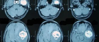
Many patients are prescribed CT and MRI. These tests help in diagnosing meningitis and its complications.
Diagnostics
Diagnosis of this group of diseases is based on analysis of the clinical picture, identification of symptoms of meningitis during a neurological examination (Kernig and Brudzinski symptoms, stiff neck, etc.), puncture and laboratory analysis of cerebrospinal fluid. In the cerebrospinal fluid during serous meningitis, an increase in the number of cells is detected (however, to a lesser extent than in bacterial purulent meningitis), mainly due to lymphocytes, a larger amount of protein is detected, and sometimes the composition of electrolytes and glucose changes. The cerebrospinal fluid pressure is significant.
With enteroviral serous meningitis, sometimes the pathogen can be identified in the cerebrospinal fluid. With tuberculous lesions, the presence of a large amount of fibrin and the presence of a fibrin film are determined in the cerebrospinal fluid.


