The human brain is covered with three membranes: hard, arachnoid and soft. Between the arachnoid and soft membranes there is a cavity - the subarachnoid space. It contains 130 ml of liquor. This space can expand under the influence of negative factors, for example, a traumatic brain injury. In this free space, a cyst can form - a benign neoplasm containing fluid inside. In other words, it is a “light” tumor with water inside. It is 4 times more common in men.
Due to the formation of a cyst in the subarachnoid space, the structure of the arachnoid membrane changes, which forms the clinical picture. Arachnoid changes of a liquorocystic nature are a collective term denoting a number of organic damage to the arachnoid membrane and the space under it.
In medicine, such a diagnosis faces several problems:
- The symptoms that appear as a result of the pathology are nonspecific, that is, they are characteristic of many other diseases.
- The diagnosis itself is controversial: there is no evidence to support the existence of the diagnosis itself.
It is generally accepted that inflammation of the arachnoid membrane develops as a result of the formation of an arachnoid cyst underneath it. However, there are problems here too: magnetic resonance imaging does not show inflammation of the membrane, but only the neoplasm itself. Whether a cerebrospinal fluid cyst causes inflammation of the membrane is also a controversial issue.
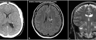
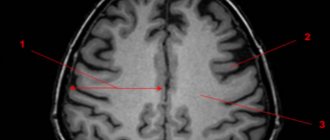
In addition, the diagnosis of arachnoid changes of a liquorocystic nature is made after examining the brain on an MRI, when the images, except for the expansion of the subarachnoid space, do not reveal anything.
Arachnoid cyst: nature of the pathology
An arachnoid cyst is a lesion of the brain in the form of a neoplasm in its membranes, filled with cerebrospinal fluid (medically defined as cerebrospinal fluid). Localization in the shell of the same name gave the name to the disease. The arachnoid membrane at the site of pathology bifurcates and cerebrospinal fluid accumulates between its walls. Basically, these are small-volume cysts, but there is a possibility of an increase in cerebrospinal fluid, which can lead to compression (pressure) on the area of the underlying cortex and the appearance of clinical symptoms.
The formation can be located in different areas of the arachnoid membrane:
- cerebellar angle;
- Sylvian fissure;
- suprassellar (on top) of the sella turcica.
According to medical forecasts, approximately 4% of humanity are carriers of arachnoid cysts, and the majority of them are men.
MR picture of arachnoid changes of a liquorocystic nature - what is it?
...cerebrospinal fluid cystic arachnoid changes are most often just an invention of radiologists...
Most often, the diagnosis of “arachnoid changes of a liquor cystic nature” is written when no cysts are reliably visible on MRI, but there is simply an uneven expansion of the subarachnoid space, that is, an increase in the amount of cerebrospinal fluid (CSF) between the brain and the skull bone. This expansion has nothing to do with arachnoid cysts, so liquorocystic arachnoid changes are most often just an invention of radiologists that do not have a reliable morphological basis. This term is absent in the scientific literature. You won’t find it on English-language sites, for example, on the world’s main radiology site, Radiopaedia.
Classification by etiology
For reasons of occurrence, the lesion may be:
- Primary (congenital) - as a result of brain abnormalities during formation and development.
- Secondary (acquired) - due to trauma, inflammation, bleeding of the meninges.
According to morphology, arachnoid cysts are divided into two categories:
- simple - brain membrane cells line the internal cavity of the cyst and produce cerebrospinal fluid;
- complex - the structural tissue of the cyst contains cells of other elements (for example, glial).
For practical neurology, the morphological type of pathology is unimportant, while the etiological origin requires mandatory consideration in the diagnostic process.
According to clinical manifestation, arachnoid cysts are classified into two types:
- progressive - with an increase in the volume of formation and severity of symptoms;
- frozen - with preservation of the latent state and size of the cyst.
Accurate determination of the type of cyst according to classification criteria is of utmost importance when prescribing therapeutic tactics.
Characteristics of the disease
A cerebrospinal fluid cyst is a formation in the brain tissue that is not a tumor, as confirmed by its morphological structure. It is a cavity isolated from other brain structures with liquid contents and dense walls. Clinically significant are large cystic formations that cause mechanical compression, compression, deformation of surrounding tissues - the brain matter, cerebrospinal fluid outflow tracts, elements of the circulatory system supplying the brain.
Arachnoid cysts are formations whose share, according to the results of intravital instrumental diagnostics, is equal to 1% of all volumetric processes occurring in the brain, which is less than that detected during autopsies (5 cases per 1000 patients). Normally, in adults and children, arachnoid cerebrospinal fluid cysts are absent in the brain tissue; if they are present, the degree of impact on health is determined by their size. Depending on the volume of the cystic cavity, the types of formations are distinguished:
- Small (volume does not exceed 30 ml).
- Medium (volume does not exceed 70 ml).
- Large (volume exceeds 70 ml).
Large formations are almost always associated with dislocation (displacement) of brain structures, which leads to neurological deficits. The suprassellar (growing deep into the skull) form is considered the most dangerous because it almost always causes occlusion or hydrocephalus. Treatment of hydrocephalic syndrome alone in this case is not enough due to significant compression of brain structures with the appearance of focal symptoms.
Causes of formation of cystic brain lesions
The causes of congenital pathology are caused by a violation of intrauterine brain formation, which is caused by:
- infectious diseases (herpes, toxoplasmosis, etc.);
- intoxication intrauterine exposure (against the background of professional characteristics, smoking, taking drugs, alcohol, teratogens, radiation);
- thermal effects (abuse of baths, saunas, solariums, hot baths, etc.);
- the presence of Morphan syndrome, hypogenesis.
Causes of acquired cyst of the arachnoid intracranial membrane:
- TBI (concussions, bruises);
- consequence of neurosurgical intervention;
- infections (meningococcal, meningoencephalitis, arachnoiditis, etc.);
- hemorrhages, meningeal hematomas.
An arachnoid cyst can also become a consequence of hyperformation of the spinal substance and its accumulation in the membrane.
Symptoms
Symptoms of an arachnoid cyst formed in the brain of newborns depend on the location, the degree of isolation from the spaces where the cerebrospinal fluid is located, and the distance from the cerebrospinal fluid circulation pathways. CSF cysts are often asymptomatic and are detected in childhood or young adulthood. Usually discovered accidentally during an instrumental diagnostic study prescribed for another reason.
Neurological symptoms appear as a result of the growth of a cystic formation, when a mass effect occurs - a tangible impact on nearby intracranial structures. Symptoms appear in 20% of patients with diagnosed pathology. More often, the manifestations of the disease are associated with hydrocephalic syndrome, which provokes the appearance of cerebral symptoms:
- Pain in the head area.
- Nausea, attacks of repeated vomiting.
- Ataxia, movement disorders.
- Hemiparesis, convulsive syndrome.
- Psycho-emotional disorders.
Less common are signs of focal damage to brain tissue, which is often associated with rupture of the cyst wall. To the main symptoms in infants, specific signs are added:
- Deformation of the skull bones.
- Dehiscence of cranial sutures.
- Protrusion of the fontanel.
- Lethargy, apathy, drowsiness.
- Lack of appetite.
- Signs of damage to the pyramidal tracts (pathological reflexes, paresis, paralysis).
- Delayed psycho-motor development.
A subarachnoid cyst formed in the brain in children occurs in 4 variants of the clinical picture. Depending on the characteristics of the manifestation of symptoms, the course of an arachnoid cyst that has arisen in the brain of a child can be:
- Lightning fast (2% of cases).
- Acute (6% of cases).
- Chronic (28% of cases).
- Remitting (2% of cases).
Symptoms may appear several weeks after birth or late in childhood and adulthood. The pathology is characterized by a pseudotumorous (resembling a tumor process) course and the absence of traces of inflammation in the meninges.
Symptoms of arachnoid cystic formations
Most often, the cyst is insignificant in volume and does not show clinical signs. Congenital pathology can be discovered completely by accident during diagnostic procedures (MRI, neurosonography) for other intracranial abnormalities. Initial symptoms may appear due to disorders of the blood vessels of the brain, trauma or infections.
The growing cerebrospinal fluid provokes an increase in the size of the cyst, and the symptoms become pronounced:
- increased pressure inside the skull;
- cephalalgia (headache);
- feeling of a “heavy” head (dizziness, noise and pounding (pulsation) in the ears and skull).
The growing volume of cystic formation provokes an increase in the intensity of permanent symptoms:
- nausea and vomiting;
- increased fundus pressure;
- bifurcation and spots of visual fields;
- hemiparesis (muscle weakness);
- dysarthia;
- numbness of the arms and/or legs;
- convulsions;
- fainting.
Sometimes the patient experiences hallucinations. Children experience mental retardation (mental retardation).
The consequence of a growing arachnoid cyst can be brain degeneration, rupture of the formation, and death.
Symptoms
If the cyst does not reach pathological sizes and does not obstruct the outflow of cerebrospinal fluid, there will be no symptoms. The clinical picture appears when the neoplasm retains cerebrospinal fluid in the subarachnoid space, which leads to increased intracranial pressure. By irritating the arachnoid membrane, the cyst also causes the clinical picture of meningeal syndrome.
General cerebral signs of arachnoid changes:
- Headache of a bursting and aching nature. Worse in the morning. In response to this, vomiting occurs, bringing relief to the patient.
- Dizziness and nausea.
- Autonomic disorders: severe sweating, hand tremors, alternating diarrhea and constipation, heart pain, difficulty urinating, feeling of lack of air, feeling of a lump in the throat, aching pain in the abdomen.
- Violation of general sensitivity: numbness, chills or a crawling sensation occurs in different parts of the body.
- Diplopia is double vision. Sometimes there are episodes of visual impairment.
- Unsteadiness of walking, lack of coordination of movements and their accuracy.
Symptoms of irritation and inflammation of the arachnoid membrane include:
- Stiff neck. In patients, the muscles of the back of the head contract, causing the patient lying on his back to throw his head back. It will not be possible to bring the head to a normal position: due to a strong spasm, the torso will follow the head.
- Lockjaw. What is it - a spasm of the masticatory muscles.
- Bickel's sign - the arms are constantly bent at the elbow joints.
- Blanket symptom: The patient will involuntarily hold the blanket if he tries to pull it off.
- Photophobia, irritation to faint smells or tastes.
- Hyperesthesia is increased sensitivity to tactile contact. Touching the patient's skin causes discomfort, even pain.
With arachnoiditis, motor and sensory disorders such as mono- and hemiparesis may occur. This means that muscle strength is weakened in one limb or on one side of the body (left arm and leg together). Sensory disturbances include hallucinations. Their content and modality are determined by the proximity of the inflammatory focus to a certain area of the cortex (occipital - visual hallucinations, temporal - auditory hallucinations).
If the cyst reaches a large size and begins to compress the cortex, the patient experiences focal epileptic seizures. In severe cases, minor seizures develop into a generalized epileptic attack, aggravated by status epilepticus.
Arachnoid changes in liquorocystosis are characteristic and can imitate an intervertebral hernia. In this case, pain occurs along the spine, which radiates to the buttocks, thighs or arms. In these same places, sensitivity is weakened and paresthesia occurs.
Diagnostics
An arachnoid cyst does not have a specific picture; its clinical picture is characteristic of many pathologies associated with brain formations of different etiologies. Diagnostic methods include:
- appointment with a neurologist;
- neurological diagnostics (rheo-, echo-, electroencephalogram);
- Magnetic resonance imaging;
- SCT (CT - computer scan).
MRI with contrast gives the most complete picture and helps to distinguish a cyst from tumor-like pathologies.
Why does dropsy of the brain occur?
In adult patients, acquired hydrocephalus is often encountered, which develops either due to any mechanical damage to the head, or as a result of the development of pathological processes. Why does cerebrospinal fluid accumulate outside the cerebral hemispheres? The explanation is simple: brain structures are disrupted, adhesions appear in the veins, arachnoid villi are destroyed, and as a result, the cerebrospinal fluid does not circulate as it should.
If we delve deeper into the question of the causes of such a disease as external dropsy of the brain, we can identify some factors:
- infectious diseases (tuberculosis, meningitis, encephalitis);
- post-stroke condition, development of sepsis, extensive hemorrhage;
- concussion, head or cervical spine injury;
- malignant tumors that develop in the stem region.
Frequent intoxication of the body leads to the appearance of external hydrocephalus. For example, alcohol abuse, which damages neurons and leads to tissue death. Those patients who suffer from metabolic disorders, diabetes mellitus, multiple sclerosis, encephalopathy, and atherosclerosis are also at risk. Another reason that deserves due attention is irreversible age-related changes that cause aging of blood vessels and brain tissue.
Main services of Dr. Zavalishin’s clinic:
- consultation with a neurosurgeon
- treatment of spinal hernia
- brain surgery
- spine surgery
Treatment
Therapeutic treatment of frozen cysts with no clinical signs is not carried out. The patient is registered with a neurologist and undergoes an MRI annually to monitor the dynamics. With a progressive course complicated by epileptic seizures, the arachnoid cyst is treated surgically:
- complete excision - for hemorrhages in the cyst area. Treatment with a high risk of injury and long-term rehabilitation;
- endoscopy - fenestration of the formation with fluid extraction;
- cystoperitoneal shunting - installation of a shunt to drain fluid into the abdominal cavity.
The choice of surgical intervention is decided by a neurologist and a neurosurgeon.
Classification of pathology
Arachnoid cysts are primary and secondary. In the first case, they appear as a congenital developmental anomaly. In the second - as a result of pathological processes affecting brain tissue. The walls of the secondary neoplasm are formed by collagen and scar tissue.
In 88% of cases, single cystic formations are observed, in 12% of cases – multiple ones. The location of multiple in 5% of cases covers both hemispheres. Arachnoid changes are classified depending on the location and nature of the growth of the cystic formation. The following forms are distinguished:
- With localization in the Sylvian (lateral) fissure (34% of cases). Congenital anomaly. Symptoms depend on the diameter of the cavity and the degree of dislocation (displacement) of nearby brain tissue. More often it manifests itself as a feeling of fullness in the head area, which is accompanied by pulsation and the occurrence of tinnitus while maintaining hearing function. Seizures and visual disturbances are common.
- Suprasellar (2% of cases). Congenital form. The cavity is most often localized in the area of the chiasm formed by the optic nerves. Manifested by dizziness, visual dysfunction, and impaired motor coordination.
- Localized in the area of the lateral ventricle (2% of cases). Congenital or acquired form. The clinical picture is represented by motor and visual disturbances, hearing impairment (sensorineural hearing loss), tinnitus, dysphagia (swallowing dysfunction).
Cerebellar (localized in the cerebellum) cyst formed in the brain occurs with a frequency of 32% of cases. It manifests itself as a violation of motor coordination, changes in muscle tone (hypotonia), nystagmus (usually horizontal). The patient experiences a change in gait, which becomes unstable and shaky.
An arachnoid cerebrospinal fluid cyst localized in the posterior cranial fossa is accompanied by oculomotor disorders (strabismus, loss of visual fields, paralysis of the optic nerves). A cystic formation in the area of the right or left frontal lobe is manifested by characteristic symptoms - deterioration of cognitive abilities, changes in gait, speech dysfunction (aphasia).
Consequences, prognosis, prevention
The patient can live his whole life with an arachnoid cyst without any symptoms. A dangerous condition occurs with progressive cystic lesions. Delayed detection of pathology can cause a person’s neurological disability and even death.
Timely diagnosis and correctly performed surgery provide a favorable prognosis for the future and recovery of the patient. Sometimes relapse and formation of new cysts are possible.
If you find yourself with the symptoms described above, then contact your doctor. Our specialists will prescribe the necessary diagnostic and treatment procedures.
Click the "SIGN UP" button and come see us.
Treatment methods
The doctor chooses treatment tactics for an arachnoid cyst formed in the brain individually, taking into account the nature of the disease and the severity of neurological symptoms. In some cases, symptomatic conservative therapy is carried out, in others surgery is indicated. Surgical methods for treating an arachnoid cyst formed in the brain include:
- Shunting. Artificial drainage of cyst contents using a drainage system.
- Endoscopic fenestration. Excision of part or all of the cyst along with the walls through a small incision in the skull bone or nasal passage.
- Drainage (needle aspiration).
Surgical intervention is performed by craniotomy (opening the skull) or endoscopically, through the introduction of traditional or valveless shunts to drain the contents of the cystic cavity. In the second case, the traumatic effect on brain structures is reduced. Craniotomy with complete excision of the walls of the cystic formation is performed if there is a volumetric effect on adjacent brain structures in the local area.
Bypass surgery involves implanting a shunt (an artificial vessel to drain cerebrospinal fluid) into the cavity of the cyst or into the ventricular system. Endoscopic operations are performed to form an anastomosis between the cystic cavity and the cisterns of the ventricular system. Among the complications, it is worth noting hemorrhages (4.5% of cases), infection, damage to vascular-neural tissues, obliteration (blockage) of ventricular catheters, which requires repeated surgical intervention. Indications for the operation:
- Increase in the size of the ventricles (according to the results of an MRI study).
- Brain edema of periventricular localization (according to neuroimaging results).
- Hydrocephalic syndrome (vomiting, intense pain in the head area, difficult to relieve with traditional painkillers, a significant increase in the diameter of the head and swelling of the fontanelle in infants).
- Increasing neurological deficit.
After surgery, more than 80% of patients experience regression of clinical symptoms. Contraindications to surgical treatment of a cerebrospinal fluid cyst formed in the brain include:
- The inflammatory process, regardless of location, occurs in the acute stage or partial remission.
- Severe anemia – low hemoglobin level.
- Severe functional state of the body (unstable hemodynamics, difficulty breathing, coma, exhaustion).
The main objectives of the operation: restoration of normal circulation of cerebrospinal fluid, reduction of the diameter of the cystic cavity, reduction of intracranial pressure.
Treatment regimen for illness
The main condition for good health in the presence of an arachnoid cyst of the brain (operated or non-operated) is compliance with the treatment regimen. The patient must adhere to the daily routine and especially the work and rest regime, abstain from alcohol, strong coffee, energy drinks and drugs that increase the tone of the nervous system.
Taking any medications that affect the nervous system and vascular tone should be agreed with a neurologist. Extreme and dangerous sports, driving, and activities related to working at heights or near moving machinery are not recommended.