Diagnostics
Questioning the patient, examination, and palpation allow us to determine a list of reasons that can cause pain. To accurately determine the etiology of cervicalgia syndrome, additional examinations are necessary:
- general blood test; blood biochemistry;
- sputum examination;
- Ultrasound of soft tissues of the neck;
- X-ray of the cervical spine;
- Ultrasound of the thyroid gland;
- electromyography;
- Ultrasound scanning of the cervical arteries;
- MRI (CT) angiography of neck vessels;
- MRI (CT) of the cervical spine, with contrast;
- MRI of soft tissues of the neck, with contrast.
The list of examinations is determined individually.
You need to see a doctor as soon as possible. Do not make sudden movements, avoid hypothermia, exercise, and make an appointment with a doctor as soon as possible. This will avoid complications. Consultation with specialists is required: surgeon, vertebrologist, endocrinologist, neurosurgeon, otolaryngologist, dermatologist, oncologist.
Causes of lumbodynia
Chronic lower back pain (lumbodynia) is most often caused by degenerative-dystrophic changes in the lumbar spine. This is osteochondrosis of the spine and its manifestations and consequences such as protrusion, intervertebral disc herniation, spondyloarthrosis (arthrosis of the facet joints of the vertebrae), and deforming spondylosis. If it is known that the cause of pain is changes in the intervertebral disc (herniation or protrusion), then the wording “Discogenic lumbodynia” can be used.
Treatment
The choice of therapy depends on the causes of cervicalgia.
It is imperative to treat the underlying disease. To relieve pain in degenerative processes of the cervical spine, non-steroidal anti-inflammatory drugs, muscle relaxants, opioid analgesics, blockades with anesthetics and a corticosteroid drug, and vascular drugs are used. For infectious and purulent processes, antibiotics are prescribed. In case of pathology associated with damage to nerve endings, B vitamins, thioctic acid, vascular drugs, and drugs that improve nerve conduction are added to therapy.
In case of injuries or pathological muscle tension, immobilization of the neck using a Shants collar is necessary. In the absence of contraindications, physiotherapy and balneotherapy are used.
For cervicalgia of vertebrogenic origin, acupuncture, massage, exercise therapy, manual therapy, and hardware traction of the cervical spine are relevant.
If the cause of pain in the cervical spine is degenerative changes in the spine (herniated discs), cysts, abscesses, tumors, surgical treatment may be indicated. Surgery is also necessary for patients who do not have a positive result from conservative therapy.
Treatment of vertebrogenic cervicalgia
Cervicalgia, the treatment of which you can receive at Belozerova’s hardware therapy clinic “M-Clinic”, requires modern and high-quality therapy using physiotherapeutic procedures.
Cervicalgia is treated with an integrated approach, which includes the prescription of painkillers, muscle relaxants, unloading of the cervical spine (Schanz collar), therapeutic exercises, as well as physiotherapy.
Chronic cervicalgia responds well to physiotherapeutic procedures carried out by a specialist, which creates the basis for the successful resolution of this disease.
Cervicalgia, which can be treated in our clinic, must be treated in order to avoid complications.
The mechanism of occurrence of lumbodynia
The mechanism of occurrence of both lumbago and lumbodynia is due to irritation of the sinuvertebral nerve. But lumbago, as a rule, occurs as a result of infringement of part of the nucleus pulposus in the cracks of the fibrous ring of the damaged spinal disc. And lumbodynia can also occur with less pronounced changes in the disc. It is also often found in patients with spondylosis of the lumbar spine, spondyloarthrosis, and spondylolisthesis. Chronic lumbodynia syndrome can also occur with anomalies in the development of the lumbar spine. These are “spina bifida”, “anomaly of tropism” and other congenital defects.
Symptoms
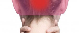
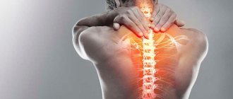
As a rule, neck pain is dull in nature. Sometimes the pain gets worse when you move your neck. Neck pain may be accompanied by numbness, tingling, sometimes the pain can be acute, there may be difficulty swallowing, enlarged lymph nodes, and dizziness.
Cervicalgia may be associated with facial headache, shoulder pain, or numbness or tingling in the shoulder (upper limb paresthesia). Such associated symptoms often result from root compression. For example, compression of a root with sensory fibers providing innervation to the occipital region can lead to neck pain radiating to the back of the head. Depending on the disease, in some cases, neck pain may be accompanied by pain in the upper back or lower back, for example with ankylosing spondylitis, when the inflammatory process covers the entire spine.
Lumbodynia - the first signs of osteochondrosis
Lumbodynia syndrome (low back pain) is often the first clinical sign of osteochondrosis of the lumbar spine. Lumbar osteochondrosis in almost half of the cases at the beginning of its course manifests itself as subacute or chronic lumbodynia.
What causes lumbodynia
Lower back pain can occur after a single severe physical fatigue, lower back injury, hypothermia, or stress. In some cases, days, weeks or even months pass before pain appears after these actions.
At first, very mild lumbodynia may appear. Some patients note that their pain developed slowly and for no apparent reason. But this does not mean at all that there was no provoking factor. It’s just that this factor is not always obvious.
Then the lower back pain may intensify. Most often this happens gradually. But the pain usually does not reach the same degree of severity as lumbar pain - lumbago (acute lumbodynia). Patients walk independently and do certain work, although it is difficult for them to bend, but even more difficult for them to straighten up. At the same time, they often place their hand on the lower back, rubbing or massaging it a little. And only after that they can straighten up. Such people try to sit with a straight back.
What is chronic vertebrogenic lumbodynia?
Lumbodynia against the background of spinal osteochondrosis in most cases is aching in nature and is more disturbing in the morning. It decreases somewhat or goes away during work. Especially if this work involves movement. When lying down, the pain subsides. This is due to a decrease in the load on the lumbar intervertebral discs. While in bed, patients try to choose the most comfortable position. They usually lie with their legs bent on their healthy or sore side.
Unlike lumbar pain (lumbago), chronic low back pain can be localized on only one side - right-sided lumbodynia or left-sided lumbodynia. It most often worries in the lower lumbar region. The pain may radiate to one or both buttocks.
What is lumboischialgia
If pain in the lower back is combined with “pulling” pain in the leg, or in other words, lumbodynia is combined with sciatica, then this is called lumbar sciatica. This is described in detail on the corresponding page.
Lumbodynia with muscular-tonic syndrome.
In some cases, the pain intensifies not only during movement, but also when talking, sneezing, straining, or bending the head forward. Moderate curvature of the spine is often detected. This is due to a stronger reflex tension of the lower back muscles on one side. In such cases, the diagnosis can be found in the following formulation: “Vertebrogenic lumbodynia with muscular-tonic syndrome,” or “lumbodynia with muscular-tonic syndrome, antalgic scoliosis.”
Pressing on the spinous processes of the lumbar vertebrae and points near the vertebrae at this level is usually painful. Patients have difficulty bending forward, but at the same time, bending the body to the sides may be less limited.
Vertebrogenic lumbodynia syndrome, as the main external manifestation of the chronic course of osteochondrosis of the lumbar spine, is more often observed in men. The duration can be from several weeks or months to 5-7 years or more.
Vertebrogenic thoracalgia - symptoms and treatment
The determining factor in examining a patient is to determine the source of pathological pain impulses, which is important when carrying out differential diagnosis and prescribing pathogenetic treatment for VT.
Differential diagnosis , as a rule, is carried out with diseases of the lungs, heart and gastrointestinal tract.[6]
The study of functional disorders of the musculoskeletal system in thoracalgia should be carried out using neurological, neuroorthopedic and manual techniques, since the use of only clinical examination greatly facilitates diagnosis, reducing the range of possible diagnoses due to the diversity of symptom complexes of the disease.[9]
Neuroorthopedic examination is a complex of techniques:
- curvimetric diagnostics;
- angular surveys;
- myotonometric examinations;
- tensoalgimetric examinations.
To interpret the data of a comprehensive neuro-orthopedic examination, it is necessary to convert them into comparable units, which is achieved by comparing each studied parameter with the corresponding norm, and their integral indicator reflects the severity of the disease and can be used as a criterion for assessing the effectiveness of treatment.
During manual testing:
- the nature, severity and localization of functional changes in the musculoskeletal system are established;
- pathologically tense or relaxed muscles, active and latent trigger points are identified;
- the degree of limitation of movements and their pain in three mutually perpendicular planes - sagittal, frontal and horizontal - is assessed;
- the symmetry of bilateral structures is assessed.
Then the identified biomechanical disorders need to be clarified. To do this, palpation, study of active and passive movements, isometric muscle tension, diagnosis of relaxed and shortened muscle groups, as well as joint play are carried out.
X-ray examination plays a leading role in the diagnosis of VT and allows:
- establish the level and degree of dystrophic damage to the spine;
- carry out differential diagnosis with other diseases of the spine;
- identify anomalies and individual characteristics of the musculoskeletal system.
Functional spondylography , carried out at maximum flexion and extension, allows us to identify the stability of the SMS, the degree of displacement of the vertebrae in relation to each other, and the condition of the ligamentous apparatus.
The most informative neuroimaging methods for diagnosing VT are computed tomography and magnetic resonance imaging (CT and MRI):
- CT scan determines the severity and nature of damage to the spinal column and spinal cord, allows you to identify the presence of a tumor or injury, determine the presence of protrusion and prolapse of discs, their size and the diameter of the spinal canal.
- MRI provides a more contrasting image of soft tissue formations, allows you to determine the presence and degree of spinal canal stenosis and sequestration (rejection of the necrotic area), changes in the ligamentum flavum, the condition of the intervertebral joints and discs, as well as the spinal cord. The advantage of the method is the absence of radiation exposure.
The use of CT and MRI allows adequate planning of treatment tactics and determination of indications for neurosurgical treatment.[7]
The functional state of the segmental reflex apparatus and peripheral nerves is determined using electroneuromyography . Stimulation electroneuromyography contributes to a qualitative assessment of the speed of nerve impulses along the motor and sensory fibers of peripheral nerves, which is very important in topical diagnostics for compression-neural syndromes.
When differentially diagnosing VT with somatic diseases and assessing the adaptive capabilities of the patient’s body, the following are used:
- bicycle ergometry (study of the cardiovascular system under increasing load);
- phonocardiography (diagnosis of heart sounds and murmurs);
- Holter monitoring (recording of cardiac signals);
- ECG (study of the electrical activity of the heart);
- spirography (measurement of breathing volume and speed);
- fibrogastroscopy (examination of the gastrointestinal tract);
- sonography (ultrasound);
- X-ray of the chest organs.