Article for the “bio/mol/text” competition: Prion diseases are a phenomenon discovered in the twentieth century, and in it they began to play a big role: an increase in life expectancy in developed countries has led to the fact that more and more people began to live to see “their Alzheimer’s” "or "your Parkinson's." The nature of neurodegenerative diseases continues to remain vague, and scientists are still studying only certain aspects of them - for example, the reason for their development in old age or the ability to be transmitted from one species of living beings to another.
“Bio/mol/text”-2012
This article was submitted to the competition of popular science works “bio/mol/text”-2012 in the category “Best News Message”.
The competition is sponsored by the visionary Thermo Fisher Scientific.
It all started when in the 20th century scientists became interested in the nature of unusual diseases in humans and animals: kuru, Creutzfeldt-Jakob, scrapie. The noticeable similarity in the pathology of these diseases gave rise to the hypothesis of their infectivity, which was subsequently confirmed experimentally. Then the question arose about the causative agent of these diseases. Before the answer was found, the extraordinary properties of the pathogens were revealed: they do not reproduce on artificial nutrient media, are resistant to high temperature, formaldehyde, various types of radiation, and the action of nucleases. Purification of the infectious material and its study made it possible to proclaim that “everything is to blame” for the protein, which 30 years ago received the name prion (from the English prion - protein infection).
Thus, famous American scientists - virologist and doctor D.K. Gaidushek, who discovered the infectious nature of prion diseases, in 1976, and biochemist S.B. Prusiner, who identified prions and developed the prion theory in 1997, were awarded Nobel Prizes. Their work became the impetus for subsequent research, thanks to which new types of prion infections were studied. But, even despite the undying interest in the “prion topic,” the formation of prions remains a mystery to this day.
A method for prolonging the life of patients with prion diseases
The invention relates to veterinary medicine, namely to the treatment of animals suffering from an infectious disease such as scrapie, which is a prion disease. To do this, the animal is injected with birch bark extract. The introduction of the extract helps delay weight loss and prolong the life of the animal. 7 tab., 5 ill.
The invention relates to the field of medicine and veterinary medicine and more specifically it relates to the search for means that could be used to at least increase the life expectancy of people and animals infected with prions.
Prions (short for the English words “protein infections” - infectious protein) are a new class of infectious pathogens, the discovery and study of which occurred and is carried out only in the last 50 years, although diseases now classified as prion diseases have been known in animals for more than 200 years, and in people - since the beginning of the 20th century.
Prions differ from all other infectious pathogens (viruses, bacteria, etc.) in that they lack any genome, that is, DNA or RNA. It is assumed that it is precisely such infectious agents that cause a group of incurable degenerative diseases of the central nervous system of humans and animals. The most famous of them in humans are Creutzfeldt-Jakob disease (CJD) and Hertsmann-Straussler-Scheinker syndrome (HSS), and in animals - sheep scrapie and bovine spongiform encephalopathy, sometimes called sheep scabies and mad cow disease in the media. or spongiform encephalitis. These diseases, regardless of whether they occur in humans or animals, are accompanied by morphological changes in brain tissue and the appearance of amyloid-like plaques containing rod-shaped protein aggregates. A unique feature of these diseases is that they arise not only as a result of infection: sporadic and hereditary CJD and HSS are known, and regardless of its origin, the disease can be transmitted further by infectious means.
According to the World Health Organization (WHO) and the International Office of Epizootics, about 200 thousand cases of bovine spongiform encephalopathy were registered between 1986 and 2000, and 85 cases of Creutzfeldt-Jakob disease were identified in young people.
Despite large-scale studies of prions, awarded two Nobel Prizes (K. Gaidushek - 1976 and S. Prusiner - 1997), many aspects of the emergence and development of prion infections are still unclear (extension of the interspecies barrier by the pathogen, pathogenic picture of the disease, etc. .). There is also no information about means of preventing and treating prion diseases.
The authors of the present invention unexpectedly discovered the ability to prolong the life of animals infected with scrapie in such a natural product as the extract of the upper layer of birch bark - birch bark, which allows us to consider it as a promising remedy for the prevention of prion infections and for the treatment of people and animals suffering from prion diseases.
The studies were carried out on model animals - Syrian hamsters, which were infected with scrapie. We used birch bark extract produced by Berezovy Mir LLC (Moscow). The main component of the extract (over 70%) is betulin.
Materials and research methods
As a laboratory model, we used 25 Syrian hamsters, 3 weeks old, of both sexes, weighing 25-30 g.
Each experimental animal was identified by color and/or dye, brilliant green. The animals were divided into 3 groups: two (1st and 2nd) - control, one (3rd) - experimental.
Group 1 (control of drug safety, 5 individuals). The animals were divided into 2 cages: group 1a - 3 hamsters (received the test drug) and group 16-2 hamsters (did not receive the drug). The animals were kept in another room separately from hamsters of other groups.
Group 2 (control of experimental infection, 10 individuals). The animals were kept in 2 cages with 5 hamsters in each. All animals are females.
3rd group - experienced (10 individuals)
All animals in this group received the test drug according to the selected treatment and prophylactic regimen. The animals were kept in 2 cages with 5 hamsters in each. One cage - 5 females, the other - 5 males.
Strain 263 K, stored at -20°C, was used as the scrapie pathogen. The pathogen underwent 2 passages on Syrian hamsters. Infectious titer - 6.5 Ig LD50/ml;
The experimental design included:
1) Administration of the test drug for prophylactic purposes.
2) Experimental infection of control and experimental animals with the scrapie pathogen.
3) Administration of the test drug for therapeutic purposes.
4) Intravital and postmortem studies of animals in the experimental and control groups.
The test drug was diluted in liquid starch and administered to hamsters orally (via a tube) at a rate of 100 μg/g of animal weight according to the following treatment and prophylactic regimen: 5 times before infection with the scrapie pathogen, 5 times 35 days after infection and 5 times in the terminal stage of the disease .
The drug was administered twice to 3 hamsters of group 1a (control of the harmlessness of the drug) and 10 hamsters of group 3. Animals of group 1a (2 hamsters) and group 3 (5 hamsters who survived), who were in the terminal stage of the disease, were additionally administered the drug (5 once every 5 days). At the same time, 2 hamsters (males) died after 2 administrations of the drug, 3 hamsters (females) received the drug in full.
Experimental infection of control and experimental animals.
Preparation of infectious material.
A 3 g sample was taken sterilely from the center of the brain of a hamster with experimental scrapie infection, ground in a sterile mortar and diluted in 2.7 ml of Hanks' solution with antibiotics (100 units of streptomycin). The material was transferred into a sterile vial and centrifuged in a benchtop angle centrifuge at 2000 rpm for 15 min. From the supernatant liquid collected after centrifugation, 0.3 ml of solution was taken and diluted in 2.7 ml of Hanks solution (10-2 dilution) and used directly for infection.
Syrian hamsters (groups 2 and 3) were infected with a prepared ex tempore suspension of hamster brain with experimental scrapie infection at a 10-2 dilution in Hanks solution in a volume of 0.03 ml per animal (approximately 100 lethal doses). Animals were infected intracerebrally by injection into the right hemisphere of the cerebrum under light ether anesthesia. Control animals (group 1) were injected with 0.03 ml of a suspension of the brain of intact animals (10-2 dilution), prepared as described above.
All work with laboratory animals was carried out in accordance with WHO recommendations.
Duration of the experiment: 4.5 months (August 9-December 24, 2004).
Intravital studies of animals.
This included monitoring the behavior, appearance and weight of the animals: Weighing the hamsters was carried out using electronic scales VE-15I (JSC "Massa", St. Petersburg, Russia), in the morning, before feeding, with a frequency of 3-4 days (2 once a week).
Experimental animals were assessed for the following characteristics:
1. Nonspecific signs:
- condition of the skin (hair, shine, dullness, presence and nature of hairless areas);
— condition of mucous membranes (eyes, oral cavity, integrity, presence of erosions, ulcers, scars, hemorrhages);
- mobility;
- appetite.
2. Specific signs:
Initial stage:
change in behavior (inadequate reaction to external stimuli (light, sound, touch, pain)
- excitation or inhibition of reactions. Stage of development of neurological symptoms of the disease:
— violation of motor functions (impaired coordination, tremor, gait disturbance, weakness in the limbs, paresis);
- sudden weight loss (cachexia).
Terminal stage (irreversible changes):
- immobility;
- paralysis of the limbs and respiratory muscles.
Post-mortem studies of animals.
1) Histological studies.
From experimental animals in the last phase of the terminal stage of the disease, under deep ether anesthesia, the brain and spleen were sterilely removed and placed either in a 4% solution of neutral formaldehyde (for histological studies) or in a freezer at -20°C.
Histological preparations were prepared according to standard methods. Pieces of tissue were isolated from the brain from its various sections: the cerebral cortex, subcortical nuclear groups, cerebellum, medulla oblongata and spinal cord. Paraffin preparations were prepared, sections were prepared in a standard manner and stained with hematoxylin and eosin and Congo red.
The results were analyzed in a Biostar light microscope (USA) at a magnification of 200-600 times.
2) Immunoblotting (IB). Immunological detection of the scrapie pathogen in the central nervous system of experimental animals was carried out using the Prionics-Check Western test system (Phonics AG, Switzerland). The positive antigen of the test system served as a positive control. Monoclonal antibodies to the PrP protein were used as primary monoclonal antibodies: mouse anti-PrP IgG1 (working dilution 1:5000), Am 308 (analog of commercial antibodies Am 3F4, titer 1:5000) and Am 6H4 (titer 1) included in the test system :5000). Work with the test system was carried out according to the manufacturer’s instructions.
Statistical processing of results.
During statistical processing of experimental data, arithmetic mean values and their confidence intervals were calculated and the reliability of differences in numerical results was assessed using the Student's t-test. The difference was considered statistically significant at p<0.05.
Intravital animal studies
Clinical picture of the disease in experimental (groups 2 and 3) and control (group 1) animals with experimental scrapie.
The development of experimental scrapie was studied in 20 Syrian hamsters (groups 2 and 3).
Daily observation of experimental animals did not reveal any changes in the coat of experimental and control animals over a period of 2-2.5 months. At 10-12 weeks of observation, the experimental animals showed some lethargy, ruffled fur and a decrease in motor activity, accompanied by loss of appetite. At week 16, experimentally infected hamsters exhibited hair loss and severe head tremors. During this period, the experimental animals differed significantly in development from the control ones: body weight decreased by 20-40%, weakness of the hind limbs and paresis gradually progressed. When moving, the animals tipped over onto their backs and had difficulty returning to their previous body position. A slow reaction to stimuli was noted. The duration of the incubation period was 3-3.5 months, the duration of the disease was 0.5-1.5 months. The main symptoms of the development of experimental scrapie were: hair loss, muscle atrophy, tremors, development of paresis of the limbs and trunk. At the same time, the mortality rate among infected hamsters by the end of the observation period was 95% (19 animals out of 20 under observation died). As a rule, the development of neurological symptoms was preceded by: weight loss (up to 40% of the maximum). There was no death observed among control animals (group 1).
Summarized data on the development of scrapie in groups 2 and 3 are presented in Table 1.
As can be seen from Table 1, the administration of the drug does not affect the incubation period and the duration of development of specific scrapie symptoms in Syrian hamsters (p>0.05). There was a difference between males and females in sensitivity to scrapie. Males died faster, so same-sex animals—females—were taken as the main criterion when comparing the performance of the control and experimental groups of animals.
Calculations of the reliability of the results obtained are presented in Tables 2-5.
Study of the body weight dynamics of Syrian hamsters with experimental scrapie.
Primary data on the dynamics of body weight of Syrian hamsters of groups 1, 2 and 3 are presented in Table 6.
Graphic changes in body weight (group average) in hamsters that did not receive (group 2) and received the test drug (group 3), in comparison with the body weight of control hamsters are shown in Fig. 1. A comparison of the curves of the dynamics of body weight loss in females and males of groups 2 and 3 is presented in Figs. 2, 3 and 4.
It can be seen that the use of the test drug according to the treatment and prophylactic regimen in hamsters of group 3 led to a delay in body weight loss by more than 2 weeks, compared with hamsters of group 2 (October 22 and October 8, respectively).
Administration of the drug for the third time to 5 hamsters of group 3, who were in the last phase of the terminal stage of development of the disease, led to a slowdown in the decline in body weight and the timing of death of 3 female experimental animals, compared with analogues in group 2.
Survival of Syrian hamsters with experimental scrapie. Summarized data on the lifespan of Syrian hamsters with experimental scrapie are presented in Table 7.
As can be seen from Table 7, the life span of hamsters of groups 2 and 3 varies widely, the average indicators are almost identical (p>0.05), if you do not take into account the sex of the animals.
However, a difference was noted in the life span of female Syrian hamsters of the 2nd and 3rd groups. Females of group 2 died on average a week earlier than females of group 3 who received the test drug. It should be emphasized that one animal (female, gray color) that received the drug continued to live for another 3 weeks after the end of the experiment.
Male Syrian hamsters are more sensitive to scrapie than females. They died on average a week earlier than infected females.
Post-mortem animal studies
Histological studies of the brain of Syrian hamsters with experimental scrapie.
For the study, we took the brains of animals in the last phase of the terminal stage of the disease. One animal from each group was studied.
In animals in the terminal stage of the disease, specific changes characteristic of scrapie infection were noted: spongioform changes in the cortex and hippocampus, neuronal death, diffuse proliferation of astroglia, subependymal gliosis, amyloidosis of the vascular walls, and others. The formation of amyloid plaques directly in the tissue of the central nervous system was noted.
Detection of PrPSc in the CNS of Syrian hamsters with experimental scrapie by immunoblotting.
PrPSc differs from normal PrPc in its resistance to protease K: after treatment with protease, the normal form of PrP is destroyed, while the molecular weight of PrPSc decreases from its original size of 32-35 kD to a smaller size of 27-30 kD. The remaining protease-resistant PrPSc fragment is detected by immunoblotting as PrP27-30 (Fig. 5).
Immunological detection of the scrapie pathogen in brain samples of experimental animals was carried out as described above.
Monochannel antibodies 308 (3F4 analogue), titer 1:5000, were used as primary antibodies.
The presented research results allow us to conclude that the tested drug (birch bark extract) is harmless and does not have a pathological effect on the body of hamsters. All animals of the 1st control group were clinically healthy throughout the entire observation period.
When Syrian hamsters are infected intracerebrally, scrapie disease develops specifically. The average incubation period in animals in group 2 was 73.3±6.21 days, in group 3 - 75.7±8.10 days (males - 72.8±2.86 days, females - 78.6± 10.31 days). The duration of the disease development until the death of animals in groups 2 and 3 was 22.1±4.68 and 19.5±9.63 (males - 15.4±1.36 and females - 23.6±12.24) days, respectively .
The main symptoms of the development of experimental scrapie were: weight loss, hair loss, muscle atrophy, tremors, and the development of paresis of the limbs. Weight loss in animals was observed 1-3 weeks before the development of clinical manifestations of the disease. Mortality among infected hamsters by the end of the observation period was 95% (19 animals died out of 20 infected).
Postmortem studies of animals using the histological method and identification of the pathogen in immunoblotting confirmed the effectiveness of experimental infection of animals of the 2nd and 3rd groups. Histological studies have confirmed the presence of specific morphological markers of scrapie development in the central nervous system of Syrian hamsters. In the cortical layer of the brain, neuronal death, spongiosis, astroglial proliferation and astrogliosis, and amyloidosis were noted. The prion protein PrPSc was detected in the CNS of Syrian hamsters with experimental scrapie in groups 2 and 3.
The use of the test drug for therapeutic and prophylactic purposes showed the following (comparison of females of the 2nd group with females of the 3rd group).
A 2-week delay in body weight loss was noted in animals treated with the test drug compared to hamsters that did not receive the drug. Moreover, females lost weight 3 weeks later than control counterparts, and males - 1 week.
It was found that the life expectancy of females of group 3 who received the test drug was on average 1 week longer than that of the analogs of group 2. Moreover, after the third administration of the test drug in the terminal stage of the disease, one female continued to live for another 21 days.
Postmortem changes were similar in animals of both groups.
Based on the results of the experiments, we can draw a general conclusion that in the acute experiment the effect of the test drug on the body of experimentally infected hamsters was noted. The effect of the intervention was to delay the onset of body weight loss in experimental animals and increase their life expectancy.
A method of prolonging the life of an animal infected with scrapie by injecting it with birch bark extract.
Biological essence of prions
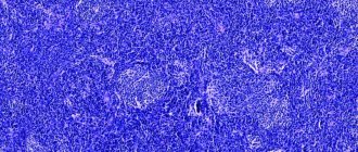
Figure 1. The metaphor for neurodegenerative brain damage is that neural tissue becomes a sponge as a result of massive neuronal death.
The prion molecule is not something exotic: in a “normal” form it is present on the surface of the nerves of each of us. At the same time, we feel great, and our nerve cells are alive and healthy. However, this is all until our normal protein “degenerates” into an abnormal form. And if this happens, it will lead to terrifying consequences: the infectious form of prions has the ability to “stick together” with other molecules and, moreover, “convert” them into the same form, causing a “molecular epidemic”. As a result of this polymerization, toxic protein plaques appear on the nerve cell, and it dies [1]. In place of the dead cell, a void is formed - a vacuole filled with liquid. Over time, one neuron after another will disappear, and more and more “holes” will form in the brain, until finally the brain turns into a sponge (Fig. 1), which will inevitably be followed by death.
There is a simplified idea that polymerized prion fibrils “puncture” the neuron, causing its death. In fact, this is not entirely true: spherical prion aggregates preceding the fibrillar stage are also toxic (at least for Alzheimer's disease): “Alzheimer's neurotoxin: not only fibrils are poisonous.” - Ed.
But how can a normal natural protein (denoted PrPC) suddenly become pathological (PrPSc; Sc - from the word “scrapie”)? What is going to happen? As in the case of a “normal” infection, such transformation requires an encounter with an infectious prion molecule. There are two ways of transmitting this molecule: hereditary (due to mutations in the gene encoding the protein) and infectious. That is, the introduction of a prion can occur unexpectedly - for example, when eating insufficiently well-fried or cooked meat (which should contain the PrPSc form), during a blood transfusion, during organ and tissue transplantation, or when administering pituitary hormones of animal origin.
And then an amazing event occurs: normal protein molecules, in contact with pathological ones, themselves transform into them, changing their spatial structure (the mechanism of transformation remains a mystery to this day) [1]. Thus, the prion, as a real infectious agent, infects normal molecules, triggering a chain reaction that is destructive to the cell.
Individual nosological units
Human prion diseases are species specific. Basically, they spread only among individuals of one species. But scientists have demonstrated interspecies transmission (overcoming the interspecies barrier): this occurred in scrapie sheep with its transmission to cows (mad cow disease). Transmission of the disease from animals to humans, despite many suspicions, has not been demonstrated.
A number of diseases are of hereditary origin, some are infectious. Sometimes the disease occurs unexpectedly, without the possibility of prevention.
Kuru
The disease existed only in Papua New Guinea in tribes of cannibals who consumed the brains of the dead. It manifests itself as tremor (“kuru” means “tremor”), speech disturbances, and balance disorders (ataxia). Death occurs a few months later. After stopping cannibalism, the kuru disease disappeared.
Creutzfeldt-Jakob disease
It is a rare disorder that affects older people (most often 50-70 year olds). Its course is relatively rapid, death occurs within a few months (up to a year) from the onset of the first symptoms. Neurons quickly die, and progressive dementia develops, which has no known cure. There is some hereditary predisposition to this disease:
- Sporadic form . The incidence is 1-2/1000000. Problems begin around age 65. The disorder occurs in the form of acute progression of dementia (over 2-3 months), ataxia, and myoclonus. There are no ways to prolong the life of neurons with this prion disease; the patient dies 5-12 months after the appearance of the first symptoms.
- Iatrogenic form . It occurred in patients receiving human growth hormone from cadaveric pituitary glands (today it is prepared recombinantly), transplantation of hard membranes, pericardium, and cornea. There is a risk of neurosurgical transmission. Infection is also possible through transfusion.
- Family form . This is a genetic form with a mutation in the PRNP gene and neuropsychiatric symptoms.
- New option . Characterized by psychiatric symptoms (anxiety, depression, behavioral changes), progressive cerebellar syndrome, myoclonus, and other neurological symptoms. Unlike the sporadic form, the new variant is characterized by a slow progression of viral infection; prion disease affects younger age groups. Transmission occurs through nutritional means (through consumption of meat from affected animals). The incubation period is more than 10 years. Around 200 people have died worldwide to date.
- The childhood version of Creutzfeldt-Jakob disease is Alper disease . Has the same manifestations as the disease in adults; in addition accompanied by hepatic steatosis.
Gerstmann-Sträussler-Scheinker disease
Gerstmann-Strüssler-Scheinker disease is a very rare autosomal dominant hereditary disease that begins in the 3rd-4th decade of life. It manifests itself as a slow slowdown in the functioning of neurons by prions, and progressive dysfunction of the cerebellum and spinal cord. Signs:
- ataxia;
- pyramidal symptoms;
- dysarthria;
- dysphagia;
- gradually developing dementia.
Article on the topic: Exercises for flat feet in children - a set of workouts at home with video
At the beginning of the disorder, apathy and depression may occur. The average duration of the disease is about 4-5 years. Several PRNP mutations associated with this disorder have been described.
Fatal familiar insomnia
This is a very rare disease that leads to degeneration of the thalamic nucleus. Main symptoms:
- progressive insomnia;
- autonomic and endocrine dysfunction;
- dysarthria;
- ataxia;
- myoclonus;
- pyramidal symptoms.
Brain dystrophy due to exposure to the prion virus
Cognitive impairment develops, confusion and hallucinations appear. In the terminal stage, complete insomnia develops. Patients die within 3 years from the onset of the first signs. The disease is associated with the PRNP D178N-129M mutation. There is also a sporadic form of the disorder.
Some information about prions
The researchers note:
- The prion protein includes 254 amino acid residues and weighs 33–35 kilodaltons [2];
- the gene encoding the PrP protein is found in humans, mammals, and birds [1];
- To completely destroy the prion protein, a temperature of at least 1000 degrees is required [1]!
- it is possible that prions take part in intercellular recognition and cellular activation [3];
- it is possible that the function of prions is to suppress age-related processes [3];
- with the development of clinical manifestations of prion diseases there are no signs of inflammation or changes in the blood;
- it is assumed that prions are involved in the development of schizophrenia and myopathy;
- The mechanism of action of prions and their transformation from a normal form to a pathological one remains unclear.
2. Reasons
Fatal familial insomnia is an inherited prion disease. Only at the turn of two centuries was it possible to establish the direct cause, which lies in the mutation of one of the genes (replacement of asparagine with an acid derived from it), as a result of which the body begins to produce a prion-type protein. Accumulating in the thalamus, prions cause avalanche-like degeneration and degeneration of functional tissue into amyloid plaques, which, in turn, grossly disrupt the conduction of neuronal impulses. The circadian rhythm (alternation of sleep and wakefulness) is disorganized, and then a number of other functions and indicators, in particular, blood pressure, the functioning of the endocrine glands, thermoregulation, etc.
Visit our Therapy page
Conditions for the occurrence of diseases
The conditions for the occurrence of prion diseases are unique. They can form according to three scenarios: as infectious, sporadic and hereditary lesions. In the latter option, genetic predisposition plays a major role [2].
Renowned prion researcher Stanley Prusiner identifies two striking features of neurodegenerative diseases such as Creutzfeldt-Jakob disease, Alzheimer's disease, and Parkinson's disease. The first is that more than 80% of cases of the disease are sporadic (that is, random, occurring “by themselves”). Second: despite the fact that a large number of mutant proteins specific to a particular disease are expressed during embryonic development, the patterns of inheritance of these neurodegenerative diseases appear later. This suggests that certain processes occur during aging that release disease-causing proteins [5]. More than 20 years ago, the author argued that this process involves the random refolding of a protein into an incorrectly folded one, which corresponds to the transition to an infectious state - a prion.
Interesting facts about Alzheimer's disease: Chronic sleep deprivation may increase the risk of Alzheimer's disease ("New step in understanding Alzheimer's disease: sleep deprivation may be a risk factor"), and Alzheimer's neuropeptide itself (amyloid-β Aβ) may be part of the innate immune system ( "Perhaps Alzheimer's β-amyloid is part of the innate immune system." - Ed.
In the last decade, interest in this topic has renewed due to the possibility of developing diagnostics and effective therapy [5]. Many different explanations have emerged for age-related neurodegenerative diseases, such as oxidative modification of DNA, lipids and/or proteins; somatic mutations; altered innate immunity; exogenous toxins; DNA-RNA mismatches; disruption of chaperone function; absence of one of the gene alleles [5]. An alternative complex explanation is that different groups of proteins can form prions. Although small amounts of prions can be eliminated through protein degradation pathways, excessive accumulation over time allows prions to self-propagate throughout the body (Figure 2), resulting in central nervous system dysfunction [5].
Figure 2. Processes of neurodegeneration caused by prions. Top: The accumulation of a “normal” prion protein increases its likelihood of transitioning to a toxic conformation, which is described by a greater content of β-structure. Prions are most pathogenic in the form of oligomers; After fibril formation, toxicity decreases. Depending on which specific prion protein we are talking about, in a pathological state it can form plaques, tangles or inclusion bodies. Possible routes of drug intervention: (I) reduction of con precursor protein; (II) inhibition of the formation of the prion form; (III) destruction of toxic aggregates. Bottom: Hereditary senile neurodegeneration is explained by two events: the presence of a mutant form of the precursor and the formation of a prion from it, ready for oligo- and polymerization to form toxic forms.
[5]
Laboratory diagnosis and treatment
Diagnosis is based on intracerebral infection of mice or hamsters, which slowly (up to 150 days) develop the corresponding disease if the patient was sick [2]. Histological examination of the brains of dead animals is often carried out [2].
Unfortunately, to date, effective methods for treating prion diseases have not yet been developed, although attempts are being made to prevent the conformational transition of a normal protein into an abnormal one. Therefore, the most reliable way to prevent the development of infectious forms is prevention [2].
The solution to the “prion issue” is becoming especially relevant in connection with the growing threat of an epidemic through invasive medical operations and even when taking medications.
Insomnia
Frequent awakenings, problems falling asleep, feeling tired in the morning are all signs of sleep disturbance.
In case of any diseases, including infectious ones, the body uses up reserves of nutrients - proteins, amino acids, vitamins and microelements. All of them are needed for the body to perform vital functions - to restore tissue and effectively cope with the disease.
All other needs are temporarily relegated to the background. This includes maintaining the required level of neurotransmitters in the brain. Neurotransmitters are biologically active chemical substances that transmit electrochemical impulses from one nerve cell to another. First of all, inhibitory neurotransmitters are affected, which have a calming, relaxing effect. This is one of the reasons why insomnia occurs after stress and illness.
The psychological aspect is also important. Any stress and illness occupy our thoughts, we get used to worrying and thinking about them. When an illness or stressful situation subsides, we are not always able to immediately switch to positive thinking.
In general, sleep disturbance may be acceptable for up to three days after illness. If the situation does not change, we can talk about a difficult course of the disease. This means that neurotransmitters cause a cascade of biochemical and hormonal reactions, causing the body's biological functions to become disrupted. A neurologist, somnologist or preventive medicine doctor can help establish a normal sleep pattern, who will prescribe a gentle treatment for the patient that can quickly restore his sleep.
Prospects
Apparently, interest in prions will not fade until assumptions about them are fully confirmed and effective methods for treating prion diseases are found. The article [6] talks about the need for modern research, which requires careful consideration of foreign prions in extraneuronal tissues.
The authors used mice as model objects: two lines that transgenically expressed sheep prion protein, and one line that expressed human prion protein (Fig. 3). The goal was to compare the effectiveness of interspecies transmission of infection through brain and spleen tissue. Intracerebral infection with a foreign prion protein resulted in the absence or small amount of the infectious agent in the brains of these mice. However, infectious foreign prions were detected in the spleen at earlier stages of infection compared to when neurotropic prions were used, suggesting that lymphatic tissue may be more permissive to the spread of foreign prions compared with the brain.
Figure 3. The ability of the Sc237 hamster prion to infect and transmit when injected into the brain or spleen of transgenic mice having the sheep (tg338; white mice) or human (tg7; gray mice) PrP prion protein. The number of diseased/injected mice is shown in parentheses; Below is the average lifetime (in days).
[6]
What causes this preferential replication of prions in lymphatic tissues is still unknown. However, the findings indicate that humans may be more sensitive to foreign prions than previously thought based on the presence of prions in the brain, and for this reason, asymptomatic carriers of prion disease may not be recognized. This once again confirms that such a powerful biomolecule as the prion is fraught with many mysteries, the disclosure of which may help in understanding a number of insoluble problems of humanity...
3. Symptoms, diagnosis
Fatal familial insomnia manifests itself in adulthood, and the trigger mechanism is still not entirely clear. The main, axial symptom of the disease is a person’s complete inability to sleep. Accordingly, the brain, deprived of the vital regime it needs and forced to constantly remain awake, becomes increasingly depleted. The spread of prions and the transformation of the brain matter also causes a number of severe neurological and mental disorders, including dementia (loss of memory and intelligence), hallucinations, affective and motor disorders, and in the terminal phase - coma. Symptoms progress and aggravate the patient’s psychosomatic status from six months to several years (there are four stages in the development of the disease), after which it results in inevitable death.
A common characteristic of all prion infections is the difficulty of diagnosis. The final diagnosis is established, as a rule, pathomorphologically during a post-mortem autopsy. If fatal familial insomnia is suspected, it is necessary, first of all, to conduct a thorough medical and genetic study of the medical history. Imaging tomographic studies are also prescribed; Sometimes a laboratory analysis of cerebrospinal fluid collected through puncture is informative.
About our clinic Chistye Prudy metro station Medintercom page!
Literature
- Abramova Z.I. Study of proteins and nucleic acids. Kazan: Kazan State University, 2006. - 157 pp.;
- Novikov D.K., Generalov I.I., Danyushchenkova N.M. Medical microbiology. Vitebsk: VSU, 2010. - 597 pp.;
- Prudnikova S.V. Microbiology with basics of virology. Krasnoyarsk: IPK SFU, 2008;
- Pozdeev O.K., Pokrovsky V.I. Medical microbiology. M.: Geotar-med, 2001. - 765 pp.;
- S. B. Prusiner. (2012). A Unifying Role for Prions in Neurodegenerative Diseases. Science
.
336 , 1511-1513; - V. Beringue, L. Herzog, E. Jaumein, F. Reine, P. Sibille, et. al.. (2012). Facilitated Cross-Species Transmission of Prions in Extraneural Tissue. Science
.
335 , 472-475; - Carolina Pola. (2012). Prion escape to spleen. Nat Med
.
18 , 360-360; - Elements: “10 facts about prions and amyloids”;
- Elements: “Geometry of protein bodies”;
- Charles Weissmann. (2012). Mutation and Selection of Prions. PLoS Pathog
.
8 , e1002582.


