Brain hemorrhages cause a lot of trouble, and 40 percent of all intracranial hemorrhages occur in subdural hematoma, which, in turn, develops after 7-10 days into a chronic form of the disease. Chronic subdural hematoma of the brain occurs most often as a result of mild traumatic brain injury, and a capsule filled with fluid forms around the hematoma. The main danger of this disease is its progression and an increase in the volume of the capsule due to new hemorrhages from the newly formed capsule vessels.
Classification
Subdural hematomas are classified according to the speed of development of clinical signs. There are the following types of hemorrhages:
- acute subdural hematoma: manifestations occur within seventy-two hours from the moment of injury;
- subacute subdural hematoma is defined when symptoms develop within four to fourteen years after injury;
- chronic subdural hematoma is characterized by the onset of symptoms several weeks or months after injury (usually more than three weeks).
Subacute and chronic types of hemorrhages occur more often as a result of vascular damage under the influence of various factors; acute – as a result of traumatic brain injury. Subdural hemorrhage occurs with equal frequency both on the side of injury and on the opposite side according to the biomeanical principle of counter-impact (the brain is displaced to the side opposite to the impact and can be injured when colliding with the bone skull on the opposite side).
Mechanism of damage formation
The appearance of subdural hemorrhage is usually preceded by trauma. At the site of bleeding, a hematoma forms, which grows rapidly. It carries a potential threat of damage to brain tissue, which is sensitive to this kind of influence. The consequence of these processes is the occurrence of neurological abnormalities. Large-scale bleeding often leads to death.
Separately, it is necessary to consider the chronic form of subdural hematoma. It occurs when the previous hemorrhage has not had time to resolve. The hematoma is covered with its own membrane, into which blood vessels can grow. Against the background of an unstable condition, these elements often burst, which contributes to the re-growth of damage. Doctors have recorded repeated cases of the formation of giant hematomas formed according to this type.
Specifying the facts presented above, it can be argued that subdural hemorrhage in the brain develops as a result of damage to the cortical and vascular vessels. In rare cases, pathology is preceded by a malfunction of the vein of Galen or a group of arteries associated with it.
Symptoms
The symptoms of a subdural hematoma are extremely variable. Manifestations of hematoma are caused by local, cerebral and brainstem disorders. A “light” period is characteristic - the time period immediately after the injury, when there are no manifestations. The duration of the “light” interval can vary from minutes and hours to several days. In chronic forms, this period can be months or years.
Subdural hematomas are characterized by an undulating course, while other patients may suddenly fall into a coma.
Focal symptoms depend on the location of the hemorrhage, general cerebral symptoms depend on its volume and the amount of compression of the brain, brainstem symptoms depend on the nature of the lesion in the brainstem and the percentage of its wedging into the foramen magnum.
Features of pathology in children
Subdural hemorrhage in newborns is quite common. It is a consequence of intracranial birth injuries and accounts for approximately 40% of the number of intrapartum pathologies. Among its main causes, doctors note the following:
- large fruit;
- use of intracavitary obstetric forceps;
- fast and rapid childbirth;
- leg/breech presentation.
It is very easy to suspect health problems in a child. All the baby’s unconditioned reflexes are depressed. He is unable to latch on or swallow. Compression of the brain stem by the hematoma provokes dilation of the pupils and convulsions. These symptoms appear in the first three days after birth.
Variants of the course of the disease
There are three main variants of the clinical picture of subdural hemorrhages:
- Classic clinic. A change in the state of consciousness occurs in three phases: loss of consciousness at the time of injury, a clear “bright” interval, and repeated loss of consciousness. During the recovery period, the patient reports severe headaches, nausea, dizziness, and possible memory loss. Focal symptoms manifest themselves later, during the period of deepening stunning. At the same time, a sharp increase in headache occurs and vomiting develops.
Focal symptoms: most often these are mydriasis, sensitivity disorders, contralateral pyramidal insufficiency (lack of brain function appearing on the opposite side from the side of the lesion).
From stem symptoms
: secondary stem syndrome (decrease in heart rate, respiratory dysfunction, tonic convulsions).The three-phase clinical pattern is more common for the subacute form than for the acute form. In these cases, euphoria and decreased criticism of one’s condition may occur.
- Option with an erased picture of the “light” gap. Primary loss of consciousness can reach the level of coma. Stem and focal symptoms are clearly expressed. Then there is a partial restoration of consciousness (usually to the point of stunning). After some time, the patient again falls into stupor or coma, and the disturbances in vital functions deepen. Epileptic seizures may develop, and hemiparesis increases.
- Option without a “light” gap. Occurs with multiple, severe brain injuries. The patient is in a stuporous state or in a coma. Moments of clarity of consciousness are either erased or absent; there is practically no positive dynamics noted.
Clinical picture
Subdural hemorrhage is characterized by the fact that symptoms appear with a precise alternation of several phases.
Immediately after the traumatic impact, the person loses consciousness. This disorder is caused by the body’s reaction to acute and sudden pain and a stress factor. Then the patient comes to his senses and begins to complain of weakness and a stunned state. In some cases, the clinical picture is supplemented by retrograde amnesia - short-term memory loss for events preceding the injury.
The second phase is characterized by improved well-being. Not every person, after receiving an injury, is attentive to their well-being in order to visit a doctor. This neglect of your own health can lead to more serious injuries. We are talking about those situations when the victim gets behind the wheel of a car or continues to work.
The third phase is determined by the appearance of cerebral, focal and meningeal symptoms. These manifestations will be discussed below.
Consequences of subdural hematoma
The occurrence of subdural hemorrhage is accompanied by rapid displacement of the brain and infringement of its stem structures. Subdural hematoma usually develops against the background of severe damage to the skull and brain, and therefore has an unfavorable prognosis.
The outcome and consequences of a subdural hematoma of the brain depend on the speed of recognition of the hemorrhage and the competently chosen treatment method. The prognosis is based on other factors: the patient’s age, the volume of hemorrhage, somatic complications. Statistics today indicate a high mortality rate among such patients and disability among survivors.
Incidence (per 100,000 people)
| Men | Women | |||||||||||||
| Age, years | 0-1 | 1-3 | 3-14 | 14-25 | 25-40 | 40-60 | 60 + | 0-1 | 1-3 | 3-14 | 14-25 | 25-40 | 40-60 | 60 + |
| Number of sick people | 0 | 0 | 0.01 | 6 | 6 | 13.8 | 17.4 | 0 | 0 | 0.01 | 3 | 3 | 9.6 | 15.3 |
Treatment
It is performed conservatively or surgically, depending on its type, volume, as well as the individual characteristics of the patient. In the acute form, removal of the subdural hematoma is often indicated. Detection of displacement and compression of brain structures is a stimulus for surgery as soon as possible from the moment of injury (or rupture of a vessel).
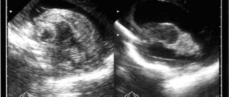
a) With MRI without contrast, the image reveals areas of fluid accumulation indicated by white arrows - subacute subdural hematomas. b) MRI visualizes foci of increased signal intensity (indicated by white arrows), as well as foci of reduced MRI signal intensity (indicated by black arrows), these signs are characteristic of acute subdural hematomas.
The absolute indication for surgical treatment of a subdural hematoma is the thickness of the accumulated blood more than one centimeter, determined by imaging studies (MSCT, MRI). The postoperative period should be accompanied by the maintenance of vital functions and control of intracranial pressure.
The operation is also indicated for subacute subdural hemorrhage, if there is an increase in focal symptoms and the appearance of signs of intracranial hypertension.
Meninges of the brain: anatomy
The structure of the membranes of the brain.
Source: golovnayabol.com Let's take a closer look at the anatomy of the meninges in order to more fully imagine the mechanism of this hemorrhage.
The dura mater (D) performs a protective function . It consists of two connective tissue sheets that are loosely connected to each other due to the loose fiber located between them. Its outer part is adjacent to the cranial bones and is the periosteum for them, and the inner part, together with the arachnoid membrane, forms a narrow and slit-like space called the subdural. It contains cerebrospinal fluid (small volume). Processes extend from the inner side of the dura mater; they separate the structures of the brain.
There are no vessels in the arachnoid membrane (AP). It is quite thin, located on top of the sulci of the brain and forms elevations (granulations) that pass into the lumen of the venous sinuses, providing drainage of cerebrospinal fluid.
The subarachnoid space separates the arachnoid space from the soft membrane (MS) (from the Latin Subarachnoidal - subarachnoid). They are held together by connective tissue bundles, which, in turn, are most developed at the tops of the convolutions (there the two membranes already form a single whole).
It is also worth noting the presence of subarachnoid cisterns - formations that arise as a result of the coating of large depressions between different parts of the brain. They communicate with the subarachnoid space.
The soft membrane is responsible for the blood supply; it is tightly connected to the structures of the brain, completely covers the gyri and penetrates deep between the grooves. Throughout its entire length, the gray matter receives blood from the vessels located in it.
Causes
In the structure of the causes of subdural hematoma, two large groups are distinguished: traumatic and non-traumatic. Why might this be important? To prevent recurrence of this type of hemorrhage. Many causes are quite specific and their elimination is the key to reducing the risk of recurrence of such life-threatening conditions.
Traumatic causes of subdural hematomas:
- Directed physical force (impact).
- Falling from height.
- Road traffic accident.
Non-traumatic causes of subdural hematomas (much less common):
- The use of devices during childbirth to extract the fetus (forceps) and, as a result, birth trauma.
- Shaken baby syndrome is a pathology that occurs when a small child is thrown and a concussion occurs in an unfixed head. In this case, the bridging veins are ruptured, since they are more stretched than in an adult due to the greater width of the subdural space in a child.
- Alcoholics and older people are also susceptible to this disease (they experience expansion of the subdural space due to brain atrophy).
- Vascular pathology (arterial hypertension, severe atherosclerosis, thromboangiitis obliterans, hemorrhagic vasculitis).
- Decreased cerebrospinal fluid pressure (for example, during a spinal tap).
- Cysts of the subarachnoid membrane of the spinal cord.
- Diseases resulting from vitamin K deficiency.
- Taking anticoagulants and antiplatelet agents (medicines that reduce blood clotting).