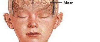
Epidural hematomas of the brain are an accumulation of blood between the cranial bones and the dura mater of the brain resulting from traumatic injury. It is considered a rare type of hematoma: according to medical statistics, it accounts for no more than one and a half percent of cases.
The Neurosurgery Department of CELT invites you to undergo diagnostics and treatment of hematomas in Moscow. Our multidisciplinary clinic has been operating in the paid medical services market for the third decade and enjoys a good reputation among patients. Our specialists have many years of practical experience and have modern techniques that allow them to carry out effective and most gentle operations.
Etiology of epidural hematomas
As already mentioned, the formation of an epidural hematoma occurs above the dura cerebral membrane, which causes its detachment from the inner cranial surface. Since under the age of two years and after sixty it is fused to the cranial bones, during these periods this type of hematoma is extremely rare. Most often it is diagnosed in male patients aged sixteen to twenty-five years who lead an active lifestyle.
An epidural hematoma occurs due to traumatic injuries. This can be a blow from a small object to a slow-moving head, or a blow to the head against a stationary object, usually when falling. Most often, the injury occurs in the area of the temples or crown, and hemorrhage occurs from the meningeal artery and its branches.
General information
Head trauma occurs as a result of a fall, a blow to the head, road traffic accidents, or accidents during sports or work. The skull bones protect the brain from damage, and the meninges provide additional protection. In this regard, most bruises do not cause brain damage.
Head injuries that damage the brain are called traumatic brain injuries (TBI or neurotrauma). The most common type of TBI is mild (this includes mild brain contusion and concussion) - it accounts for 80% of cases. If there is damage to the substance of the brain, meninges, cranial nerves and blood vessels, the injury is regarded as moderate to severe and they account for 15% of cases. Not always even a mild traumatic brain injury goes away without a trace, and moderate and severe injuries cause disability and even death of the patient.
Severe neurotrauma includes severe brain contusion , intracranial hematomas (epidural, subdural and intracerebral), which occur in 40% of neurotraumas, and diffuse axonal damage. With hematomas and brain injuries, the mortality rate is 55%, and 29% of victims are disabled.
Epidural hematoma clinic
The diameter of the hematoma formed above the hard shell does not exceed eight millimeters, the volume is one hundred and twenty milliliters. Its peculiarity is that its thickness increases from the periphery to the center, and the danger is that it causes an increase in intracranial pressure and compression of brain tissue. The classic picture of hematoma formation is characterized by the presence of a clearly visible light interval, which lasts about half an hour and manifests itself as follows:
- The victim loses consciousness for a short period of time and then comes to, often maintaining an insignificant stunned state;
- He complains that he is dizzy, has a feeling of weakness and a headache of moderate intensity;
- He may experience a lack of symmetry in the nasolabial folds, as well as involuntary oscillatory movements of the eyeballs.
After this, the victim becomes worse, which is manifested by the following:
- Increased headaches;
- Depressed state of consciousness (sometimes comatose);
- Increased blood pressure;
- Facial nerve paralysis;
- Pupil dilation and lack of reaction to light.
Symptoms
| Occurrence (how often a symptom occurs in a given disease) | |
| Severe headache spreading over the entire head | 100% |
| Vomiting of various types, including indomitable | 90% |
| Fainting (loss of consciousness) | 80% |
| General weakness (fatigue, tiredness, weakness of the body) | 75% |
| Weakness in one side of the body | 70% |
| Redness of the facial skin | 60% |
| Motor restlessness | 50% |
| Difficulty speaking (speech disorder, speech disorder, speech problems) | 50% |
| Numbness of one half of the body | 50% |
| Memory loss (memory impairment, poor memory, memory impairment, forgetfulness) | 40% |
| Attacks of seizures with or without loss of consciousness (convulsions, convulsive seizures, convulsions, convulsions) | 25% |
| Unilateral throbbing headache | 0% |
Diagnosis of epidural hematomas
In order to correctly make a diagnosis, CELT specialists conduct comprehensive studies to accurately determine the location, diameter and volume of the hematoma, as well as to determine its consequences. Instrumental diagnostic tests prescribed for the patient are presented as follows:
- X-ray of the skull to detect fractures;
- Cerebral angiography (study of cerebral vessels);
- Echoencephalography;
- Computer and magnetic resonance imaging, incl. and with contrast.
Pathogenesis
With any brain injury, hydrodynamic forces and a shock wave act on an important hypothalamic-reticular part of it. Therefore, the synthesis of catecholamines , autonomic and vascular disorders develop, as well as liquor hypertension . The acute period of brain injury is accompanied by oxidative stress, neuronal depolarization, autoimmune inflammation and cell damage. All these mechanisms cause the death of neurons and loss of connections between different parts of the central nervous system.
Primary brain damage entails secondary changes, which are divided into intracranial ( hemorrhage , ischemia , increased intracranial pressure , swelling and displacement of the brain, compression of cerebral vessels and their spasm , neuroinfection ) and extracranial (high or low blood pressure, disturbances in potassium and sodium levels). All severe bruises and compression of the brain are accompanied by ischemia of varying severity, which plays a significant role. It develops due to deterioration of blood flow in the brain and lack of oxygen in the blood. Vascular spasm aggravates ischemia , hypoxia and cerebral edema . Ischemia can be regional (due to brain dislocation and herniation) and local (occurs in the compression zone).
If we consider the mechanism of hematoma formation, they are formed when cerebral vessels rupture. Subdural hematomas are associated with venous damage and venous bleeding. The subdural veins, which connect the cerebral cortex with the venous sinuses, are damaged. With epidural hematomas, the source is the arteries of the membranes or veins damaged by the bones of the skull during their fractures. In young children, ruptures of the sinuses of the dura mater occur.
Treatment of epidural hematomas
When developing treatment tactics for hematomas, neurosurgeons and neuropathologists are based on the results of diagnostic studies and individual patient testimony. It may involve the use of conservative or surgical techniques.
| Hematoma treatment methods | What are they? |
| Conservative | Conservative treatment is carried out for hematomas with a volume of up to 50 mm if there is no progressive clinical picture and there are no symptoms of brain compression. It involves the use of drugs to prevent cerebral edema, relieve pain, vomiting and convulsions. |
| Neurosurgical | Surgery is the more common treatment option. Removal of the hematoma is carried out using endoscopic techniques through a specially created hole in the skull. Through it, part of it is aspirated, after which they resort to trepanation and completely remove the hematoma. The neurosurgeon also locates and ligates the damaged blood vessel using various techniques. |
The Neurosurgery Department of CELT is staffed by neurologists and neurosurgeons with many years of experience in scientific and practical work. You can make an appointment with them online on our website or by contacting our operators: +7 (495) 788 33 88.
Make an appointment through the application or by calling +7 +7 We work every day:
- Monday—Friday: 8.00—20.00
- Saturday: 8.00–18.00
- Sunday is a day off
The nearest metro and MCC stations to the clinic:
- Highway of Enthusiasts or Perovo
- Partisan
- Enthusiast Highway
Driving directions
3.Diagnostics and treatment methods
To make an accurate diagnosis, the doctor will definitely conduct a detailed diagnosis, based on the results of which a treatment strategy and tactics will be selected. The presence of compression of the brain due to the formation of a hematoma is an indisputable indication for urgent (urgent) surgical intervention. In this case, trepanation (opening) of the skull is performed, followed by removal of blood fluid and clots using a special aspirator, and the source of bleeding is neutralized. Then the bone flap is put in place and the wound is sutured.
About our clinic Chistye Prudy metro station Medintercom page!
Forecast
The outcomes of traumatic brain injury can be:
- death;
- lifelong vegetative state;
- severe disability;
- average degree of disability;
- recovery.
The outcome of the injury can be assessed 6 months after the injury. With severe traumatic brain injury, 68% survive and only 8% of patients experience recovery, the rest have neurological disorders of varying severity.
The degree of recovery is influenced by the severity of neurotrauma (duration of loss of consciousness and amnesia) and age. Depending on this, recovery of brain function can be expected from 2 weeks to 9 months. Unfavorable factors for epidural hematomas are: age, anisocoria (or mydriasis), depression of consciousness , hemorrhage volume greater than 30 cm3, intracranial hypertension and displacement of structures greater than 10 mm. When the hemorrhage volume is more than 50 cm3, postoperative mortality increases. The outcome of chronic subdural hematoma also depends on the neurological status before surgery.
The mortality rate for patients who were conscious/stunned before surgery is 5%, and for patients in a state of stupor and coma - 13%. The following treatment outcomes for chronic subdural hematoma are possible: good recovery is achieved in 82% of cases, moderate disability is 14% of cases, severe disability is 2%, and a vegetative state is 1%.
Consequences of traumatic brain injury and complications
Considering TBI, its consequences and complications, it should be noted that their severity varies from headache to severe neurological symptoms. At different periods of time after a head injury, the following are possible:
- Disorders of consciousness up to coma .
- Meningeal syndrome.
- Post-traumatic arachnoiditis.
- Post-traumatic hydrocephalus .
- Post-traumatic epilepsy .
- Cerebral ischemia.
- Pathological reflexes and paresis .
- Asthenovegetative syndrome.
- Post-traumatic amnesia .
- Post-concussion syndrome , which was mentioned above.
- Akinetic mutism : the patient is awake, but there are no attempts to speak or move.
- Chronic disorders of consciousness - a state of minimal consciousness and a vegetative state.
- Primary post-traumatic dementia manifests itself after recovery from a coma due to severe traumatic brain injury with contusion of the frontal and temporal lobe.
- Persistent neurosis-like syndrome.
- Disorientation and disintegration of speech, behavioral disorders.
- Most patients do not become disabled immediately after receiving an injury, but the consequences of a head injury develop over time - after several months or even years. The long-term period lasts up to 2 years and 70% of victims develop traumatic brain disease and dementia. Post-traumatic dementia can progress over time and resembles Alzheimer's disease . Such complications occur especially often with persistent subdural hematomas.
The operations performed are also accompanied by complications and consequences. The following consequences of removal of a cerebral hematoma can be mentioned:
- Postoperative recurrence of hematomas (especially in chronic subdural hematomas).
- Pneumocephalus (accumulation of air in the skull).
- Secondary hemorrhages into brain tissue.
- The appearance of seizures .
- After surgery, bacterial meningitis (especially after drainage of the ventricles of the brain).
- Pulmonary embolism.
- Thrombophlebitis.
- Septic condition.
- Severe cerebral edema .
Approach to therapy
Once a diagnosis is made, immediate action is required. The operation to relieve a hematoma is carried out in stages:
- removal of a blood clot using an aspirator;
- finding the source of hemorrhage and stopping the bleeding;
- closing the hole with a flap of skin and suturing.
The effectiveness of surgical intervention directly depends on the time that has passed since the injury and on the general well-being of the patient. Sometimes there are cases when a hematoma forms a second time, then the operation is performed again.
Conservative treatment is used only when the hematoma is small, does not increase, and there are no signs of compression of brain tissue. Therapeutic procedures in this case will be carried out in a hospital setting with strict bed rest and constant medical examination.
Warning, shocking photo! Click to open
For conservative treatment, the following drugs are used:
- diuretics;
- hemostatics - to stop bleeding;
- agents that promote the resorption of hematoma.
If a person's condition begins to deteriorate, surgery is performed immediately.
Be careful, video of the operation! Click to open
List of sources
- Zhulev N.M., Yakovlev N.A. Mild traumatic brain injury and its consequences. – M., 2004; 127.
- Zotov Yu. V. Complex treatment of severe TBI, taking into account the nature of brain damage and the severity of hypertensive-dislocation syndrome // Bulletin of Surgery, 1996, No. 1, p. 53-56.
- Clinical Guide to Traumatic Brain Injury / Ed. A. N. Konovalov, L. B. Likhterman, A. A. Potapov. T. 1. M.: Antidor, 1998.
- Dzhindzhikhadze R. S., Dreval O. N., Lazarev V. A. Emergency neurosurgical interventions in patients with intracranial tumors // Problems of neurosurgery. – 2011. – Vol. 3. – pp. 62–71.
- Fraerman A.P., Kravets L.Ya., Sheludyakov A.Yu., Trofimov A.O., Balyabin A.V. Compression of the brain in isolated and combined traumatic brain injury. N. Novgorod; 2008; 324 pp.