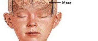
Cephalohematoma on the head of a newborn
Cephalohematoma is a birth injury in newborns when hemorrhage occurs in the periosteum of the skull on one or both sides.
As a result, a lump is formed on the head, the diameter of which, depending on the degree of injury, can reach from several centimeters to twenty. Often the diagnosis is not made immediately, since in many babies a birth tumor can be observed at birth; passing through the birth canal, the baby’s head experiences pressure, resulting in swelling that subsides after 1-2 days.
But cephalohematoma in newborns does not go away in two days, but, on the contrary, can even increase in size, since during this period of life the blood is not yet able to clot quickly and continues to flow into the periosteum, the swelling grows. It is also distinguished from a generic tumor by clear boundaries - the hemorrhage is localized strictly within the affected bones of the skull.
Reasons for appearance
The formation of cephalohematoma in a newborn child is facilitated by many provocative factors:
- Large child size;
- Severe complications causing post-term pregnancy;
- Incorrect position of the child in the womb of the mother - longitudinal oblique or transverse;
- Hydrocephalic shape of the fetal head, outstripping its growth, in infectious pathologies;
- Deviations in fetal development caused by multisystem, endocrine and metabolic dysfunctions;
- Entwining the baby with the umbilical cord, which can cause hypoxia and will not allow the fetus to pass freely through the birth canal.
The cause of hemorrhages in a baby can be anomalies in the structure of the mother's pelvis - narrow or flat, the presence of bone or cartilaginous growths on the pelvic bones. The age of a woman is also of great importance, since in adulthood there is often incomplete divergence of the pelvic bones, which creates an obstacle to the free exit of the fetus.
There may also be weakness in labor - weakened uterine muscles and weakness of their contractile functions will not be able to help the child to the proper extent.
The formation of hemorrhage can also be provoked by the rapid progress of labor. The rapid pushing of the fetus out not only leads to a sharp drop in pressure (barotrauma effect), which can affect the blood vessels, but also provoke injury to the baby’s head, causing vascular damage.
Treatment of hematoma
If hematomas bleed heavily, it becomes necessary to use medications or remove them. This also applies to hematomas that quickly increase in size (especially those located on the child’s face).
There are two treatment methods: medication and surgery. In the first case, steroids are prescribed, which stop the growth of the vessels that make up the hematoma. Steroids are taken for several weeks or even months. They can have side effects on the child’s body: he may have changes in appetite, mood, a slight decrease in growth rate, the appearance of swelling (especially on the face), stomatitis, and the inability to receive vaccinations while taking medications. All side effects go away when you stop taking the medication.
Another type of treatment for hematomas is surgery. It is performed using a laser or scalpel. The laser can destroy the growing vessels that make up the hematoma. This leads to the cessation of its growth. Using a scalpel, the hematoma can be cut out entirely.
The use of a particular treatment method depends directly on the size, shape, location and type of hematoma. In order to choose the most suitable treatment method, you need to consult a dermatologist or surgeon.
Classification
Based on the combination of cephalohematoma with other possible injuries, the following are distinguished:
- cephalohematoma with skull fracture;
- cephalohematoma with brain damage (epidural hematoma, cerebral edema, or cerebral hemorrhage);
- combination of cephalohematoma with neurological manifestations (focal and general brain symptoms).
Based on the size of the subperiosteal hemorrhage, three degrees of cephalohematoma are distinguished:
- 1st degree – hemorrhage diameter is 4 cm or less;
- 2nd degree – diameter of cephalohematoma 4.1 – 8 cm;
- Grade 3 – the diameter of the hemorrhage is more than 8 cm (in the case of multiple cephalohematomas, the total area of hemorrhage is assessed).
Based on the location of the hemorrhage, cephalohematomas are distinguished: parietal (most common), frontal, occipital (less common) and temporal bone (very rare localization).
Symptoms of cephalohematoma
The first symptoms of cephalohematoma (see photo) become noticeable on days 2–3, when the birth tumor subsides.
The size of the hemorrhage tends to increase from the first day of birth, due to a deficiency of coagulation factors in the newborn’s blood - the blood remains liquid for a long time, so it is not possible to clot damaged vessels with blood clots.
The cephalohematoma is elastic to the touch; when you press on the area of hemorrhage, you can feel the movement of fluid. If the cephalohematoma is small in size, it begins to shrink on the 7th – 8th day and disappears without a trace. If the hemorrhage is significant, the process of its resorption may take several months. Often, a bone fracture (crack) is observed in the area where the cephalohematoma is located.
Cephalohematoma always has clear boundaries in the form of a compacted ridge around the circumference of the hemorrhage. The delimitation of the cephalohematoma is associated with the tight fusion of the periosteum with the bones of the skull in the area of the sutures, so the hemorrhage is located in the area of one bone.
Causes and symptoms of hematomas
There are three types of hematomas: simple, having a bright red tint, tricky, penetrating deeper under the skin and having a purple or blue tint, and mixed, which are characterized by signs of the first two types.
Some hematomas are already present on the newborn’s body, some appear during the first months of his life. Initially, all three types of hematomas may appear the same color: white, blue-gray, or pink. After a few weeks, strawberry hematomas turn red, while deep, tricky ones turn blue. The blood vessels of a hematoma grow faster than normal ones, so in the first years of a child’s life it will appear that the hematoma is increasing in size.
The hematoma usually stops growing in size by the first year of a child’s life, sometimes by the second. The first sign of stopping the growth of strawberry hematoma is the appearance of a white “edge” around the red spot. Then white spots will begin to appear on it. This indicates that it is beginning to dissolve. Over the next few years, the hematoma will begin to take on a pinkish-gray color. After this, it will begin to fade. 50% of hematomas become invisible by 5 years, 75% by 7 years, and by 9 years about 90% of hematomas disappear. Sometimes the only traces of a former hematoma are a pink tint to the skin and slight swelling if the hematoma protruded above the skin. Usually there are no traces of hematomas.
Treatment of cephalohematoma in newborns
The disease is treated by a neonatologist and, according to indications, a pediatric surgeon. Therapy for cephalohematoma involves:
- Feeding the baby with donor or expressed breast milk for 4 to 6 days.
- Calcium preparations (Calcium gluconate) for a course of 3 - 5 days.
- Vitamin K course 3-5 days.
In case of suppuration of hemorrhage or large size (more than 8 cm) of the tumor, puncture of the cephalohematoma in the newborn is indicated. Also, a puncture is performed if the tumor does not shrink within 10 days.
The operation can be performed from the 10th day of the child’s life. At the site of the hematoma, hair is removed, the area is treated with an antiseptic and a puncture is performed. Then apply a tight bandage for 3 hours. If the operation is performed in case of suppuration of the hematoma, the contents are drained, followed by the application of an antiseptic bandage (with antibacterial drugs).
In case of ossification of the hematoma, to correct the deformation of the skull, resection of the periosteum to the native bone is performed.
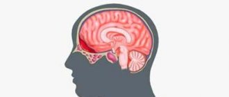
Cerebral hemorrhage due to birth trauma
The most typical manifestations of any intracranial hemorrhage in newborns are: 1. sudden deterioration in the general condition of the child with the development of various variants of the depression syndrome with periodically occurring signs of hyperexcitability; 2. change in the nature of the cry; 3. bulging of the large fontanel or its tension, 4. abnormal movements of the eyeballs; 5. violations of thermoregulation (hypo- or hyper.1 army); 6. vegetovisceral disorders (regurgitation, pathological loss of body weight, flatulence, unstable stools, tachypnea, tachycardia, peripheral circulatory disorders); 7. pseudobulbar and motor disorders, convulsions; 8. progressive posthemorrhagic anemia; 9. acidosis, hyporbilirubgnemia and other metabolic disorders; 10. addition of somatic diseases (meningitis, sepsis, pneumonia, cardiovascular and adrenal insufficiency, etc.). Subdural hemorrhages . Depending on the location, there are: tentorial hemorrhages with damage to the direct and transverse sinuses of the vein of Galen or small infratentorial veins; occipital osteodiastasis - rupture of the occipital sinus; rupture of the falciform process of the dura mater with damage to the inferior sagittal sinus; rupture of the connecting superficial cerebral veins. Subdural hematomas can be unilateral or bilateral; they may be combined with parenchymal hemorrhages resulting from hypoxia.
Tentorial rupture with massive hemorrhage, occipital osteodiastasis, damage to the inferior sagittal sinus is characterized by an acute course with the rapid development of symptoms of compression of the upper parts of the brain stem such as stupor, eye deviation to the side, anisocoria with a sluggish reaction to light, the “doll’s eyes” symptom, muscle rigidity back of the head, opisthotonus pose; unconditioned reflexes are suppressed, the child does not suck or swallow, attacks of asphyxia and convulsions are observed. If the hematoma grows, symptoms of compression of the lower parts of the brain stem appear: coma, dilated pupils, pendulum-like eye movements, arrhythmic breathing. In the subacute course of the pathological process (hematoma and smaller rupture), neurological disorders (stupor, excitability, arrhythmic breathing, bulging of the large fontanelle, oculomotor disorders, tremor, convulsions) occur at the end of the first day of life or after several days and persist for several minutes or hours. Death, as a rule, occurs in the first days of a child’s life from compression of the vital centers of the brain stem.
Convexital subdural hematomas caused by rupture of the superficial cerebral veins are characterized by minimal clinical symptoms (restlessness, regurgitation, vomiting, tension of the large fontanelle, Graefe's symptom, periodic increase in body temperature, signs of local brain disorders) or their absence and are detected only during an instrumental examination of the child.
Primary subarachnoid hemorrhages are the most common. They occur when vessels of various sizes are damaged within the subarachnoid space, small veins of the leptomeningeal plexuses or connecting veins of the subarachnoid space. They are called primary in contrast to secondary subarachnoid hemorrhages, in which blood enters the subarachnoid space as a result of intra- and periventricular hemorrhages and aneurysm rupture. Subarachnoid hemorrhages are also possible with thrombocytopenia, hemorrhagic diathesis, and congenital angiomatosis. With primary subarachnoid hemorrhages, blood accumulates between separate areas of the brain, mainly in the posterior cranial fossa and temporal regions. As a result of extensive hemorrhages, the entire surface of the brain is covered with a red cap, the brain is swollen, and the vessels are filled with blood. Subarachnoid hemorrhages can be combined with small parenchymal hemorrhages.
The clinical picture of neurological disorders depends on the severity of the hemorrhage and its combination with other disorders (hypoxia, hemorrhages of other locations). More common are mild hemorrhages with clinical manifestations such as regurgitation, hand tremors, anxiety, and increased tendon reflexes. Sometimes neurological symptoms may appear only on the 2-3rd day of life after the baby is put to the breast. With massive hemorrhages, children are born with asphyxia, they experience anxiety, sleep disturbances, general hyperesthesia, stiffness of the neck muscles, regurgitation, vomiting, nystagmus, strabismus, Graefe's symptom, tremor, convulsions. Muscle tone is increased, tendon reflexes are high with an expanded zone, all unconditioned reflexes are clearly expressed. On the 3-4th day of life, Harlequin syndrome is sometimes noted, manifested by a change in the color of half the newborn’s body from pink to light red; the other half is paler than normal. This syndrome is clearly detected when the child is positioned on his side. A change in body color can be observed within 30 seconds to 20 minutes; during this period the child’s well-being is not disturbed. Harlequin syndrome is considered a pathognomonic sign of traumatic brain injury and asphyxia of the newborn.
Intraventricular and periventricular hemorrhages are most typical for premature babies born with a body weight of less than 1500 g. The morphological basis of these hemorrhages is the immature choroid plexus located under the ependyma lining the ventricles (germinal matrix). Until the 35th week of pregnancy, this area is richly vascularized, the connective tissue framework of the vessels is insufficiently developed, and the supporting stroma has a gelatinous structure. This makes the vessel very sensitive to mechanical stress, changes in intravascular and intracranial pressure. High risk factors for the development of hemorrhages are prolonged labor, accompanied by deformation of the fetal head and compression of the venous sinuses, respiratory disorders, hyaline membrane disease, various manipulations performed by the midwife (suction of mucus, replacement blood transfusion, etc.). In approximately 80% of children with this pathology, periventricular hemorrhages break through the ependyma into the ventricular system of the brain and blood spreads from the lateral ventricles through the foramina of Magendie and Luschka into the cisterns of the posterior cranial fossa. The most typical localization of the forming thrombus is in the region of the occipital cistern magna (with limited spread to the surface of the cerebellum). In these cases, abliterative arachnoiditis of the posterior cranial fossa may develop, causing obstruction through cerebrospinal fluid circulation. Intraventricular hemorrhage can also involve the periventricular white matter of the brain and be combined with cerebral venous infarctions, which are caused by compression of the venous outflow pathways by the dilated ventricles of the brain.
Hemorrhage usually develops in the first 12-72 hours of life, but may subsequently progress. Depending on the extent and speed of spread, three variants of its clinical course are conventionally distinguished: fulminant, intermittent and asymptomatic (low-symptomatic). With lightning-fast hemorrhage, the clinical picture develops over several minutes or hours and is characterized by deep coma, arrhythmic breathing, tachycardia, and tonic convulsions. The child’s eyes are open, the gaze is fixed, the reaction of the pupils to light is sluggish, nystagmus, muscle hypotonia or hypertension, bulging of the large fontanel are observed; metabolic acidosis, decreased hematocrit, hypoxemia, hypo- and hyperglycemia are detected.
The intermittent course is characterized by similar, but less pronounced clinical syndromes and an “undulating course, when a sudden deterioration is followed by an improvement in the child’s condition. These alternating periods are repeated several times over 2 days until the condition stabilizes or death occurs. With this variant of the pathological process, pronounced metabolic disorders are also observed.
Asymptomatic or low-symptomatic course is observed in approximately half of children with intraventricular hemorrhage. Neurological disorders are transient and mildly expressed, metabolic changes are minimal.
Cerebellar hemorrhages occur as a result of massive supratentorial intraventricular hemorrhages in full-term infants and hemorrhages in the germinal matrix in preterm infants. Pathogenetic mechanisms include a combination of birth trauma and asphyxia. Clinically, they are characterized by a rapid progressive course, as with subdural hemorrhages in the posterior cranial fossa: respiratory distress increases, hematocrit decreases, and death quickly occurs. A less acute course of the pathology, manifested by atony, areflexia, drowsiness, apnea, pendulum-like eye movements, and strabismus, is also possible.
Epidural hemorrhages - occur between the inner surface of the skull and the dura mater and do not spread beyond the cranial sutures. These hematomas are formed when there are cracks and fractures of the cranial vault with rupture of the vessels of the epidural space. Epidural hemorrhages are often combined with external cephalohematomas. In the clinic, the sequence of development of symptoms is characteristic: after a short “bright” interval (from 3 to 6 hours), brain compression syndrome develops, which manifests itself as severe anxiety 6-12 hours after the injury, progressive deterioration of the child’s condition, up to the development of coma after 2 hours-36 hours. Typical symptoms are pupil dilation (in 3-4 grooves) on the affected side, clonic-tonic convulsions, hemorrhoids on the opposite side of the hematoma, asphyxia, bradycardia, arterial hypotension, congestive optic nerve nipples.
Parenchymal (intracerebral) hemorrhages occur more often when the terminal branches of the cerebral arteries are damaged. With small-point hemorrhages, the symptoms are atypical and mild: lethargy, regurgitation, decreased muscle tone and reflexes, nystagmus, Graefe's symptom, etc. With large hematomas, the clinic is clear and manifests itself with symptoms characteristic of PIVC.
Atypical intracranial hemorrhages in newborns can be caused by vascular abnormalities, tumors, coagulopathies, and hemorrhagic infarction. The most common types of hemorrhagic diathesis are vitamin K-deficiency hemorrhagic syndrome, hemophilia A, and isoimmune thrombocytopenic purpura of newborns. Hemorrhagic disorders in newborns can also be caused by congenital thrombocytopathy due to the mother's prescription of acetylsalicylic acid and sulfonamide drugs before birth, while the hemorrhages are mainly subarachnoid and mild. Neonatal intracranial hemorrhages can cause congenital arterial aneurysms, arteriovenous anomalies, coarctation of the aorta, brain tumors (teratoma, glioma, medulloblastoma).
Flow. The following periods of the course of birth brain injury are distinguished: acute - 7-10 days, sometimes up to 1 month; subacute (early recovery up to 3-4-6 months and late - from 4 months to 2 years). During the acute period in full-term infants, phases of excitation and depression of the central nervous system are distinguished. Manifestations of intracranial hemorrhage are gradually eliminated. The prognosis depends on the severity of the process, the gestational age of the child and concomitant diseases. The survival rate is 50-70%, the rest retain various neurological disorders (hemisyndrome, hydrocephalus, cysts, etc.).
In premature infants, ICH may be asymptomatic or have a poor atypical clinical picture; with dominance of signs of respiratory disorders, apnea attacks; with the prevalence of general depression syndrome or hyperexcitability syndrome, etc.
Consequences of cephalohematoma
Why is this hemorrhage dangerous? Most often nothing. In the vast majority of cases, there are no consequences of cephalohematoma in a newborn, and at an older age it is impossible to say that the child had this complication of childbirth in the past.
However, with cephalohematoma, the following complications may occur:
- Anemia - develops with severe blood loss;
- Jaundice - due to the breakdown of hemoglobin with the formation of bilirubin;
- Suppuration - when bacteria enter the tumor through damaged skin;
- Ossification (ossification) of cephalohematoma - due to the deposition of calcium salts in the cavity.
Since blood accumulates above the bones of the child's skull, it cannot damage his brain and other parts of the nervous system. A festering hematoma, with timely and adequate treatment, will also not affect the condition of the body. The only consequence of cephalohematoma in the future can be detected when it is organized. If the blood is not absorbed, but is replaced by connective tissue, bone deformation occurs. However, with a small volume of hemorrhage (up to 70 ml), detecting this defect in the form of a small protrusion will be extremely difficult at an older age. As a rule, it does not have much cosmetic significance.
Let us emphasize once again that mental retardation, speech impairment, paralysis and other neurological diseases do not occur after cephalohematoma. A cephalohematoma is just a collection of blood that is located above the bone. The brain is located inside the cranium. The blood clot cannot deform the bone so much that it damages the organs contained in the baby's skull.
It is quite easy to recognize this pathology - there are characteristic external signs for this, so instrumental and laboratory diagnostics do not have any significance. Treatment for cephalohematoma is chosen by the doctor together with the child’s mother. Regardless of the tactics (surgical or conservative), hemorrhage is successfully treated and, most often, does not leave any consequences for the child.
Forecasts
The prognosis for cerebral hematoma largely depends on the extent of the lesion, the speed and correctness of the treatment and the adequacy of subsequent rehabilitation therapy. The child’s body has high compensatory capabilities, so the chances of a full life after removal of the hematoma are high, excluding severe damage.
The consequences of the disease can be very long-term, appearing months and years after surgery. The child may experience hydrocephalus, cerebral palsy, epilepsy, hyperactivity, various speech disorders and general developmental delay.
The consequences of a cerebral hematoma are varied and depend on many factors.
To prevent the development of hematoma in the brain, the course of pregnancy should be monitored, special attention should be paid to the prevention of fetal hypoxia.
Prevention
Periosteal hemorrhage can be avoided if the following conditions are met:
- the expectant mother should lead a healthy lifestyle and not take medications without consulting a doctor;
- The doctor and midwife must conduct the birth carefully, choose the method of delivery correctly and quickly.
To prevent recurrent hemorrhage, mothers should not rock the baby until 6 months of age. You cannot refuse surgical treatment of a hematoma. If your doctor recommends a cesarean section, it is best to choose this method of delivery. If these recommendations are followed, the child will quickly recover from the injury.
A cephalohematoma on the head of a newborn noticed in time, the treatment of which is quite easy, does not pose a threat to the child’s life.