Perinatal encephalopathy is a number of diseases (deviations) of the central nervous system in newborns. The disease can manifest itself in different ways, and therefore it is very difficult to diagnose, especially in infants. Related to this is the fact that the symptoms of encephalopathy are often regarded by pediatricians as signs of completely different diseases. As a result, treatment of the disease is not given the necessary attention at an early age, when the likelihood of a complete recovery is greatest. A progressive disease as the child grows up is often also diagnosed symptomatically, and appropriate treatment is prescribed.
Getting rid of PEDs without osteopathic influence on the root causes of the disease is, if not completely impossible (after all, the body is a self-regulating system that is quite capable of coping with a number of serious diseases), then it is significantly difficult. Of course, other treatment methods should not be neglected - as practice shows, complex procedures are the most effective.
Symptoms and consequences of perinatal encephalopathy
Symptoms of the disease manifest themselves differently at different periods of a child’s life. To simplify classification and improve diagnosis, it is customary to distinguish three main periods of PEP: acute (during the first month of life), recovery (up to 1 year, less often up to 2 years - mainly in premature infants) and outcome of the disease. There is a possibility that the child’s body - as a self-regulating system, from the point of view of osteopathy - can fully recover and neutralize the symptoms of the disease by adapting to them. This does not mean complete recovery, since the effects of AEDs may appear at a later age. Therefore, if you suspect a disease, you must immediately show the child to an osteopathic doctor, who can make the correct diagnosis and prescribe competent, adequate treatment aimed at ridding the body not of the consequences, but of the causes of the disease.
Early symptoms that should be addressed to a pediatric neurologist
- sluggish breastfeeding, choking during feeding, leakage of milk through the baby's nose - weak cry of the child, nasal or hoarse voice - frequent regurgitation and insufficient weight gain - decreased motor activity of the child, drowsiness, lethargy or severe anxiety - trembling of the chin, upper and/or or lower extremities, frequent startlings - difficulty falling asleep, frequent awakenings during sleep - throwing back the head - slowing or rapid increase in head circumference - low (flabby muscles) or high tone of the muscles of the limbs and torso - decreased activity of movements of the arm or leg on any side , limited hip separation or the presence of a “frog” position with pronounced hip separation, unusual position of the child - strabismus, torticollis - birth of a child by caesarean section, in breech presentation, with an anomaly of labor or with the use of obstetric forceps, extrusion, with the umbilical cord entwined around the neck - prematurity of the child - presence of convulsions during childbirth or in the postpartum period
Main symptoms of perinatal encephalopathy
During the acute period of disease development, the following are observed:
- CNS depression syndrome. Characterized by general lethargy of newborns, reduced response to external stimuli, and the presence of spontaneous motor reflexes;
- comatose syndrome. It usually develops quickly and suddenly, causing unconsciousness in the child. May manifest itself in acute disruption of vital body functions;
- increased neuro-reflex excitability. Manifests itself in the form of tremor of the limbs and an abnormally excited reaction to external stimuli;
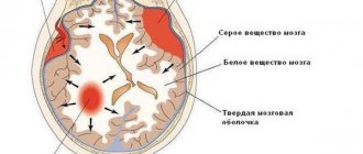
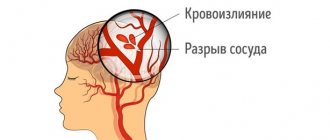
- increased cranial pressure with subsequent hypertensive-hydrocephalic syndrome and a disproportionate increase in skull size;
- convulsions.
During the recovery period, these symptoms may be added to:
- motor disorders. They manifest themselves in different ways, but the common thing is that the child does not fully or partially control his movements;
- psychomotor development delay (PDD). Manifests itself in delayed development, speech problems, memory impairment, problems with attention, etc.;
- epileptic seizures.
Signs of the disease can appear both in combination and individually. And at different ages. If at least one symptom is detected, it is recommended to contact an osteopathic specialist for a more detailed examination.
The outcome of the disease can be:
- recovery;
- minimal brain dysfunction;
- mental retardation;
- cerebral palsy;
- neurotic diseases;
- epilepsy;
- hydrocephalus.
As practice shows, with mild forms of perinatal encephalopathy, the likelihood of complete recovery is quite high. Especially with the right osteopathic treatment, which can be combined with other methods of influencing the child’s body.
In more severe forms, longer treatment will be required, which, due to various factors, may not be 100% effective. Some problems with memory, attention, and coordination of movements in a child can persist both for a long period and throughout life. But osteopathy contributes, if not to a complete cure, then to a significant minimization of the symptoms of perinatal encephalopathy.
Course of PEP and possible prognosis
During PEP, three periods are distinguished: acute (1st month of life), recovery (from 1 month to 1 year in full-term infants, up to 2 years in premature infants) and outcome of the disease. In each period of PEP, various syndromes are distinguished. More often there is a combination of several syndromes. This classification is appropriate, since it allows us to distinguish syndromes depending on the age of the child. For each syndrome, appropriate treatment tactics have been developed. The severity of each syndrome and their combination make it possible to determine the severity of the condition, correctly prescribe therapy, and make prognoses. I would like to note that even minimal manifestations of perinatal encephalopathy require appropriate treatment to prevent adverse outcomes.
Let us list the main syndromes of PEP.
Acute period:
- CNS depression syndrome.
- Comatose syndrome.
- Syndrome of increased neuro-reflex excitability.
- Convulsive syndrome.
- Hypertensive-hydrocephalic syndrome.
Recovery period:
- Syndrome of increased neuro-reflex excitability.
- Epileptic syndrome.
- Hypertensive-hydrocephalic syndrome.
- Syndrome of vegetative-visceral dysfunctions.
- Movement impairment syndrome.
- Psychomotor development delay syndrome.
Outcomes:
- Full recovery.
- Delayed mental, motor or speech development.
- Attention deficit hyperactivity disorder (minimal brain dysfunction).
- Neurotic reactions.
- Autonomic-visceral dysfunctions.
- Epilepsy.
- Hydrocephalus.
- Cerebral palsy.
All patients with severe and moderate brain damage require hospital treatment. Children with mild impairments are discharged from the maternity hospital under outpatient supervision by a neurologist.
Let us dwell in more detail on the clinical manifestations of individual PEP syndromes, which are most often encountered in outpatient settings.
The syndrome of increased neuro-reflex excitability is manifested by increased spontaneous motor activity, restless shallow sleep, prolongation of the period of active wakefulness, difficulty falling asleep, frequent unmotivated crying, revival of unconditioned innate reflexes, variable muscle tone, tremor (twitching) of the limbs and chin. In premature infants, this syndrome in most cases reflects a lowering of the threshold for convulsive readiness, that is, it indicates that the baby can easily develop convulsions, for example, when the temperature rises or when exposed to other irritants. With a favorable course, the severity of symptoms gradually decreases and disappears within a period of 4-6 months to 1 year. If the course of the disease is unfavorable and there is no timely treatment, epileptic syndrome may develop.
Convulsive (epileptic) syndrome can manifest itself at any age. In infancy it is characterized by a variety of forms. An imitation of unconditioned motor reflexes is often observed in the form of paroxysmal bending and tilting of the head with tension in the arms and legs, turning the head to the side and straightening the arms and legs of the same name; episodes of shuddering, paroxysmal twitching of the limbs, imitations of sucking movements, etc. Sometimes it is difficult even for a specialist to determine the nature of the convulsive conditions that arise without additional research methods.
Hypertensive-hydrocephalic syndrome is characterized by excess fluid in the spaces of the brain containing cerebrospinal fluid (CSF), which leads to increased intracranial pressure. Doctors often call this disorder to parents exactly that - they say that the baby has increased intracranial pressure. The mechanism of occurrence of this syndrome can be different: excessive production of cerebrospinal fluid, impaired absorption of excess cerebrospinal fluid into the bloodstream, or a combination of both. The main symptoms of hypertensive-hydrocephalic syndrome, which doctors focus on and which parents can control, are the rate of increase in the child’s head circumference and the size and condition of the large fontanel. For most full-term newborns, the normal head circumference at birth is 34 - 35 cm. On average, in the first half of the year, the monthly increase in head circumference is 1.5 cm (in the first month - up to 2.5 cm), reaching about 44 cm by 6 months. In the second half of the year, the growth rate decreases; by one year, head circumference is 47-48 cm. Restless sleep, frequent profuse regurgitation, monotonous crying combined with bulging, increased pulsation of the large fontanel and throwing the head back are the most typical manifestations of this syndrome.
However, large head sizes often occur in absolutely healthy babies and are determined by constitutional and family characteristics. The large size of the fontanel and the “delay” in its closure are often observed with rickets. The small size of the fontanel at birth increases the risk of intracranial hypertension in various unfavorable situations (overheating, increased body temperature, etc.). Carrying out a neurosonographic examination of the brain makes it possible to correctly diagnose such patients and determine treatment tactics. In the vast majority of cases, by the end of the first six months of a child’s life, normal growth of head circumference is noted. In some sick children, hydrocephalic syndrome persists by 8-12 months without signs of increased intracranial pressure. In severe cases, the development of hydrocephalus is noted.
Comatose syndrome is a manifestation of the serious condition of the newborn, which is assessed by 1-4 points on the Apgar scale. Sick children exhibit severe lethargy, decreased motor activity up to its complete absence, and all vital functions are depressed: breathing, cardiac activity. Seizures may occur. The severe condition persists for 10-15 days, with no sucking or swallowing reflexes.
The syndrome of vegetative-visceral dysfunctions, as a rule, manifests itself after the first month of life against the background of increased nervous excitability and hypertensive-hydrocephalic syndrome. Frequent regurgitation, delayed weight gain, disturbances in cardiac and respiratory rhythm, thermoregulation, changes in the color and temperature of the skin, marbling of the skin, and dysfunction of the gastrointestinal tract are noted. Often this syndrome can be combined with enteritis, enterocolitis (inflammation of the small and large intestines, manifested by stool upset, impaired weight gain), caused by pathogenic microorganisms, with rickets, aggravating their course.
The syndrome of movement disorders is detected from the first weeks of life. From birth, a violation of muscle tone can be observed, both in the direction of its decrease and increase, its asymmetry can be detected, and there is a decrease or excessive increase in spontaneous motor activity. Often the syndrome of motor disorders is combined with a delay in psychomotor and speech development, because disturbances in muscle tone and the presence of pathological motor activity (hyperkinesis) interfere with purposeful movements, the formation of normal motor functions, and mastery of speech.
With delayed psychomotor development, the child later begins to hold his head up, sit, crawl, and walk. A predominant disorder of mental development can be suspected in the presence of a weak monotonous cry, impaired articulation, poor facial expressions, late appearance of a smile, and delayed visual-auditory reactions.
Cerebral palsy (CP) is a neurological disease that occurs as a result of early damage to the central nervous system. In cerebral palsy, developmental disorders usually have a complex structure, combining motor disorders, speech disorders, and mental retardation. Motor disorders in cerebral palsy are expressed in damage to the upper and lower extremities; fine motor skills, articulatory muscles, and oculomotor muscles suffer. Speech disorders are detected in most patients: from mild (erased) forms to completely unintelligible speech. 20 - 25% of children have characteristic visual impairments: convergent and divergent strabismus, nystagmus, limitation of visual fields. Most children have mental retardation. Some children have intellectual impairments (mental retardation).
Attention deficit hyperactivity disorder is a behavioral disorder associated with the child's poor control of his attention. It is difficult for such children to concentrate on any task, especially if it is not very interesting: they fidget and cannot sit still calmly, and are constantly distracted even by trifles. Their activity is often too violent and chaotic.
Advertising
Risk factors
Risk factors that increase the risk of developing the disease include:
- chronic maternal illnesses. Often they are not transmitted directly to the child, since they are not hereditary, but at the genetic level they cause the manifestations of various pathologies and abnormalities in the development of the fetus and the already born child;
- infectious diseases suffered during pregnancy. Untreated pathologies that can manifest themselves against the background of a general weakening of the body’s immunity due to pregnancy are very dangerous;
- poor nutrition. During the period of gestation, the mother's body must receive the entire necessary set of proteins, vitamins and minerals. Nutrition should be balanced and agreed with a doctor to avoid food allergies and digestive disorders;
- mother's age is too young. A girl’s body may simply be unprepared to bear a full-fledged and healthy child. If the expectant mother is too young or has insufficient physical development, she should be under the constant supervision of a specialist throughout the entire period of pregnancy and after childbirth;
- metabolic disorder in the mother's body. Since her body is closely interconnected with the body of the unborn child, any disturbances affect the nutrition and health of the fetus. Therefore, proper nutrition, which was mentioned earlier, is so important, as well as the trouble-free operation of the organs responsible for metabolism in the body;
- pathologies during pregnancy. Early and late toxicosis, stress, physical activity and other factors are very dangerous, which can lead to interruption and abnormal course of pregnancy;
- unfavorable environmental conditions. Unfortunately, today this factor is more dangerous than many others, because it is often impossible to avoid the effects of harmful external manifestations. It is recommended to contact specialists (including an osteopath) who will help to significantly neutralize the adverse effects of the environment;
- prematurity or postmaturity of the fetus.
- Thus, most risk factors are associated
specifically with the health of the mother. Therefore, during pregnancy, she should carefully monitor her well-being, listening to the advice of a good doctor. In parallel with other specialists, it is recommended to regularly visit an osteopath, who will also monitor the progress of pregnancy and will be able to correct possible deviations using osteopathic methods, without the use of drugs that can harm the unborn child.
Treatment
The central nervous system of newborns is plastic, capable of development and recovery, so treatment of encephalopathy must begin as early as possible. It depends on the severity of the disease and specific symptoms.
If brain dysfunction is mild or moderate, the child remains on home treatment. In this case, use:
- individual regime, calm atmosphere at home, balanced nutrition, lack of stress;
- assistance from correctional teachers, psychologists, speech therapists for alalia and dysarthria
- massage and physical therapy to normalize tone, develop motor functions and coordination of movements
- physiotherapy;
- herbal medicine (various sedatives and herbs to normalize water-salt metabolism).
For severe motor and nervous disorders, delayed development of the child and other PEP syndromes, medications are used. The doctor prescribes medications, as well as other treatment methods, based on the manifestations of the disease:
- For movement disorders, dibazole and galantamine are most often prescribed. With increased muscle tone - Baclofen and Mydocalm to reduce it. These drugs are introduced into the body, including using electrophoresis. Massage, special exercises, and physiotherapy are also used.
- If PEP is accompanied by convulsive syndrome, the doctor prescribes anticonvulsant drugs. For seizures, physiotherapy and massage are contraindicated.
- Delayed psychomotor development is a reason for prescribing drugs to stimulate brain activity and increase blood circulation in it. These are Actovegin, Pantogam, Nootropil and others.
- For hypertensive-hydrocephalic syndrome, herbal medicine is used, and in severe cases, Diacarb is used to accelerate the outflow of cerebrospinal fluid. Sometimes part of the cerebrospinal fluid is removed through a puncture of the fontanel.
For the treatment of PEP of any severity, B vitamins are prescribed, as they are necessary for the normal development and functioning of the nervous system. In many cases, swimming, baths with salt or herbal remedies, and osteopathy may be recommended.
Perinatal encephalopathy is one of the most common diagnoses among pediatric neurologists. This is due to the fact that PEP is a collective term denoting disorders of the child’s brain in the perinatal period, which have various causes, including the health of the mother, the course of pregnancy, the absence of congenital diseases, complications during childbirth, ecology and other circumstances.
Symptoms can be different, relating to disorders of the nerves, muscles, internal organs, metabolism, therefore, for an accurate diagnosis, the doctor must not only examine the child, but also collect the entire history regarding the health of the mother and father, complications during pregnancy, labor, and also order additional examinations.
An untimely or incorrectly treated disease can lead to complications including cerebral palsy and epilepsy.
The danger of perinatal encephalopathy
Like many other infant diseases, if not cured in the early stages, perinatal encephalopathy progresses with age and can manifest itself in the form of a wide variety of disorders that at first glance seem unrelated:
- frequent morbidity syndrome. These can be either frequent colds or periodic ailments associated with poor health, increased fatigue, headaches, etc.;
- chronic diseases of the respiratory system. They can manifest themselves against the background of frequent acute respiratory infections, gradually turning into a chronic form. In the absence of competent osteopathic treatment, with age they can develop into asthma and other equally dangerous diseases;
- developmental delay. They may manifest themselves in a less obvious form as learning difficulties, especially in the exact sciences and creative disciplines;
- posture disorders. Unnoticeable birth injuries and disturbances in the functioning of the central nervous system, which arose during pregnancy, often later lead to curvature of the spine and the appearance of such serious diseases as scoliosis, with all the ensuing consequences, leading to the prolapse of internal organs and their partial or complete dysfunction;
- disturbances in the functioning of internal organs, the metabolic system and the circulatory system. Typically, these disorders invariably lead to other diseases, since due to impaired blood flow, various parts of the body begin to receive less oxygen, which leads to cell death and dysfunction.
Diagnostics:
It is necessary to conduct an ultrasound examination of the fetus, use cardiotocography (recording of the fetal cardiac activity) and Dopplerometry (study of the blood flow of the vessels of the uterus and umbilical cord of the fetus) with the frequency prescribed by the attending physician. Auscultation (listening) of the fetal heartbeat with a stethoscope is also used. It should be noted that not every pregnancy occurs against the background of the above diseases, complicating intrauterine fetal hypoxia. To prevent the possible occurrence of hypoxia, special attention is paid to its prevention: long walks in the fresh air, mandatory dosed physical activity (gymnastics, exercises, exercises for pregnant women and breathing exercises, swimming, yoga). It is possible to use hyperbaric oxygen therapy (HBO) as prescribed by the attending physician. It must be remembered that treatment must be prescribed by a gynecologist, be comprehensive and take into account an individual approach to each expectant mother.
How do osteopathic specialists evaluate perinatal encephalopathy?
From the point of view of osteopathy, the entire complex of central nervous system diseases, united under this term, is a consequence of mechanical disorders in the body of the fetus of a newborn child. Moreover, they can occur during pregnancy or later - during difficult childbirth and/or caesarean section. The reasons leading to the appearance of the disease are:
- dysfunction of the spinal column or its individual sections;
- disturbances in the functioning of internal organs;
- prolonged oxygen starvation;
- circulatory disorders.
But even if the fetus developed normally during pregnancy, there is a high probability of injury during childbirth, since, passing between the pelvic bones, the fetus rotates 360 degrees, which can cause disturbances in the skeletal structure and muscle tissue. The most common disturbances observed are in the position of the cervical vertebrae. In most cases, in the first weeks of a child’s growth, the effects of damage are completely neutralized by the body on its own. But this is not always the case; any changes or deviations in development can lead to complications, which subsequently provoke disturbances in the blood supply to the brain and the manifestation of perinatal disorder.
The main reasons for the development of CNS HIP:
- anemia in the mother (a decrease in the amount of hemoglobin, which reduces the delivery of oxygen and nutrients to the tissues of the baby’s body) - any chronic diseases and defects: congenital heart defects, lungs of other organs, kidney disease, the presence of diabetes mellitus, which contribute to poor circulation - complications pregnancy and childbirth (gestosis, threat of premature birth, pathology of the placenta and umbilical cord, prematurity and post-term pregnancy, multiple pregnancies, polyhydramnios and oligohydramnios, various anomalies of labor) - fetal diseases (hemolytic disease of the newborn, intrauterine infection, bleeding) Tangible signs of fetal hypoxia mainly are expressed in changes in its motor activity (sudden increase, increased heart rate and movements (movements) of the fetus during acute hypoxia, or a decrease and weakening of them during chronic hypoxia). When a diagnosis of intrauterine fetal hypoxia is made, the expectant mother needs to carry out procedures to identify the causes, followed by comprehensive treatment of their source, with possible hospitalization in a hospital, adherence to bed or home rest and daily routine.
How is perinatal encephalopathy diagnosed in newborns and infants?
There are a number of safe brain imaging techniques that newborns can undergo. They allow you to create a complete clinical picture, determine the presence of the disease, its degree, and the nature of its course. The most informative and effective is neurosonography, which accurately detects the presence of damaged areas in the baby’s brain. In addition to this, Doppler ultrasound can be performed to assess blood flow in the vessels of the brain.
If developmental abnormalities are detected, it is recommended to undergo an electroencephalographic examination aimed at determining the degree of developmental delay. This procedure also allows you to identify foci of epileptic lesions and assess the degree of their activity and danger to the body.
In some cases, an examination by an ophthalmologist is also recommended, who will help determine the extent of the damage by assessing the condition of the optic nerves and analyzing the fundus.
Treatment of encephalopathy with osteopathic methods
A good osteopathic doctor is able to diagnose early symptoms of the disease and create a complete picture of its course. This makes it possible to prescribe competent treatment that helps the body adapt to new conditions, launch and stimulate its self-regulatory functions.
The effects of an osteopath - depending on the course and complexity of the disease - are directed primarily to the affected areas of the body in order to relieve pain and spasms resulting from improper development and disturbances in the circulatory system. In parallel with this, the specialist carries out a set of procedures to normalize brain function. Usually, various techniques of muscular-energetic influence are used for this, which allow manually restoring the balance of tension in the intracranial membranes. This helps restore micropulsations in the bones of the skull and the brain itself, and normalize blood circulation.
After normalization of brain activity, the body receives a powerful incentive to self-regulate and get rid of encephalopathy. The osteopath's further actions are to help the baby's body cope with the disease. For this purpose, manual therapy, massage, physical therapy and other techniques are used, which, in the opinion of a specialist, can help a speedy recovery.
As practice shows, perinatal encephalopathy in newborns is completely cured in 30% of cases - with early detection and proper treatment. In another 20-30% - usually in severe and moderate cases - newborns are not completely cured. Some symptoms remain, but often some of them can be corrected with complex therapy, including exercise therapy, osteopathy, massage techniques, and special education, training, etc.
Unfortunately, in many cases when perinatal encephalopathy manifests itself in the form of serious damage to the cells of the cerebral cortex, it is impossible to get rid of the disease. The reason is the death of brain cells and disruption of neural connections, which cannot be restored due to the lack or absence of physical carriers in the child’s brain. Even in these cases, osteopathy can help relieve some neurological symptoms, but complete restoration of brain activity is impossible.
It is important that possible neonatal encephalopathy is diagnosed at the earliest stages of development - in the first months of a child's life. To facilitate this, it is recommended to visit an osteopath at 3 months of life for a full examination. The specialist will prescribe the necessary tests and conduct an independent examination, which together will help detect symptoms and identify the possible presence of the disease.
Often, when PEP is detected, pediatricians advise starting drug treatment. Moreover, this must be done from early infancy. Every parent should be aware that this approach is not always effective and can most likely harm the child. The fact is that the newborn’s body is quite weak, but at the same time it is balanced and capable of adaptation and self-regulation. Exposure to potent drugs leads to irreversible changes that even an osteopath cannot neutralize. Therefore, if a child is diagnosed with perinatal encephalopathy or there is serious suspicion of its presence, it is recommended to also undergo examination by an osteopath. Osteopathic techniques are more subtle and delicate, and therefore more effective. They have a targeted effect on areas of the body and brain, the nervous system of the child, bringing it to a normal state.
More on the topic “What is PEP and brain damage in a newborn”:
PEP, movement disorder syndrome
We are seeing a neurologist, and at 7.5 months we went to another doctor for another routine examination (our neurologist quit). The new one made the following diagnosis (before that there was PEP, myodystonia). The doctor prescribed an injection of cortexin with novocaine, electrophoresis with calcium chloride on the thoracic spine and drinking Calcium Gluconate. She wrote in the chart that she doesn’t group well when picked up by the arms, sits leaning forward, slides, when standing on her legs she spreads them wide (the child was forcibly awakened before the examination), orally she said that there was hypotonia in her legs.
perinatal encephalopathy
Girls, don’t you know if it’s possible to do DPT and polio in case of perinatal encephalopathy? otherwise one neurologist told us that only polymyelitis is possible, and the second one resolved everything... thanks in advance..
PEP
I have a question again. My child’s chart says “risk of PEP (perinatal encephalopathy)”, no one has ever seen anything like that, eh? what does this threaten? tell.
Urgently! Brain edema in a newborn!
The birth of the baby was difficult; there were frequent contractions for three days, but there was no dilation and the water did not break. On the fourth day overnight, he was finally born, quite quickly! But he didn’t breathe, they pumped him out, sucked out meconium and water from his lungs and stomach - in the first three days he managed to swallow, and he also received severe swelling of the head due to the non-standard configuration of his mother’s birth canal. The bones were very close to each other.
PEP
“Among diseases of the nervous system of newborns, hypoxic vascular damage is in first place. One of the most serious consequences of impaired oxygen metabolism in the fetus and newborn is perinatal encephalopathy. Perinatal brain damage accounts for more than 60% of all pathology of the nervous system in childhood and is directly involved in the development of diseases such as cerebral palsy, epilepsy, minimal brain dysfunction...”
About PEP
A lot has been said here about the diagnosis of PEP. PEP is placed and treated from the age of 2, until the child is two years old, and if a child sleeps very little at half a year, this does not mean that he has PEP, children are very different, he may simply be overexcited. Therefore, any child can take calming herbs, but do not try to treat PED at this age, there is no point. And some children become calm after treatment, this is from sedative herbs.
perinatal encephalopathy (PEP),
What are the symptoms of this disease? If my child’s Apgar score was 7-8, there was a risk of asphyxia. Now it seems almost not, but before he had trouble falling asleep, stayed awake a lot, and shuddered. The doctors didn't tell me anything. Is this really what it is? What to do then? Please share your experience. Thanks in advance to everyone who responded.
Once again PEP Scientifically - 2
Chapter from the book Palchik A.B., Shabalov N.P. Hypoxic-ischemic encephalopathy of newborns: a guide for doctors. St. Petersburg: “Peter”, 2000
Once again PEP Scientifically
Chapter from the book Palchik A.B., Shabalov N.P. Hypoxic-ischemic encephalopathy of newborns: a guide for doctors. St. Petersburg: “Peter”, 2000
PEP
I am looking for mothers and fathers whose children are diagnosed with PEP. I would like to communicate with them about the treatment of this disease. Maybe I can advise them on something, maybe they will give me some advice. I'm tired of fighting alone like a fish against ice.
PEP
Girls, can someone explain in understandable language what PEP is. What does it threaten in the future? My son was born 5 days ago, my wife is very worried, and the doctors cannot clearly explain what’s what. They only say that you need to be seen by a neurologist.
PEP!! I asked this question
PEP!! I asked this question with my comments (no one can give an exact description of what a PEP is) on the website of a neurological clinic. And here is the answer: You are right, PEP is not a diagnosis, but an indication that the child has a brain pathology. As a rule, this “diagnosis” is given to infants as an intermediate one, i.e. until an accurate diagnosis is made - delayed psycho-speech development, cerebral palsy, epilepsy, hydrocephalus, etc., or the absence of any disease. Many diseases of the nervous system in young children can be accurately recognized only after some time has passed, when the features of the transformation of brain disorders can be assessed.
Has anyone been given M after being diagnosed with PEP?
Has anyone been diagnosed with MMD (minimal cerebral dysfunction) after being diagnosed with PEP? Please respond. What did you do after that and were there any problems with development, behavior, etc.?