Author: Sozinova A.V., obstetrician-gynecologist, has been in continuous practice since 2001.
Neonatal hypoxia is not considered a separate disease, but refers only to pathological conditions, that is, it is a manifestation of some congenital/acquired pathology or unfavorable course of pregnancy and childbirth.
Hypoxia necessarily accompanies respiratory distress syndrome, which often develops in premature infants. Moreover, the shorter the gestational age, the more severe this syndrome manifests itself.
So, hypoxia of the newborn is called oxygen starvation of the brain, which leads to its dysfunction, as well as other systemic disorders. Brain hypoxia poses a huge danger to a newborn and can lead to disability and even death.
Causes
The factors that provoke the development of hypoxia in the newborn are very numerous. Conventionally, they can be divided into 4 large groups:
Antenatal causes (acting during pregnancy)
These include:
- severe somatic diseases of the mother (cardiovascular, respiratory, endocrine pathology),
- chronic intoxication of a pregnant woman (smoking, drinking alcohol, using drugs, occupational hazards, disturbed ecology).
This list also includes:
- premature and post-term pregnancy,
- gestosis,
- severe pronounced anemia,
- bleeding during pregnancy (previa, placental abruption),
- intrauterine infection of the fetus with chronic maternal infections and acute infection suffered during pregnancy,
- polyhydramnios and oligohydramnios,
- multiple pregnancy.
In addition, hypoxia may be to blame for:
- Rhesus conflict pregnancy and antiphospholipid syndrome,
- permanent threat of miscarriage and development of fetoplacental insufficiency,
- constant stress, unfavorable living conditions, poor nutrition.
Intrapartum causes (complicated labor)
This group includes:
- protracted or, conversely, rapid labor,
- birth injury to the fetus (damage to the brain or spinal cord),
- labor stimulation with oxytocin,
- surgical delivery (application of obstetric forceps, caesarean section).
This group also includes:
- drop in blood pressure during childbirth,
- preeclampsia and eclampsia during childbirth,
- placental abruption during childbirth,
- hypoxia of a woman during general anesthesia,
- uterine ruptures,
- anomalies of labor (discoordination of labor forces).
Pathology from the umbilical cord
- true nodes and their tightening of the umbilical cord,
- rupture of the umbilical vessels,
- umbilical cord entanglement,
- umbilical cord compression.
Fetal causes (from the fetus).
These reasons include:
- hemolytic disease of the fetus and newborn (anemia due to hemolysis of red blood cells),
- fetal malformations (anomalies of the cardiovascular and pulmonary systems),
- infectious intrauterine diseases,
- hemorrhages in the brain, adrenal glands.
Asphyxia of the newborn
Asphyxia, which subsequently turns into hypoxia of the newborn, develops as a result of blockage of the respiratory tract (ingestion of amniotic fluid and meconium by the child, blockage of mucus, tight entanglement of the umbilical cord, prolonged and problematic birth of the head, and others).
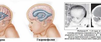
Acute form
An acute form of oxygen deficiency can occur at any time and even during delivery. With such a diagnosis, urgent surgical action is required. If this happens late in pregnancy, the woman will most likely be induced into labor or have a caesarean section.
Complications depend on the severity of hypoxia, the individual characteristics of the woman’s health and the course of pregnancy:
- With increased blood density, a woman in labor, her tissues and the tissues of the child are exposed to a large deficiency of oxygen and nutrients. This provokes hemorrhage and the death of some cells in the baby after birth. The nervous system and brain are most often affected;
- The pathology causes premature aging and placental abruption, which is dangerous for the child and mother. Premature labor may begin, bleeding may occur, and the child may experience hemorrhagic shock and bleeding.
It is difficult to predict what consequences a child will have during his subsequent life from oxygen starvation. But statistics show that such children have a weakened immune system, suffer from frequent illnesses and find it difficult to concentrate on something; it is difficult for them to study.
Signs of hypoxia. Apgar score
The baby's condition is assessed immediately after birth, in the first minute and 5 minutes later. For this purpose, a scale developed by Virginia Apgar is used, taking into account and summing up the following indicators, each of which is scored from 0 to 2 points:
- skin coloring;
- breathing rate;
- reflex activity;
- heart rate;
- muscle tone.
Based on the score obtained, the absence or presence of hypoxia and its degree are determined:
- norm – number of points 8-10;
- mild hypoxia – 6-7 points;
- moderate hypoxia – 4-5 points;
- severe hypoxia – 0-3 points.
Mild hypoxia is detected in almost all newborns in the first minute of life and disappears within 5 minutes on its own.
Moderate hypoxia in a newborn requires certain treatment; the child’s condition returns to normal after a few days. In case of severe hypoxia or asphyxia, immediate resuscitation measures are carried out, complex treatment and further monitoring of the child are prescribed.
The clinical picture of neonatal hypoxia is usually pronounced and the diagnosis is established immediately after the birth of the child. Signs of this condition include tachycardia, with gradual replacement by bradycardia (less than 100 beats per minute), irregular heartbeat, auscultation of heart murmurs, pallor of the skin and cyanosis of the nasolabial triangle and extremities.
Irregular breathing or its absence is noted, motor activity is reduced or absent (the child is lethargic or does not move), and the presence of meconium (green water) in the amniotic fluid. Blood clotting rates increase, which leads to thrombosis in the vessels and hemorrhage in the tissue.
Subsequently, if hypoxia was missed in the first minutes of the child’s life, the following signs are added:
- constant drowsiness;
- restless sleep, trembling;
- marbled skin tone of the extremities;
- the child freezes quickly (when bathing, changing clothes);
- restless, capricious behavior, causeless crying;
- trembling of the facial muscles while crying or at rest.
Hypoxic encephalopathy
Hypoxia in a newborn leads to the development of hypoxic encephalopathy (brain damage), which is divided into degrees of severity:
mild – drowsiness or agitation of the newborn, disappearing after 5-7 days;
moderate - in addition to drowsiness and/or agitation, there is crying for no reason, convulsions, aversion to being carried, rapid freezing;
severe - severe drowsiness and lethargy, development of psychomotor agitation or coma with ongoing convulsions.
How to detect fetal hypoxia?
You can try to identify fetal hypoxia yourself by observing changes in fetal motor activity. The initial stage is characterized by fetal restlessness, increased frequency and strength of movements.
Prolonged or progressive fetal hypoxia leads to a weakening of its movements.
The reason for urgent consultation with a doctor is a decrease in fetal movements up to 3 times within an hour. This indicates the suffering of the fetus. In this case, additional examination of amniotic fluid, cardiotocography, Doppler measurements, etc. will be carried out.
The most accurate and informative methods for assessing the condition of the fetus are cardiotocography and Doppler.
The main symptom of fetal hypoxia during childbirth is a violation of its cardiac activity. Therefore, cardiac monitoring of the fetal condition is widely used during childbirth. If the amniotic fluid is stained with meconium, i.e. turn green, which means the fetus may have hypoxia. However, this sign is only significant if the fetus lies head first.
If a pregnant woman has chronic intrauterine fetal hypoxia, then rest is important for her, because Bed rest greatly contributes to improving the blood supply to the uterus. Chronic fetal hypoxia is treated comprehensively, paying special attention to the underlying disease that led to it. Therapy is carried out to improve the oxygen supply to the fetus and normalize metabolic processes. If there is no effect from complex therapy, and the gestational age exceeds 28 weeks, then a decision is made about an emergency birth by cesarean section.
It is important to monitor the course of pregnancy very carefully in order to prevent or promptly diagnose and treat fetal hypoxia.
Therapeutic tactics of hypoxia
If fetal hypoxia occurs during the period of pushing or during contractions (decrease in heartbeat), a decision is made to complete the labor as soon as possible: a cesarean section or the application of obstetric forceps (in the case of labor stimulation with oxytocin, the administration of the drug is stopped). After the birth of the child, immediate medical care begins:
- clearing the respiratory tract from mucus, meconium and water (suction with a special aspirator);
- supplying a mixture of oxygen with air or pure humidified oxygen through a mask, nasal catheter or mechanical ventilation device (in case of severe hypoxia, the newborn is placed in an incubator, intubated and mechanical ventilation is started);
- heating the baby with radiant heat (on a special changing table), and in case of severe hypoxia, placing the baby in an incubator;
- administration of drugs that stimulate blood circulation and increase blood pressure (camphor, dopamine) and drugs that stimulate the respiratory center (etimizole);
- intravenous infusions solution, sodium bicarbonate (neutralization of carbon dioxide in the blood), glucose to restore the reduced volume of the vascular bed;
- transfusion of blood products if necessary (hemolytic disease of the newborn);
- prescribing antibiotics to prevent pulmonary infections in case of severe hypoxia or intrauterine infection of the fetus, as well as in respiratory distress syndrome during premature birth;
- prescription of anticonvulsants (phenobarbital, phenazepam);
- To reduce intracranial pressure, administration of diacarb and veroshpiron (diuretics with the effect of reducing cerebrospinal fluid production) is indicated.
Brain hypoxia in children
Hypoxia is a pathological process that occurs when there is insufficient supply of oxygen to the body’s tissues or a violation of its utilization in the process of biological oxidation; an important component in the pathogenesis of many diseases.
Depending on the causes and mechanisms of development, the following types of hypoxia are distinguished: exogenous (hypo- and normobaric), respiratory (respiratory), cardiovascular (circulatory), blood (hemic), tissue (primary tissue) and mixed.
Hypobaric exogenous hypoxia occurs when rising to altitude, when the total atmospheric pressure decreases and, accordingly, the partial pressure of oxygen decreases. Normobaric exogenous hypoxia develops under normal general barometric pressure, but a reduced partial pressure of oxygen in the inhaled air, for example, when staying in small enclosed spaces, working in mines, wells, or when there is a malfunction of the oxygen supply systems in the cabins of aircraft or submarines.
Respiratory (respiratory) hypoxia occurs as a result of insufficient gas exchange in the lungs due to alveolar hypoventilation, disturbances in ventilation-perfusion relationships, excessive intrapulmonary shunting of venous blood or difficulty in diffusion of oxygen in the lungs. The pathogenetic basis of respiratory hypoxia, as well as exogenous one, is arterial hypoxemia, in most cases combined with hypercapnia.
Cardiovascular (circulatory) hypoxia develops with circulatory disorders leading to insufficient blood supply to organs and tissues. A decrease in the amount of blood flowing through the capillaries per unit time may be due to general hypovolemia, i.e. a decrease in blood volume in the vascular bed (with massive blood loss, dehydration, etc.), and disturbances in the functioning of the cardiovascular system. Hypoxia of this type is local in nature when there is insufficient blood flow to a particular organ or tissue area or obstruction of blood outflow.
Blood (hemic) hypoxia occurs as a result of a decrease in the oxygen capacity of the blood during anemia, hydremia and when the ability of hemoglobin to bind, transport and release oxygen to tissues is impaired. Hemic hypoxia is characterized by a combination of normal oxygen tension in arterial blood with a reduced volume content. The tension and oxygen content in the venous blood are reduced.
Tissue (primary tissue) hypoxia develops due to a violation of the ability of cells to absorb oxygen or due to a decrease in the efficiency of biological oxidation as a result of the uncoupling of oxidation and phosphorylation. With tissue hypoxia, the tension and oxygen content in arterial blood can remain normal for a certain moment, while in venous blood they can exceed normal values; the arteriovenous difference in oxygen in these cases is reduced.
Mixed hypoxia occurs when two or more main types of hypoxia are combined. Often, primary hypoxia of any type, having reached a certain degree, causes dysfunction of other organs and systems involved in ensuring biological oxidation, giving hypoxia a mixed character.
In clinical practice, based on the speed of development and duration of the course, fulminant hypoxia is distinguished, developing within tens of seconds, acute hypoxia, occurring in several minutes or tens of minutes, and chronic hypoxia, lasting for weeks, months and years.
According to prevalence, local and general hypoxia are distinguished. Local hypoxia is associated with local disturbances in blood supply; general hypoxia of any type is common, but different organs and tissues are affected to different degrees due to significant differences in their resistance to hypoxia.
In acute hypoxia, dysfunctions of the nervous system usually begin with disorders of the most complex analytical and synthetic processes. Euphoria is often noted, and the ability to adequately assess the situation is lost. As hypoxia deepens, severe disturbances of higher nervous activity occur. Already in the early stages, there is a loss of coordination, first of complex and then of simple movements, which turns into adynamia. Circulatory disorders can be expressed in tachycardia, weakened contractility of the heart, arrhythmias up to atrial and ventricular fibrillation. Blood pressure may initially rise, then progressively fall until collapse develops; microcirculation disorders occur. In the respiratory system, after the activation stage, dyspneic phenomena are observed (various disturbances in the rhythm and amplitude of respiratory movements). After an often short-term stop, terminal (agonal) breathing develops - rare convulsive sighs, gradually weakening until it stops completely.
With fulminant hypoxia, most of the clinical changes are absent, because a complete cessation of vital functions quickly occurs and clinical death occurs.
Chronic hypoxia, which occurs with prolonged circulatory and respiratory failure, with blood diseases and other conditions accompanied by persistent disturbances of oxidative processes in tissues, is manifested by increased fatigue, shortness of breath, palpitations with little physical exertion, general discomfort, gradually developing dystrophic changes in various organs and tissues .
Rehabilitation after discharge
Further treatment after discharge from the maternity hospital (the child’s condition is satisfactory) is carried out by a local neonatologist, who prescribes massage, therapeutic exercises, feeding regimen and explains to the woman the rules for caring for a child who has experienced hypoxia after birth.
Medicines prescribed include drugs that improve blood circulation and nutrition of the brain (vinpocetine, piracetam, cerebrolysin), drugs that reduce intracranial pressure (diacarb, asparkam, potassium-containing drugs), and anticonvulsants according to indications.
The child is being monitored by a pediatrician and must be regularly examined by a neurologist.
Prevention
It is impossible to completely prevent the development of one or another pathology during pregnancy, but by following preventive measures, you can improve the condition of the body and prepare for gestation:
- eliminate bad habits completely, no alcohol and cigarettes;
- proper balanced nutrition;
- spend a lot of time outdoors;
- quality sleep and rest;
- physical activity;
- register with the consultation in a timely manner.
Ideally, doctors strongly recommend monitoring the condition of the reproductive system from an early age and undergoing gynecological examinations, and planning pregnancy. It is natural that when carrying a child, lead a correct lifestyle and control your well-being.
Consequences and prognosis
Mild and moderate hypoxia with timely and high-quality treatment does not lead to serious consequences, but severe hypoxia is dangerous due to the development of the following conditions:
- anxiety, restlessness of the child and mental lability;
- retardation in physical and mental development, headaches and development of neurocirculatory dystonia;
- increased intracranial pressure;
- development of epileptic syndrome and hydrocephalus;
- formation of brain cysts;
- damage to the cranial nerves and loss of their functions.
Rare consequences of severe hypoxia in a newborn include disability of the child or death.
The prognosis depends on the degree of hypoxia. With mild and moderate hypoxia and adequate therapy, the prognosis is favorable; with severe brain hypoxia, the prognosis is questionable.
Osteopathic treatment of hypoxia during childbirth
Osteopathic treatment rightfully occupies one of the leading places in the rehabilitation of children who have suffered from hypoxia. Balancing the bones of the skull allows you to relieve tension from the dura mater and the cerebral hemispheres, thereby ensuring the most complete functioning of the central nervous system.
Osteopathic techniques allow for drainage of the venous sinuses, as a result of which cerebrospinal fluid resorption is improved and intracranial pressure is normalized. Freeing the cervical spine and eliminating torticollis promotes adequate blood supply to the brain.
Early osteopathic treatment in the first year of life allows children not only to keep up with their peers in development, but sometimes even to develop ahead of schedule. It is very important to carry out this treatment from the first months of life, as this will help get rid of long-term clinical pathological symptoms. Osteopathy sessions are conducted for children starting from the first month of life. Osteopathic treatment, creating optimal functioning of the central nervous system, stops the process of post-hypoxic changes in neurons, thereby somehow reprogramming the brain for full development.
There is no need to be afraid of hypoxia; you need to take the entire range of measures for rehabilitation after it. Moreover, modern medicine gives us many tools for this.
Oxygen starvation of the fetus in pregnancy pathologies
Chronic diseases can provoke complications during pregnancy, so women suffering from hypertension, anemia or other systemic diseases need especially careful attention from a doctor. They must strictly follow the specialist’s recommendations and register with the antenatal clinic as early as possible.
But even for a seemingly healthy expectant mother, pregnancy may not proceed as desired. Previously undetected vascular diseases can lead to the formation of blood clots during pregnancy. Serious and severe complications of pregnancy are:
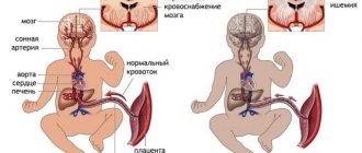
- placental abruption, in which part of the vessels supplying the placenta “switches off” and bleeding begins;
- placenta previa is its pathologically low location, disrupting the blood supply to the uterus. The lower the placenta is located, the thinner the walls of the uterus become and the fewer vessels nourish the placenta and the fetus in it;
- preeclampsia is a pathological condition that causes a sharp increase in blood pressure in a woman, which can lead to dangerous consequences, including the death of the mother.
All these conditions require mandatory medical monitoring. They are dangerous not only due to fetal asphyxia, but also pose a direct threat to the life of a woman. Treatment of the mother during their diagnosis is carried out strictly in a hospital setting. And if preeclampsia is detected, the doctor will recommend a set of mandatory measures that will reduce the risk of transition from mild to moderate and severe pathology, and also reduce the severity of oxygen starvation of the fetus.
“Observations show that as the mother’s age increases, the risk of pathologies during pregnancy increases,” notes obstetrician-gynecologist Evgeniy Petreikov. — Today, more and more women decide to become mothers for the first time at the age of 30–35. At the same time, many may develop chronic diseases that in ordinary life they simply do not notice and do not contact doctors about. Thus, hypertension is often detected after pregnancy, which becomes a risk factor for complications.”
pixabay.com/