Plegia and paresis are different stages of the same disease. Plegia (paralysis) involves a complete lack of movement, and with paresis the patient has partial impairments or slightly reduced activity. Paralysis can develop in the lower and upper limbs.
As a rule, plegia and paresis are symptoms of severe neurological disorders, the level of development of which is determined by the degree of development of muscle paralysis. Various muscles may be at risk (smooth muscles of the stomach, extraocular muscles, etc.).
This is weakness of some muscles caused by a disruption in communication with the nervous system and disorders of brain activity. Another cause of pathology is dysfunction of peripheral nerves, which lose the ability to transmit brain impulses to muscles.
Classification of violation
According to the number of limbs affected, experts divide paralysis into several categories:
Disorders are also classified according to the level of motor neuron damage. When the peripheral motor neuron is damaged, the disease is called flaccid (peripheral) paralysis; when the corticospinal tract is damaged, it is called spastic or central.
Causes and forms of development of deviations
Plegia cannot appear on its own; its development requires a trigger mechanism, that is, pathological processes, including:
- bulbar palsy - can have a progressive or acute form, affecting the muscles of the larynx and tongue;
- Bell's palsy - damages the face;
- familial myoplegia - can occur simultaneously in several relatives.
The development of the syndrome is also observed against the background of damage to the shoulder joint and can occur when the baby moves incorrectly along the birth canal.
Any paresis, like plegia, is associated with disorders of the nervous system, depending on the degree of its development, the level of complexity of the pathology will depend.
If the damage has spread to the roots, anterior horns and peripheral vessels, this causes loss of reflex and voluntary activity. This also affects tendon mobility.
With flaccid paresis, there are no protective reflexes or synkinesis; due to disorders in the functioning of the nerves, the muscle group that ensures their functioning atrophies.
Symmetrical paresis of the legs or feet can develop due to polyneuritis; if the plexuses are disturbed, unilateral plegia, localized in the muscles of the pelvic girdle, is caused.
Complete paralysis occurs with complete damage, when not only one segment of the brain space is affected, but also adjacent ones. The development of such a disease is possible in severe illness (amyotrophic lateral sclerosis, poliomyelitis).
Bell's palsy is caused by malignant tumors, infections, damage or hypothermia of the brainstem area, unsuccessful injury or surgery.
Familial paralysis develops for unknown reasons, and the appearance of bulbar palsy extends to the medulla oblongata.
A common cause of monoplegia is damage to the cortex in the cerebral hemispheres, vascular damage, tumors, abscess and trauma. It rarely occurs in lesions of the corticospinal tract, spinal cord, or brainstem due to the close proximity of fibers to the lower and upper extremities.
Paraplegia can develop when the spinal roots, spinal cord, or peripheral nerves are affected. Sometimes leg weakness can develop with hydrocephalus and tumors of the parasagittal zone.
Subacute and chronic paraplegia can develop with spinal cord tumors, multiple sclerosis, syphilitic meningomyelitis, diseases of the musculoskeletal system, herniated intervertebral discs in the cervical region, etc.
Possible causes of tetraplegia include the same factors as paraplegia. But the disease often occurs with lesions of the spinal cord in the cervical, thoracic and lumbar regions.
Other causes of plegia include: poisoning with salts of heavy metals, alcohol and poisons;
Mechanism of disease development
When various parts of the pyramidal tract are damaged, the natural path of impulse transmission is disrupted - from the cells of the cerebral cortex to the motor neurons, the cells responsible for the regulation of movement.
Coming from the base of the skull, the nerve pathways intersect, which gives grounds to assume damage to the structures of the right hemisphere in case of damage to the left side of the body, and in the case of dysfunction of the right limbs and face - in the left. But if changes in the nerve plexuses are localized below the overlap, then the manifestations of the disease are found on the affected side.
Plegia can be temporary and disappear after treatment, or become a persistent lifelong pathology.
Symptoms of pathology
Each age category has its own manifestations of the disease. Also, the signs differ due to their appearance:
- Bell's palsy is characterized by the lack of movement of the facial muscles on one side. At the same time, the patient’s speech is incomprehensible, he cannot open his eyes freely and eat without support.
- With bulbar palsy, patients complain of severe and sharp headaches, rapid heartbeat and difficulty breathing. Patients may experience dizziness, chills or fever, lack of clarity of speech, and problems with swallowing and breathing. Eating becomes impossible as all muscles are affected.
- The symptoms of pseudobulbar plegia are similar to the previous disease, but there is no atrophy of the facial muscles with sudden contractions. Sometimes all limbs may be affected simultaneously.
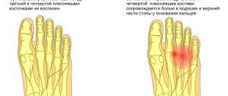
Weakness of any muscle or group leads to changes in gait, foot drop when raising the leg, weakness of the arms and legs when moving, and head droop.
If any symptom appears, you should immediately contact a specialist. The possibility of effective therapy and the duration of the rehabilitation period will depend on this.
Degrees of neurological deficit
In clinical practice, to identify the severity of the lesion and quickly determine the prognosis, a classification according to the degrees of neurological deficit is used, based on certain clinical symptoms:
1st degree:
accompanied by pain, stiffness of gait, disruption of typical motor reactions such as running and jumping, a slight proprioceptive deficit is possible, i.e. incorrect positioning of the limbs due to decreased/absent skin sensitivity.
2nd degree:
characterized by increased clinical signs, gait disturbance, and incoordination of movements. The pain may persist or be mild. Paraparesis (partial absence of motor function in the pelvic limbs) is possible. In this case, the animal is able to move independently.
3rd degree:
manifested by paraparesis of the pelvic limbs. At this stage, the ability to support is lost, the animal no longer moves independently, but the ability to control urination is retained.
4th degree:
characterized by paralysis of the limbs with preservation of deep pain sensitivity; the animal’s independent urination is not controlled. It is possible to urinate when the bladder is full, which is often mistaken by owners for normal controlled urination.
5th degree:
manifested by paralysis with the absence of deep pain sensitivity for less than 24 hours, there is no control of independent urination.
Assessment of deep pain sensitivity - consists of testing the dog’s ability to respond to a strong painful stimulus (compression of the periosteum of the fingers with a clamp) with a conscious reaction. If the test result is positive, the dog yelps or licks its lips, turns its head towards the source of pain, tries to bite, etc. Important!
The presence of a withdrawal reflex (the dog does not turn its head, but actively withdraws the irritated paw) is not a sign of the presence of deep pain sensitivity. 6th degree:
manifested by paralysis, lack of deep pain sensitivity for more than 24 hours, control of independent urination is also absent. The prognosis for restoration of limb function at grade 6 is unfavorable.
Making a diagnosis and conducting research
Typically, otolaryngologists, psychoneurologists, psychiatrists, neurosurgeons and pulmonologists are involved in the diagnosis of plegia. When identifying the causes of the syndrome, a detailed history taking and identifying each patient’s propensity for such psychogenic reactions is important.
When collecting anamnesis, the patient must answer several questions:
- duration of decrease in muscle strength;
- approximate reasons that caused the complaints (fever, diarrhea, headache, ingestion of canned food);
- whether harmful substances are used in the work;
- Do relatives have such phenomena?
A neurological examination is also carried out with an assessment of muscle strength using 5 points and a search for other manifestations (loss of reflexes, facial asymmetry, muscle atrophy).
The patient is prescribed a blood test to determine the erythrocyte sedimentation rate and the number of leukocytes. A toxicological analysis is carried out to identify poisoning.
Additional studies include:
- electroneuromyography (to assess the electrical activity of muscles, the time of nerve impulse conduction);
- test with Proserin (to determine myasthenia gravis);
- magnetic resonance angiography (to assess the integrity and patency of arteries in the skull, detect brain tumors);
- MRI, computed tomography of the spinal cord and head to study their structure, identify tissue disorders, tumors, hemorrhages, ulcers, etc.;
- electroencephalography to assess the electrical activity of areas of the brain.
A neurosurgeon may be needed.
What to do if your dog has paralysis
Paresis (paralysis) may occur suddenly or develop gradually over several days.
In any case, to make an accurate diagnosis, it is necessary to contact a veterinarian - a surgeon or neurologist - as soon as possible and conduct a series of studies.
Diagnosis should always begin with a neurological examination. During this procedure, the degree of neurological deficit in the patient and the location of the spinal cord damage are determined (in which particular segment of the spinal cord the compression occurred), which directly determines the method of further treatment and the prognosis for the restoration of the dog’s motor activity.
Approach to therapy
As a rule, the primary appearance of plegia is muscle discomfort. If the therapy is incorrectly chosen or absent, the pathology can cause complete paralysis.
Usually, with partial immobility, severe pain and acute illnesses appear, on the basis of which treatment is prescribed. Primary therapy is usually aimed at initially identifying and eliminating the underlying cause.
There are several methods for treating pathologies:
- surgery to remove a tumor of the spinal cord or brain, removal of hematomas, ulcers and antibacterial treatment;
- treatment with antibiotics for infectious damage to the nervous system;
- normalization of blood pressure , prescribing medications to improve blood flow in the brain and metabolism;
- to eliminate poisoning, vitamins A, B, C and solutions ;
- the use of drugs to improve neuromuscular reactions in myasthenia gravis;
- For botulism, antibotulinum serum .
In addition to drug therapy, massage courses are prescribed to increase muscle tone. An important role in the treatment of various types of pathology is played by the determination and willpower of the patient, who must strive for recovery to activate internal vital forces.
Neurological examination
Neurological examination includes:
- collecting a history of the dog’s life;
- establishing the time of onset of paralysis (less than 24 hours, more than 24 hours ago, more than 48 hours);
- assessment of the mental status (degree of consciousness) of the patient;
- assessment of motor activity, presence or absence of support ability, skin sensitivity, determination of muscle tone, testing of tendon reflexes and assessment of deep pain sensitivity.
Based on the results of the examination, additional research methods may be prescribed, such as radiography, myelography, CT or MRI of the spinal cord, blood tests, virological or bacteriological studies, cytology and biochemical analysis of cerebrospinal fluid.
Complications and prevention
In the absence of proper treatment, complications may occur in the form of impaired labor and social adaptation, persistent neurological defect and decline in muscle activity.
Preventive measures include:
- giving up alcohol and smoking;
- timely detection and treatment of infections, contacting a doctor when symptoms appear;
- checking blood pressure;
- healthy lifestyle, walks in the fresh air, exercise, sleep at least 8 hours;
- compliance with the diet and diet, adding foods with vitamins.
You should also normalize the load after restoration of muscle functions, avoid hypothermia and the manifestation of neuroses.