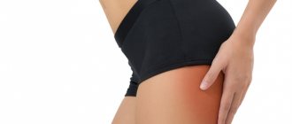
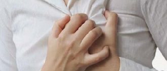
Pain in the buttock often appears as a result of pinching of the sciatic nerve by the piriformis muscle - a group of muscle fibers that begin in the sacrum area inside the pelvis and are attached by the lower tendon end to the femur. This muscle is responsible for external rotation of the hip and foot. Piriformis syndrome often accompanies degenerative changes in the discs of the lumbosacral region with the formation of a picture of radiculitis.
Doctors at the Osteomed Clinic in St. Petersburg almost daily observe manifestations of piriformis muscle syndrome, since lumbar osteochondrosis is the most common spinal problem in patients, regardless of their gender or age. And although pain in the buttock itself is not dangerous, it signals other disorders that need immediate correction. Experienced specialists conduct examinations and advise patients on this matter after making an appointment in advance.
Causes of piriformis syndrome
The cause of the syndrome can be various pathological changes in the piriformis muscle - damage, inflammation, fibrotic changes, spasms and an increase in muscle volume. Sometimes intramuscular injections lead to the disease, causing abscesses and the formation of infiltrate.
The main etiological factors of SHM: • Trauma. Piriformis syndrome can be associated with muscle overextension, muscle fiber tears, and fibrosis. In the latter case, the piriformis muscle shortens and thickens. • Post-traumatic hematomas and inflammatory processes – myositis, sacroiliitis, cystitis, prostatitis, endometriosis, prostate adenoma. • Vertebrogenic pathologies. This group includes spondyloarthrosis, osteochondrosis, intervertebral hernia in the lumbar region, spinal and vertebral tumors. As a result of irritation of the fibers of the sacral plexus and spinal roots, a reflex spasm occurs. • Muscle overload associated with prolonged forced position of the lumbopelvic segment. Increased loads occur in cases where, with radiculopathy, the patient tries to take an antalgic position (body position in which pain is minimal). Various sports can cause impairment, including weightlifting and running. • Oncological diseases of the tissues of the sacral region and proximal femur. They cause anatomical changes in structures. Neoplasia can cause spasm of the piriformis muscle. • Asymmetry, which occurs with shortened legs or scoliosis. Sometimes a hip amputation can put the muscle into a permanent spastic state that causes phantom pain.
PRINCIPLES OF THERAPY
In most cases, correction is required by the primary condition that caused the formation of muscular-tonic syndrome. When the primary source of pain impulse is eliminated, reflex muscular-tonic syndrome can regress. In cases where muscular-tonic disorders become the main or independent source of pain, both local and general effects are used. Stretching, massage of the affected muscle, warming physiotherapy, and manual therapy techniques aimed at mobilizing the affected spinal motion segment are performed. It is advisable to correct the motor stereotype, avoid provoking loads and poses
Block of the piriformis muscle. The point of infiltration of the piriformis muscle is found as follows. The greater trochanter of the femur, the superior posterior spine of the ilium and the ischial tuberosity are marked. These points are connected and a bisector is drawn from the superior posterior spine to the base of this triangle. The required point is located on the border of the lower and middle parts of this bisector. A needle is inserted here vertically to a depth of 6–8 cm and the muscle is infiltrated with a solution of an antiseptic and a steroid.
Then a massage will be performed with post-isometric relaxation of the muscles that rotate the thigh outward.
Gymnastic exercises that are recommended to relax the piriformis muscle and activate its antagonists can be performed in the following order. In the supine position with half-bent legs, the soles resting on the couch, the patient makes smooth movements of connecting and spreading the knees. Then, connecting the bent legs, the patient energetically pushes the other with one knee for 3-5 s. The next exercise, the “cradle,” is performed, if possible, without the help of hands while actively flexing the hips. Then, in a sitting position, they spread their soles wide apart, connect their knees and, leaning on the couch with the palm of their outstretched hand, begin to get up from the couch. By the time the palm comes off the couch, the other hand is offered to the instructor, who helps complete the straightening of the body. At this point, the connected knees are freely separated. When the condition improves, during the regression stage and during the period of remission, it is recommended to sit often (but not for long) in the cross-legged position.
Mechanism of development of the syndrome
The piriformis muscle (Musculus piriformis) is attached with its narrow end to the greater trochanter of the femur, and its wide end to the sacrum. It is involved in external rotation and during internal hip abduction, passing through the greater sciatic foramen. The inferior gluteal, sciatic, pudendal and posterior cutaneous nerves, gluteal arteries and veins also pass through this opening. This makes piriformis syndrome a disease that requires a comprehensive systemic approach. Persistent contraction of the piriformis muscle reduces the size of the infrapiriformis foramen. This leads to compression of passing vessels and nerves, primarily the sciatic one. Compression impairs the blood supply to the nerve trunk, being an additional pathogenetic component of sciatica.
Osteopathy offers an alternative to classical medicine and complements it, expanding the understanding of the disease. Osteopaths at the Quality of Life clinic can offer a solution to the problem with the piriformis muscle. Painful sensations that do not have a clear localization often arise as a result of spasm of the muscular-ligamentous apparatus, the causes of which are very diverse. When muscles contract, they move internal organs from their place, disrupt their function and cause pain. This is due to tension, compression and twisting of nerve, blood and lymphatic communications.
Read also
Rotational subluxation of the C1 vertebra
Dislocation or subluxation of a vertebra is a partial or complete disruption of the shape of the connecting articular surfaces of two adjacent vertebrae, and the overlying ones are considered dislocated.
The reason for the rotational... Read more
Humeroscapular periarthritis
Humeral-scapular periarthritis - (humeral-scapular periarthrosis, glenohumeral periatropathy) is the most common cause of shoulder pain. The disease is associated with pathology of the soft tissues around the joints.…
More details
Schmorl's hernia
When a person undergoes magnetic resonance imaging of the spine for the first time and sees the phrase “Schmorl’s hernia” in the conclusion, he is naturally overcome by a feeling of anxiety. After all, according to the patient, the terms...
More details
Pain in the neck
Pain in the cervical spine is somewhat less common than pain in the lower back. According to statistics, every fifth person suffers from such pain, and this problem occurs more often among ladies than among gentlemen. In patients with pathology...
More details
Radiculopathy
Radiculopathy is pain in various parts of the body caused by damage to the nerve roots of the spinal cord. Causes of radiculopathy This condition occurs due to...
More details
Classification of piriformis syndrome
Piriformis syndrome has only two forms: • Primary, which occurs due to direct damage. Primary SHM occurs as a result of injuries, myositis, and excess stress. • Secondary. This is the result of prolonged pathological impulses from the sacral or lumbar spine, sacroiliac joint, and small pelvis. Secondary SHM is formed by neoplasms in the spine, pelvic organs and hip joint.
Primary piriformis syndrome is caused by anatomical factors, variations of which may be related to separation of the piriformis muscle or sciatic nerve, as well as abnormal development of this nerve. Among patients with this disorder, less than 15% of cases are due to primary causes. Conventional medicine does not have precise data regarding the prevalence of FMS and does not offer evidence linking the disorder to sciatic nerve abnormalities or other types of sciatica.
The occurrence of secondary piriformis muscle syndrome is associated with the action of aggravating factors, including macro- and microtrauma, long-term accumulation of ischemia and the existence of local ischemia. • The development of piriformis syndrome most often (in about half of all cases) is promoted by trauma to the buttocks, which inflames the soft tissues and causes muscle spasm. This eventually causes compression of the nerve. • Spasms may be associated with muscle shortening caused by changes in leg and lumbosacral biomechanics. This causes compression or irritation of the sciatic nerve. When the piriformis muscle stops contracting normally, it can cause a variety of symptoms in the sciatic nerve area, including pain in the gluteal region and/or the hamstrings, calf, and lateral foot. Microtrauma can be caused by overuse, such as long walking or long distance running, as well as direct compression.
Osteopaths work with the root cause of the disorder, eliminating the problems that caused the development of BMS.
Butt stretch
Lying on your back, legs bent at the knees. Place your right ankle on your left knee. Grab your left thigh and pull it towards your chest. You will feel a stretch along your buttock and outer thigh. Hold the achieved position for 15-30 seconds. Then repeat the exercise with the other leg. 3 reps on each leg.
The role of spasms in the pathogenesis of piriformis syndrome
Spasm is a protective reaction of the body in the form of sustained tension and muscle contraction. It occurs as a response to threats associated with physical influences (blows, pain) or mental stress (fear, anxiety). When the threat passes, the tension gradually subsides, the tone of the muscles and ligaments is restored. However, this does not always happen.
If the muscle is constantly tense, this leads to compression of the vessels of the circulatory and lymphatic systems, because the tissues do not receive the required amount of oxygen and nutrients. As a result, their hypoxia develops and local immunity decreases, which leads to chronic inflammation. With constant stress over several weeks, fibrosis develops - a hardening of the tissue, which is usually accompanied by severe pain. But even at this stage the situation can still be corrected: the osteopath, having worked with the genitourinary system, the lumbopelvic region, and the dura mater, will eliminate spasms of muscles and ligaments and return them to their original tone. However, with very long spasms (more than a year), irreversible tissue scarring processes begin, resulting in ossification (calcification) of the muscle or ligament. Considering that spasm can be caused even by a visit to the gynecologist, not to mention childbirth or abortion, the scale of this problem in women is amazing.
The role of this muscle in ensuring the normal functioning of a woman’s body is great. Spasms and fibrotic phenomena often cause dull aching pain of different localization: • sacral; • lumbar; • gluteal.
They often intensify during squatting. In addition, spasms can be an additional risk factor for the development of a number of other pathologies - in particular, hemorrhoids and arthrosis of the hip joint.
Muscle spasms and fibrosis can also cause difficulties during labor. This is due to the fact that the birth canal fits tightly to the sacrum, so the contractions of the Musculus piriformis play an important role in the birth of the child, pushing and turning the fetal head. If the muscle is fibrotic, the fetal head stands in one position for a long time. This is fraught with damage to soft tissues and a number of other complications.
On the other hand, Musculus piriformis itself is injured during childbirth, which can cause fibrosis in the postpartum period. Therefore, every woman should visit an osteopath before and after childbirth. Voltage is easily determined during examination. Osteopathic techniques gently and painlessly eliminate spasm, and exercise therapy doctors will teach the patient the correct technique of exercises to strengthen the hips, back, buttocks and abs, and normalize the position of the lumbopelvic region.
We have considered only a small part of the situations and problems associated with internal spasms of the piriformis muscle. Each organ is supported by several muscles and ligaments, each of which, in turn, can go into spasm.
Moreover, the consequences are far from limited to pain, but can be much more serious. For example, forced displacement of the uterus during an abortion causes deep spasms of all the ligaments that attach the organ to the skeleton.
Since it does not return to its original anatomical position, sometimes twisting of blood vessels and nerves occurs. In case of a new pregnancy, this can lead to fetal hypoxia and developmental delay.
Osteopathic correction is a soft and delicate effect not only on the source of pain, but also on the true causes of the syndrome. A competent osteopath will help set the body up for self-healing, rehabilitate muscles and internal organs.
Symptoms
In approximately 70%, the gluteal-sacral area is affected first. The pain is constant, often nagging and aching. They intensify during walking, squats, and hip adduction. To reduce discomfort, the patient has to spread his legs to the sides. Over time, pain along the sciatic nerve - sciatica - is added to the symptoms. Shooting marks appear running from the foot to the buttocks. In the area where the SGM is located, pain sensitivity decreases and a burning sensation occurs.
Hypotonia of the muscles of the foot and lower leg gradually develops. With total compression of nerve fibers, “dangling foot” may develop. The main manifestations include intermittent claudication, which is a consequence of vascular compression. The same disorder leads to a decrease in leg temperature, pale skin and numbness of the fingers.
Clinical picture
Manifestations of the syndrome include a whole range of disorders:
- local symptoms;
- signs of sciatic nerve compression;
- clinical picture of vascular compression.
Local manifestations include the following:
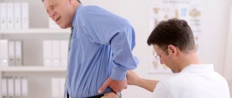
- Drawing and aching pain. It is located in the buttock, sacrum, and hip joint. The pain increases in a vertical position, with movement, with adduction of the hip. It progresses when a person squats.
- Vilenkin sign. In this case, discomfort appears during percussion. It is localized in the area of the piriformis muscle.
- Reducing pain in a horizontal position. It also decreases if a person sits with his legs apart.
- Bonnet-Bobrovnikova sign. When the large gluteal muscle is completely relaxed, a compacted piriformis muscle is felt underneath it. When pulled, it causes pain.
- Discomfort in the ischial spine. It is this area that the finger feels when quickly moving from the ischial tuberosity straight up.
Quite often, tonic tension of the piriformis muscle is complemented by the same condition of other muscle tissues of the pelvic floor. Almost always the deviation is accompanied by unexpressed sphincter deviations. In this case, there is a delay in the onset of urination.
When the sciatic nerve and vessels of the infrapiriform region are compressed, the following symptoms occur:
- Dull pain. In this case, a vegetative coloration is observed, which manifests itself in the form of chilliness, decreased sensitivity or burning in the affected area.
- Distribution of pain throughout the lower extremities. It mainly passes through the area of innervation of the peroneal and tibial nerves.
- Weakening of the Achilles reflex. A person experiences a deterioration in surface sensitivity.
- Symptoms worsen when exposed to stressors or heat. Also, the clinical picture may intensify when the weather changes.
If the abnormal process predominantly involves the fibers that form the tibial nerve, discomfort affects the posterior group of muscle tissues of the lower leg. It occurs when walking.
When the blood vessels are compressed, a sudden spasm of the lower limb occurs, which provokes lameness. In such a situation, a person is forced to stop while moving. Dermanogi acquires a pale tint. After resting, the person can continue to move, but soon the attack recurs.
Possible risks and complications of piriformis syndrome
Constant debilitating pain reduces the patient's ability to work. It can lead to: • emotional lability; • insomnia; • increased fatigue.
Peripheral paresis of the leg and foot caused by the syndrome develop muscle atrophy. With prolonged progression of the syndrome, the changes become irreversible. Persistent paresis leads to disability. Secondary spasm of the pelvic floor muscles is also possible. This causes discomfort when urinating, dyspareunia (pain during sexual intercourse) in women.
Osteopathic treatment
Osteopathic treatment does not just act on the sore spot, but treats the entire body as a whole; the doctor “listens to the body with his hands.” All organs and systems constantly pulsate, and spasms and tensions distort the picture characteristic of the body of a healthy person. The magnitude of these pulsations is insignificant, but is accessible to the sensitive hands of an osteopath.
Having examined these micropulsations, the doctor determines the cause of the syndrome and eliminates muscle tension. Muscle tissues and ligaments are stretched and relaxed, and the doctor uses gentle movements to move organs and tissues to return them to their normal anatomical position.
Osteopathic correction of the bones, muscles and ligaments of the pelvis, as well as the lumbosacral spine, plays a special role in osteopathic treatment. Soft muscular-fascial relaxation of the Musculus piriformis quickly restores the normal state and eliminates the cause of the syndrome - compression of the sciatic nerve and blood vessels. In especially severe cases, the treatment program is supplemented with drug blockade of the brain.
Plank
Lying on your stomach, lean on your elbows at shoulder level. Lift your hips off the floor, balancing on your forearms. It is important that your hips and shoulders are in line. Try to hold this position for 15 seconds. Then slowly lower yourself to the floor and relax. In the first stages, you can simplify the exercise by resting on the floor with your knees instead of your toes. Gradually increase the time you hold the plank to 1 minute.
Exercise therapy in the treatment program
After the acute pain syndrome is relieved, the results of osteopathic treatment are consolidated with the help of physical therapy. This allows you to avoid re-exacerbation of the syndrome. Exercises are performed under the guidance of an instructor. The program includes the implementation of special complexes aimed at strengthening: • the muscular corset of the lower back; • buttocks; • lower extremities. Work is also carried out with other parts of the musculoskeletal system.
Treatment of BMS is not aimed at relieving symptoms, but at eliminating the root cause of the syndrome. Therefore, any pain relief without treating the provoking factor is pointless. The exercises that are prescribed to patients are aimed at fully relaxing the muscular-ligamentous system, as well as activating antagonists in the movement of the lumbosacral region
Contraindications
In some clinical situations, blockade is not recommended. Contraindications are:
- blood diseases with an increased tendency to bleed;
- the patient is taking blood thinning medications;
- active infectious process on the skin in the area of the planned injection;
- acute infectious and inflammatory disease, accompanied by an increase in body temperature and a deterioration in the general condition of a person;
- history of allergies to local anesthetics (contraindication for the use of novocaine or lidocaine);
- diabetes mellitus, adrenal disease (contraindication for the use of corticosteroids);
- myasthenia gravis;
- obvious tendency to hypotension.