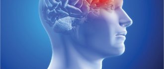
A hemorrhagic stroke is a bleeding in the brain. The disease develops due to damage to blood vessels. People are susceptible to pathology in the second half of life, mainly the elderly. The attack is accompanied by a characteristic neurological picture and requires urgent medical attention. In most cases, patients have a history of blood pressure surges or chronic diseases.
How does pathology develop?
Translated from Greek, “hemorrhagic” means “bleeding”, and stroke is translated as “stroke”. During an attack, the liquid part of the blood leaves the vascular bed, accumulates and forms a hematoma, which compresses the neurons of the brain, which are injured and die.
During a stroke, there are two types of bleeding in the brain:
- Epithelial damage - usually occurs due to weakness of the vessel wall and increased pressure. The artery “bursts” under stress, the blood forms a pulsating hematoma, which presses on the brain tissue.
- Diapedesis is a pathological increase in the permeability of the vascular wall. The artery itself is not damaged, but its lining allows blood cells to pass through, which accumulate and form a hematoma.
Clinical picture
The clinical manifestations of brain tumors are determined by their location in a limited volume of the cranial cavity. Compression or destruction of brain tissue in the tumor area (due to tumor growth) causes the so-called primary, or focal, symptoms. As the disease progresses, so-called cerebral symptoms appear, caused by impaired hemodynamics and intracranial hypertension.
Focal symptoms
Focal symptoms are largely determined by the location of the tumor. The following groups can be distinguished:
Sensory impairment
The ability to perceive external stimuli acting on the skin - thermal, painful, tactile - decreases or disappears. The ability to determine the position of parts of one's body in space may be lost. For example, with his eyes closed, the patient is unable to tell whether he is holding his hand with his palm up or down.
Memory impairment
When the cerebral cortex responsible for memory is damaged, complete or partial memory loss occurs. From not being able to recognize your loved ones to not being able to recognize or recognize letters.
Motor disorders (paresis, paralysis)
Muscle activity decreases due to damage to the pathways transmitting motor impulses. Depending on the location of the tumor, the pattern of the lesion differs. Both lesions of individual parts of the body and complete or partial lesions of the limbs and torso can develop. When the transmission of motor impulses from the cerebral cortex is disrupted, central type paralysis occurs, that is, a signal is received from the spinal cord to the muscles, they are in hypertonicity, but control signals from the brain do not enter the spinal cord, voluntary movements are impossible. When the spinal cord is damaged, peripheral paralysis develops; the signal from the brain enters the spinal cord, but the spinal cord cannot transmit it to the muscles, and the muscles are hypotonic.
Epileptic seizures
Convulsive seizures appear due to the formation of a focus of congestive excitation in the cortex.
Hearing and speech recognition impairment
When the auditory nerve is damaged, the ability to receive signals from the hearing organs is lost. When the area of the cortex responsible for recognizing sound and speech is damaged, for the patient all audible sounds turn into meaningless noise.
Impaired vision, object and text recognition
When the tumor is located in the area of the optic nerve or quadrigeminal region, complete or partial loss of vision occurs due to the inability to deliver a signal from the retina to the cerebral cortex. When areas in the cortex responsible for image analysis are damaged, a variety of problems occur, ranging from the inability to understand incoming signals to the inability to understand written language or recognize moving objects.
Speech impairment, oral and written
When areas of the cortex responsible for written and oral speech are damaged, their complete or partial loss occurs. This process, as a rule, is gradual and intensifies as the tumor grows - at first the patient’s speech becomes slurred (like that of a 2-3 year old child), his handwriting gradually changes, then the changes increase until it is completely impossible to understand his speech and handwriting in the form of a jagged line.
Autonomic disorders
Weakness and fatigue appear, the patient cannot get up quickly, he becomes dizzy, and fluctuations in pulse and blood pressure occur. This is due to impaired control of vascular tone and the influence of the vagus nerve.
Hormonal disorders
The hormonal background changes, the level of all hypothalamic-pituitary-dependent hormones may fluctuate.
Loss of coordination
When the cerebellum and midbrain are damaged, coordination is impaired, gait changes, and the patient is unable to make precise movements without vision control. For example, he misses when trying to reach the tip of his nose with his eyes closed. The patient is unstable in the Romberg position.
Psychomotor and cognitive impairment
Memory and attention are impaired, the patient becomes distracted, irritable, and his character changes. The severity of symptoms depends on the size and location of the affected area. The spectrum of symptoms ranges from absent-mindedness to complete loss of orientation in time, space and self. Episodes of psychomotor agitation and confusion are possible.
Intellectual and emotional disorders
When the brain is damaged due to space-occupying formations, intellectual functions and those personal characteristics that reflect the characteristics of social interaction are most affected: social conformity and psychoticism. The lateral features of this effect are that when the functions of the left hemisphere are impaired due to the development of a tumor, psychoticism increases to a greater extent, and when there is a deficit in the functioning of the right hemisphere, social conformity increases. Hemispheric features in changes in cognitive status in cases of space-occupying brain lesions are less pronounced than changes in personal characteristics. It should be noted, however, that damage to the left hemisphere causes a relatively greater decrease in intelligence, and to the right hemisphere - in creativity indicators.
Hallucinations
When areas in the cerebral cortex responsible for image analysis are damaged, the patient begins to hallucinate (usually simple: the patient sees flashes of light, a solar halo). The patient hears monotonous sounds (ringing in the ears, endless knocking).
General cerebral symptoms
General cerebral symptoms are symptoms that occur when intracranial pressure increases and compression of the main structures of the brain.
Headache
A distinctive feature of oncological diseases is the constant nature and high intensity of headaches, and their poor response to non-narcotic analgesics. Reducing intracranial pressure brings relief.
Vomiting (regardless of food intake)
Vomiting of central origin usually occurs due to an effect on the vomiting center in the midbrain. Nausea and vomiting constantly bother the patient; changes in intracranial pressure trigger the gag reflex. Also, the patient is unable to eat food and sometimes drink water due to the high activity of the vomiting center. Any foreign object that lands on the root of the tongue causes vomiting.
Dizziness
May occur as a result of compression of the cerebellar structures. The functioning of the vestibular analyzer is disrupted, the patient has central type dizziness, horizontal nystagmus, and often has the feeling that he, while remaining motionless, is turning or moving to one side or another. Dizziness can also be caused by tumor growth, leading to a deterioration in blood supply to the brain.
Why is a stroke not always fatal?
With hemorrhage, the risk of mortality is very high - about 70%. The survival of the remaining patients is explained by the following points:
- Area of damage - much depends on where the hematoma is located (vital centers, peripheral areas or lining of the brain). In some cases, a person’s lungs may fail, in others, coordination, vision or hearing will only decrease.
- Features of cerebral circulation - intracranial vessels are designed in a unique way, they reduce pulsation and ensure uniform blood flow. This slightly reduces the trauma to neurons caused by the growing hematoma.
- The presence of anastomoses is a bypass for blood circulation. When one vessel ruptures, some neurons receive nutrition through collaterals.
In most cases, the stroke clinical picture is the same, but with varying degrees of symptom severity. This is explained by the localization of hemorrhage and the death of specific neurons.
Classification
For a full perception of the disease, gradation by localization is common. Depending on which parts of the brain are affected, there are several variants of the disease:
- A subarachnoid stroke is a hemorrhage in the membranes of the brain. In this case, the hematoma puts pressure on the hemispheres from the outside.
- Hemispheric - one of the lobes is damaged, the hematoma can be located superficially on the outer, inner or lower side, in the ventricles of the brain.
- Stem - there may be hemorrhage in the medulla oblongata or midbrain, pons, cerebellum. These centers conduct impulses from certain receptors to the hemispheres and are responsible for coordination, breathing, auditory and visual reflexes.
- A combined option is when a hematoma compresses several areas of the brain. These varieties are the most common.
General cerebral symptoms and syndromes
Method of neurological diagnosis
Main neuropathological syndromes
Topical diagnosis of nervous system lesions
Additional research methods
Making a diagnosis in neurology has its own characteristics. A doctor examining a patient with damage to the nervous system always looks for an answer to two main questions:
— Where is the pathological focus?
- What is the nature of the pathological process?
In most cases, it is not possible to see the pathological focus with the eyes or palpate it, since the brain is enclosed in a bone case - the bones of the skull, and the peripheral nervous system is located deep in the soft tissues. In order to answer these questions, a neurologist must first of all be able to identify symptoms and syndromes of damage to the nervous system.
Thus, a three-stage scheme of neurological diagnosis emerges:
1. First, a syndromic diagnosis is made based on a carefully conducted clinical neurological examination;
2. Based on the analysis of identified symptoms and syndromes, a topical diagnosis is made, i.e. The location of the lesion in the nervous system is determined. At this stage, additional research methods are widely used. They allow you to confirm the doctor’s working hypothesis regarding the essence of the disease and play a key role in differential diagnosis.
3. At the final stage, after a differential diagnosis, a nosological diagnosis is made. A correctly established nosological diagnosis allows you to prescribe appropriate treatment.
Such a three-stage diagnostic scheme in neurology is quite logical, worked out over many decades by several generations of neurologists. Strict compliance with it greatly facilitates diagnostic work, especially for young doctors.
Main neuropathological syndromes
General cerebral symptoms and syndromes
They arise due to increased intracranial pressure, increased brain volume, difficulty in the outflow of cerebrospinal fluid through the cerebrospinal fluid pathways (brain aqueduct, foramina of Luschka, Magendie), irritation of blood vessels and meninges, and liquorodynamic disorders.
General cerebral symptoms include disturbances of consciousness, headache, dizziness, vomiting, and generalized seizures. These symptoms can sometimes occur as a manifestation of local brain damage.
Impaired consciousness. Coma
– complete loss of consciousness, lack of active movements, loss of sensitivity, loss of reflex functions, reactions to external irritation, respiratory and cardiac dysfunction.
With stupor
Separate elements of consciousness are preserved, the reaction to violent pain and sound stimulation is preserved.
Somnolence
– mild degree of impairment of consciousness. Characterized by lethargy, drowsiness, disorientation, lethargy, and indifference to the environment.
With brain tumors, “congestion” may occur,
expressed in lethargy, depression. Patients are indifferent, interest in the environment is reduced; Having answered the question, the patient withdraws again.
In infectious diseases accompanied by high intoxication, there may be disturbances of consciousness in the form of confusion of thinking (amentia).
The patients are inadequate, periods of psychomotor agitation are replaced by depression;
delusions and hallucinations (infectious delirium) are possible.
Headache
– the most common symptom of diseases of the nervous system. There are circulation headaches - with disorders of blood and liquor circulation, mechanical - with volumetric intracranial processes, toxic - with general infections, reflex - with diseases of the eyes, ears and other organs, psychogenic - with neuroses. Depending on the cause, the headache can be acute or dull, squeezing, bursting, throbbing, constant or paroxysmal, and can intensify with sudden turns of the head, walking, or shaking.
Dizziness
often observed in anemia, cerebral vascular diseases, cerebral hemodynamic disorders, volumetric processes. Systemic vertigo occurs when the vestibular apparatus is damaged - there is a clear direction of rotation of surrounding objects.
Vomit
– one of the most common cerebral symptoms. It should be remembered that “cerebral vomiting” does not always occur without preceding nausea and can improve the patient’s well-being. It is important to take into account the connection between vomiting and headaches and the simultaneity of their occurrence.
Seizures
may be focal symptoms. As a general cerebral symptom, they are more often observed in hypertension syndrome and cerebral edema.
Hypertension syndrome
accompanied by headache
, vomiting (often in the morning), dizziness, meningeal symptoms, stupor, and congestion in the fundus.
Hypertensive-hydrocephalic syndrome is caused by increased intracranial pressure and an increase in the amount of fluid in the cranial cavity. Hydrocephalus can be congenital or acquired. With acquired hydrocephalus, hypertension syndrome predominates in the clinic. Hydrocephalus can be internal (fluid accumulates in the ventricular system of the brain), external (fluid in the subarachnoid spaces of the brain) and mixed. Dislocation syndrome
develops when the brain stem or hemisphere is displaced during edema-swelling of the brain during space-occupying processes in the brain.
Meningeal syndrome occurs when the soft and arachnoid membranes are damaged due to increased intracranial pressure, an inflammatory or toxic process, or subarachnoid hemorrhage. It includes headache, nausea, vomiting, general hyperesthesia of the skin, hyperacusis, photophobia, and a specific meningeal posture (the “kicking dog” or “cocked hammer” pose - the stomach is retracted in a scaphoid manner, the arms are pressed to the chest, the legs are pulled up to the stomach). The result of the tonic reflex of the meninges is the following meningeal symptoms: stiff neck, Kernig's symptom (inability to straighten the leg at the knee joint when the hip is bent), Brudzinski's symptoms (bending of the legs when trying to bend the head with the chin to the sternum, when pressing on the pubis, when examining the symptom Kernig is on one leg, the other is bent).
Cortical lesion syndrome includes symptoms of loss of function or irritation of the cortical parts of various analyzers. Cortical movement disorders with damage to the anterior central gyrus are characterized by motor Jacksonian epilepsy and the presence of cortical monoplegia. Corticocerebellar ataxia, disturbances in standing and gait, and gaze paralysis in the opposite direction may occur.
Cortical sensitivity disorders are characterized by sensory Jacksonian epilepsy, hemi- or monohypesthesia. With cortical disorders, disorders of complex types of sensitivity predominate.
Visual cortical disorders with damage to the “visual” cortex are manifested by visual agnosia, homonymous half or quadrant hemianopsia, visual hallucinations, micro- and macropsia, photopsia. Auditory cortical disorders are manifested by auditory hallucinations, auditory agnosia (damage to the “auditory” cortex), taste, olfactory disorders - taste, olfactory hallucinations, agnosia (damage to the limbic system). A complex type of cortical disorders are apractical disorders (constructive, ideational, motor, oral, total apraxia).
Cortical speech disorders that arise from damage to the cortical speech centers are characterized by local types of aphasia (afferent, efferent, motor, sensory, amnestic, semantic) or total aphasia. Aphasic disorders are combined with reading, arithmetic, and writing disorders (alexia, acalculia, agraphia). Cortical hemispheric disorders may also be accompanied by mental disorders (“frontal psyche”, euphoria, manic, depressive state, decreased memory, attention, and other intellectual abilities).
Etiology
Provoking factors create conditions for damage to the vessel wall and the release of the liquid part of the blood beyond the vascular bed. There are many causes of hemorrhagic stroke. Let's note those that occur most often:
- Hypertension - a malignant form of the disease leads to cerebral hemorrhage in most cases. When pressure surges, the vessels are damaged and do not have time to recover.
- Atherosclerosis - arteries lose their strength and stretch worse when pressure changes.
- Congenital pathologies can cause rupture or dissection of the artery with subsequent hemorrhage.
- Chronic diseases - the cause of vessel rupture can be pathologies of an inflammatory or dystrophic nature. They can also provoke diapedesis.
Less common causes of hemorrhagic stroke include vitamin deficiencies, stress, and heart disease. Usually they aggravate the impact of the main factor and provoke hemorrhage.
There are risk groups that increase the likelihood of developing a stroke several times. Let's list them in descending order:
- hypertension;
- obesity;
- increased cholesterol;
- lack of leukocytes;
- eating disorders;
- stress;
- diabetes;
- hemorrhage in relatives.
Causes of cerebral disorders
- Increased intracranial pressure
- Increased brain volume
- Disturbance of cerebrospinal fluid dynamics
- Irritation of blood vessels and membranes of the brain
General cerebral symptoms include
- Impaired consciousness
- Headache
- Dizziness
- Nausea and vomiting
- Seizures
Impaired consciousness
Stunned
Loss of coherence of thoughts and actions. The basis is a violation of attention. It can be observed both with lesions of the cortex and with lesions of the stem structures of the reticular formation. It is observed with toxic, metabolic lesions of the brain, as well as with focal lesions of the cortex (especially the right parietal lobe). The patient is awake, but cannot perform a task requiring sustained attention (may be accompanied by severe writing disorder)
Delirium
- Stunned
- Increased activity of the sympathetic nervous system
- Hallucinations and delusions
Characterizes conditions accompanied by an increase in the content of catecholamines in the blood, alcohol withdrawal.
Pathological drowsiness
Constantly being in a state of drowsiness, sleep, from which the patient can be easily aroused. Without violating instructions and answering questions.
Sopor
It is impossible to completely awaken the patient even with the help of painful stimuli. Purposeful defensive movements are preserved. Speech contact is extremely difficult or impossible.
Coma
Superficial coma
- simple, erratic movements in response to a painful stimulus.
It is not possible to wake the patient. Deep coma
- there is no reaction to a painful stimulus.
Decerebrate rigidity
Extension, adduction and internal rotation of the arms with extension of the legs (focus in the upper parts of the brain stem between the red and vestibular nuclei).
Decorticate rigidity
Flexion and adduction of the arms with extension of the legs (focus above the midbrain, deep in the cerebral hemispheres).
Pseudocomatose states
Psychogenic unresponsiveness
While awake, the patient does not respond to examination and spoken speech.
An attempt to open your eyes encounters active resistance. During a cold test, the fast and slow phases of nystagmus are determined. EEG is not changed. Isolation syndrome (defferentation)
When the corticobulbar and corticospinal tracts are damaged.
Lack of motor functions while maintaining blinking and vertical eye movements. Extensive bilateral damage
to the prefrontal cortex Apathy, abulia, akinetic mutism.
Headache
Circulatory (for disorders of blood and liquor dynamics) Mechanical (for the occurrence of a volumetric process in the cranial cavity) Toxic (for general infectious diseases) Reflex (for pathology of the sensory organs) Psychogenic (for neuroses, including headaches of muscle tension) Headaches are divided into dull and sharp, squeezing and expanding, pulsating, pressing. There are constant and paroxysmal headaches
Dizziness
They can develop not only with neurological pathology, but also with somatic disorders. Dizziness, as a cerebral symptom, is distinguished by the absence of a clear direction of rotation of objects, while with damage to the vestibular apparatus, dizziness has a clear direction.
Vomit
Usually has a clear association with headache or dizziness. Although it is believed that vomiting during intracranial processes does not bring relief, in a sufficient number of cases this statement is very controversial, and patients sometimes feel relief in their well-being after an attack of vomiting.
Seizures
Usually a consequence of increased intracranial pressure or cerebral edema. More often they are generalized; local convulsions (especially in children) are often of a “flickering” nature, followed by convulsions of various parts of the body.
Symptoms of intracranial liquorodynamic disorders
Intracranial hypertension syndrome (hypertension syndrome)
Accompanied by headache, vomiting (often in the morning), dizziness, often the presence of meningeal symptoms and congestion in the fundus (with a long course of the process). An X-ray of the skull (with a long course of the process) reveals a widening of the entrance to the sella turcica, thinning of the sphenoid processes, an increase in the pattern of digital impressions and diploetic veins, and the phenomena of local osteoporosis in the bones of the skull can be determined.
Hydrocephalus (hydrocephalic syndrome)
Develops with increased intracranial pressure, impaired absorption of cerebrospinal fluid or increased production of cerebrospinal fluid.
Congenital hydrocephalus
A progressive increase in the size of the skull Divergence of the cranial sutures Thinning of the skull bones Bulging and tension of the large fontanelle Strengthening the venous pattern of the head Accompanied by the phenomena of stagnation and atrophy of the optic nerves in the fundus (usually on both sides) When performing a lumbar puncture, a decrease in the amount of protein is noted (less than 0.099 percent) and an increase in cerebrospinal fluid pressure of more than 180 millimeters of water.
Congenital hydrocephalus is often accompanied by severe neurological impairment of psychomotor development. Acquired hydrocephalus
Internal hydrocephalus is characterized by dilation of the ventricles of the brain due to the accumulation of large amounts of cerebrospinal fluid in them.
External hydrocephalus is characterized by an increase in the amount of cerebrospinal fluid in the subarachnoid space. Mixed hydrocephalus
is characterized by a combination of signs of external and internal hydrocephalus.
Occlusion syndrome
It develops as a result of blockage of the cerebrospinal fluid pathways at the level of the cerebral aqueduct, the foramina of Magendie, Luschka and Monroe. It often develops acutely and is called Bruns syndrome.
Occlusion at the level of the cerebral aqueduct
“Quadrigeminal” syndrome, characterized by nausea, vomiting, oculomotor disturbances, vertical nystagmus, upward or downward gaze paresis, “floating” gaze and cerebellar disorders.
Occlusion at the level of the foramina of Magendie and Luschka
It is characterized by dilation of the fourth ventricle and is manifested by dizziness, vomiting, nystagmus, severe bradycardia, “floating” gaze, ataxia and incoordination of movements of the eyeballs.
Occlusion at the level of the foramen of Monroe
It is characterized by dilation of the lateral ventricles and is manifested by general cerebral symptoms in combination with symptoms of damage to the hypothalamic-pituitary region
Bruns syndrome
Develops suddenly and is manifested by nausea, vomiting, dizziness, severe headache and impaired breathing and cardiac activity. Often occurs with sudden turns of the head or body.
Dislocation syndrome
Syndrome of displacement of the brain stem or hemisphere, which occurs with edema and swelling of the brain or with the development of an intracranial space-occupying process. With subtentorial changes, the clinic of forebrain lesions comes to the fore. With supratentorial changes, the clinic of damage to the brain stem (damage to the posterior cranial fossa) comes to the fore.
Meningeal syndrome (meningeal syndrome)
Meningeal syndrome is caused by damage to the soft and arachnoid membranes of the brain and develops due to increased intracranial pressure, inflammatory or toxic damage, and subarachnoid hemorrhage. The syndrome is based on irritation of the receptors of the vessels of the membranes, choroidal plexuses and sensory endings of the trigeminal, vagus nerves and sympathetic fibers.
Headache
Diffuse, most pronounced in the frontal or occipital region.
Vomit
Repeatedly recurring and independent of food intake and medications.
General cutaneous hyperesthesia and increased sensitivity to light and sound stimuli (hyperacusis and photophobia)
Cocked Pose
The head is thrown back, the torso is extended, the stomach is pulled in, the arms are pressed to the chest, the legs are pulled up to the stomach. Occurs due to involuntary reflex tonic contraction of muscles.
Meningeal symptoms
Stiff neck
Increased tone of the neck extensors (detected when trying to bend the head to the chest)
Kernig's sign
Inability to straighten the leg at the knee joint, previously bent at an angle of 90 degrees at the knee and hip joints. The symptom is involuntary.
Brudzinski's symptoms (provocation of meningeal posture)
The upper Brudzinski's symptom
is expressed in bending the legs at the knee joints in response to an attempt to bring the head to the chest.
The zygomatic Brudzinski symptom
is expressed by flexion of the legs at the knee joints in response to tapping on the zygomatic arch.
Brudzinski's buccal sign
is expressed by raising the shoulders and flexing the forearms when pressing on the cheek.
Brudzinski's pubic symptom
is expressed in the bending of the legs at the knee joints when pressing on the pubic symphysis.
The inferior Brudzinski sign
is examined together with the Kernig sign. When trying to straighten a leg bent at the knee joint, the second leg involuntarily bends at the knee and is brought to the stomach.
Gillen's sign
When the quadriceps muscle of the thigh is compressed, the leg involuntarily bends at the knee and is brought to the stomach.
In young children, tonic muscle tension is physiological, therefore the following symptoms are used to determine the presence of meningeal syndrome.
Lessage's sign of suspension
With the armpits raised, the child pulls his legs toward his stomach.
Tension and bulging of the large fontanel
(with increased intracranial pressure).
Bekhterev's symptom
Upon percussion of the zygomatic arch, an increase in headache is noted and an involuntary grimace of pain is detected on the corresponding half of the face.
Tripod symptom
The child sits, leaning on his hands located behind the buttocks.
Fanconi's sign
Inability to stand with the knee joints extended and fixed.
The “kiss the knee” symptom
You cannot touch the child's face to his knee due to the extension posture.
Meitus symptom With fixed knee joints, the child cannot sit up in bed (the back and legs form an obtuse angle).
Symptoms
In the stroke clinic, several periods are distinguished; symptoms differ depending on the location and type of lesion. It is important to know the characteristic signs of the disease, which will help provide first aid and determine the future prognosis.
Precursor period
The warning period is not observed in all patients; in some cases, the attack occurs acutely. Sometimes the symptoms indicate a second attack. Patients at risk should know the main warning signs:
- weak aching headaches;
- nausea and vomiting;
- dizziness;
- impaired sensitivity in the limbs;
- feeling of heat and redness of the skin on the face;
- painful reaction to light and sharp sounds;
- unilateral decrease in muscle tone.
Usually this period lasts several minutes, sometimes hours. Patients may feel fear of death. Sometimes behavioral disorders occur and increased aggression appears.
Attack
During this period, hemorrhage occurs, a growing hematoma appears, and the patient’s condition greatly deteriorates. The degree of loss of basic body functions depends on the nature of the brain damage.
Hemorrhagic stroke clinic - main symptoms:
- disturbance of consciousness, coma;
- shallow and rare breathing;
- convulsions on the affected side;
- Generalized spasms may occur;
- change in facial geometry;
- turning the eyes towards the hemorrhage and dilating the pupil;
- decreased reflexes on the opposite side of the body;
- meningeal signs - overstrain of the neck muscles, inability to bend the head.
An attack is the most dangerous period in a stroke clinic. According to statistics, more than 90% of deaths occur at this stage.
Recovery period
After a hemorrhagic stroke, patients experience characteristic consequences - these symptoms indicate the death of brain tissue:
- constant headaches;
- speech disorder;
- decreased vision and hearing;
- paresis and paralysis;
- lack of coordination;
- mental disorders, inappropriate behavior;
- serious impairment of motor activity.
If the patient does not die within the first 2 weeks, the likelihood of death is minimal. Further restoration is carried out throughout the year.
General cerebral and focal symptoms
Clinicians divide stroke symptoms into two large groups:
- General cerebral - characteristic of most forms. They cannot be used to determine the location of the hematoma; they only indicate damage to the central neurons.
- Focal signs are specific signs; based on their severity, the doctor can guess the location of the lesion.
More detailed symptoms are given in the table:
| General cerebral signs | Focal signs |
|
|
Important! In hemorrhagic stroke, general cerebral symptoms prevail over focal ones. This is explained by the fact that a hematoma can compress several lobes of the brain that regulate various functions.
Neurology of acute traumatic brain injury
There are several main types of interrelated pathological processes:
- direct damage to the brain substance at the time of injury;
- cerebrovascular accident;
- violation of liquor dynamics;
- disorders of neurodynamic processes;
- formation of scar-adhesive processes;
- autoneurosensitization processes.
The basis of the pathological picture of isolated brain injuries are primary traumatic dystrophies and necrosis, circulatory disorders and organization of tissue defects. Concussions are characterized by a complex of interconnected destructive, reactive and compensatory-adaptive processes occurring at the ultrastructural level in the synaptic apparatus, neurons, and cells. Brain contusion is an injury characterized by the presence in the substance of the brain and in its membranes of macroscopically visible foci of destruction and hemorrhage, in some cases accompanied by damage to the bones of the vault and base of the skull. Direct damage to the hypothalamic-pituitary, brainstem structures and their neurotransmitter systems during TBI determines the uniqueness of the stress response. Impaired metabolism of neurotransmitters is the most important feature of the pathogenesis of TBI. Cerebral circulation is highly sensitive to mechanical influences. The main changes that develop in the vascular system are expressed by spasm or dilation of blood vessels, as well as increased permeability of the vascular wall. Directly related to the vascular factor is another pathogenetic mechanism for the formation of the consequences of TBI—a violation of liquor dynamics. Changes in the production of cerebrospinal fluid and its resorption as a result of TBI are associated with damage to the endothelium of the choroid plexuses of the ventricles, secondary disorders of the microvasculature of the brain, fibrosis of the meninges, and in some cases, liquorrhea. These disorders lead to the development of liquor hypertension, and less commonly, hypotension.
In TBI, hypoxic and dysmetabolic disorders play a significant role in the pathogenesis of morphological disorders, along with direct damage to nerve elements. TBI, especially severe, causes respiratory and circulatory disorders, which aggravates existing cerebral dyscirculatory disorders and collectively leads to more pronounced brain hypoxia.
Currently (L.B. Likhterman, 1990) there are three basic periods during traumatic brain disease: acute, intermediate, and long-term.
The acute period is determined by the interaction of the traumatic substrate, damage reactions and defense reactions and is the period of time from the moment of the damaging effects of mechanical energy until the stabilization at one level or another of impaired cerebral and general body functions or the death of the victim. Its duration ranges from 2 to 10 weeks, depending on the clinical form of TBI.
The intermediate period is characterized by the resorption and organization of areas of damage and the deployment of compensatory and adaptive processes until complete or partial restoration or stable compensation of impaired functions. The length of the intermediate period for non-severe TBI is up to 6 months, for severe TBI – up to a year.
The long-term period is the completion or coexistence of degenerative and reparative processes. The length of the period for clinical recovery is up to 2-3 years, for a progressive course it is unlimited.
Classification of acute traumatic brain injury
All types of TBI are usually divided into closed, open and penetrating brain injuries. Closed TBI is a mechanical damage to the skull and brain, resulting in a number of pathological processes that determine the severity of the clinical manifestations of the injury. Open TBI should include injuries to the skull and brain in which there are wounds to the integument of the skull (damage to all layers of the skin). Penetrating injuries involve disruption of the integrity of the dura mater.
Classification of traumatic brain injury (Gaidar B.V. et al., 1996):
- brain concussion;
- brain contusion: mild, moderate, severe;
- compression of the brain against the background of a bruise and without a bruise: hematoma - acute, subspinal, chronic (epidural, subdural, intracerebral, intraventricular); hydro wash; bone fragments; edema-swelling; pneumocephalus.
It is very important to determine:
- condition of the intrathecal spaces: subarachnoid hemorrhage; cerebrospinal fluid pressure - nolimotension, hypotension, hypotension; inflammatory changes;
- condition of the skull: no bone damage; type and location of the fracture;
- condition of the skull: abrasions; bruises;
- associated injuries and diseases: intoxication (alcohol, drugs, etc., degree).
It is also necessary to classify TBI according to the severity of the victim’s condition, the assessment of which includes the study of at least three components:
- state of consciousness;
- state of vital functions;
- state of focal neurological functions.
There are five gradations of the condition of patients with TBI.
Satisfactory condition . Criteria:
- clear consciousness;
- absence of violations of vital functions;
- absence of secondary (dislocation) neurological symptoms;
- absence or mild severity of primary focal symptoms.
There is no threat to life (with adequate treatment); the prognosis for recovery is usually good.
Moderate condition . Criteria:
- state of consciousness - clear or moderate stupor;
- vital functions are not impaired (only bradycardia is possible);
- focal symptoms - one or another hemispheric and craniobasal symptoms may be expressed, often appearing selectively.
The threat to life (with adequate treatment) is insignificant. The prognosis for restoration of working capacity is often favorable.
Serious condition . Criteria:
- state of consciousness - deep stupor or stupor;
- vital functions are impaired, mostly moderately according to 1-2 indicators;
- focal symptoms:
- stem - moderately expressed (anisocoria, decreased pupillary reactions, limited upward gaze, homolateral pyramidal insufficiency, dissociation of meningeal symptoms along the body axis, etc.);
- hemispheric and craniobasal - clearly expressed both in the form of symptoms of irritation (epileptic seizures) and loss (motor disorders can reach the degree of plegia).
The threat to life is significant and largely depends on the duration of the serious condition. The prognosis for restoration of working capacity is sometimes unfavorable.
Extremely serious condition . Criteria:
- state of consciousness - coma;
- vital functions - gross violations in several parameters;
- focal symptoms:
- stem - expressed roughly (plegia of upward gaze, gross anisocoria, divergence of the eyes along the vertical or horizontal axis, a sharp weakening of the pupils' reactions to light, bilateral pathological signs, hormetonia, etc.);
- hemispheric and craniobasal - pronounced.
The threat to life is maximum and largely depends on the duration of the extremely serious condition. The prognosis for restoration of working capacity is often unfavorable.
Terminal state . Criteria:
- state of consciousness - terminal coma;
- vital functions - critical impairment;
- focal symptoms:
- stem - bilateral fixed mydriasis, absence of pupillary and corneal reflexes;
- hemispheric and craniobasal - blocked by general cerebral and brainstem disorders.
Survival is usually impossible.
Clinical picture of acute traumatic brain injury
Brain concussion. Clinically, it is a single functionally reversible form (without division into degrees). With a concussion, a number of general cerebral disorders occur: loss of consciousness or, in mild cases, a short-term blackout from several seconds to several minutes. Subsequently, a stunned state persists with insufficient orientation in time, place and circumstances, unclear perception of the environment and narrowed consciousness. Retrograde amnesia is often detected - loss of memory for events preceding the injury, less often anterograde amnesia - loss of memory for events subsequent to the injury. Speech and motor agitation are less common. Patients complain of headache, dizziness, nausea. An objective sign is vomiting. A neurological examination usually reveals minor scattered symptoms: oral automatism (proboscis, nasolabial, palmomental); unevenness of tendon and skin reflexes (as a rule, there is a decrease in abdominal reflexes and their rapid exhaustion); moderately expressed or unstable pyramidal pathological signs (Rossolimo, Zhukovsky, less often Babinsky symptoms). Cerebellar symptoms are often clearly manifested: nystagmus, muscle hypotonia, intention tremor, instability in the Romberg position. A characteristic feature of concussions is the rapid regression of symptoms; in most cases, all organic signs disappear within 3 days. Various vegetative and, above all, vascular disorders are more resistant to concussions and mild bruises. These include fluctuations in blood pressure, tachycardia, acrocyanosis of the extremities, diffuse persistent dermographism, hyperhidrosis of the hands, feet, and armpits.
Brain contusion (CBM) is characterized by focal macrostructural damage to the brain substance of varying degrees (hemorrhage, destruction), as well as subarachnoid hemorrhages, fractures of the bones of the vault and base of the skull.
Mild brain contusion is clinically characterized by a short-term loss of consciousness after the injury, up to several tens of minutes. Upon its recovery, typical complaints are headache, dizziness, nausea, etc. As a rule, retro-, con-, anterograde amnesia, vomiting, and sometimes repeated are noted. Vital functions are usually without significant impairment. Moderate tachycardia and sometimes arterial hypertension may occur. Neurological symptoms are usually mild (nystagmus, mild anisocoria, signs of pyramidal insufficiency, meningeal symptoms, etc.), mostly regressing in 2-3 weeks after TBI. With mild UHM, in contrast to concussion, fractures of the calvarial bones and subarachnoid hemorrhage are possible.
Moderate brain contusion is clinically characterized by loss of consciousness after injury lasting up to several tens of minutes or even hours. Con-, retro-, anterograde amnesia, headache, often severe, are expressed. Repeated vomiting may occur. Mental disorders occur. Transient disorders of vital functions are possible: bradycardia or tachycardia, increased blood pressure; tachypnea without disturbances in the rhythm of breathing and patency of the tracheobronchial tree; low-grade fever. Meningeal symptoms are often prominent. Brainstem symptoms are also detected: nystagmus, dissociation of meningeal symptoms, muscle tone and tendon reflexes along the body axis, bilateral pathological signs, etc. Focal symptoms are clearly manifested, determined by the localization of the brain contusion: pupillary and oculomotor disorders, paresis of the limbs, sensitivity disorders, etc. . Organic symptoms gradually smooth out over 2-5 weeks, but some symptoms can persist for a long time. Fractures of the bones of the vault and base of the skull, as well as significant subarachnoid hemorrhage, are often observed.
Severe brain contusion is clinically characterized by loss of consciousness after injury lasting from several hours to several weeks. Motor agitation is often pronounced, and severe, threatening disturbances in vital functions are observed. The clinical picture of severe UHM is dominated by brainstem neurological symptoms, which in the first hours or days after TBI overlap focal hemispheric symptoms. Paresis of the limbs (up to paralysis), subcortical disorders of muscle tone, reflexes of oral automatism, etc. can be detected. Generalized or focal epileptic seizures are noted. Focal symptoms regress slowly; gross residual effects are frequent, primarily in the motor and mental spheres. Severe UHM is often accompanied by fractures of the vault and base of the skull, as well as massive subarachnoid hemorrhage.
An undoubted sign of fractures of the base of the skull is nasal or auricular liquorrhea. In this case, a “spot symptom” on a gauze napkin is positive: a drop of bloody cerebrospinal fluid forms a red spot in the center with a yellowish halo along the periphery.
Suspicion of a fracture of the anterior cranial fossa arises with the delayed appearance of periorbital hematomas (a symptom of glasses). With a fracture of the temporal bone pyramid, Battle's symptom (hematoma in the mastoid region) is often observed.
Compression of the brain is a progressive pathological process in the cranial cavity, arising as a result of injury and causing dislocation and infringement of the brainstem with the development of a life-threatening condition. With TBI, compression of the brain occurs in 3-5% of cases, both with and without UHM. Among the causes of compression, intracranial hematomas come first - epidural, subdural, intracerebral and intraventricular; This is followed by depressed fractures of the skull bones, areas of brain crushing, subdural hygromas, and pneumocephalus.
The clinical picture of brain compression is expressed by a life-threatening increase in a certain period of time (the so-called light interval) after the injury or immediately after it of general cerebral symptoms, progression of impaired consciousness, focal manifestations, brain stem symptoms.
Complications of traumatic brain injury
Violations of vital functions - a disorder of the basic life support functions (external respiration and gas exchange, systemic and regional circulation). In the acute period of TBI, the causes of acute respiratory failure are dominated by deterioration in pulmonary ventilation associated with impaired airway patency caused by the accumulation of secretions and vomit in the nasopharynx with their subsequent aspiration into the trachea and bronchi, and retraction of the tongue in comatose patients.
Dislocation process: temporotentorial inclusion, representing a displacement of the mediobasal sections of the temporal lobe (hippocampus) into the fissure of the tentorium of the cerebellum and herniation of the cerebellar tonsils into the foramen magnum, characterized by compression of the bulbar sections of the trunk.
Purulent-inflammatory complications are divided into intracranial (meningitis, encephalitis and brain abscess) and extracranial (pneumonia), hemorrhagic (intracranial hematomas, cerebral infarctions).
Scheme of examination of victims with traumatic brain injury
- Identification of injury history: time, circumstances, mechanism, clinical manifestations of injury and extent of medical care before admission.
- Clinical assessment of the severity of the victim’s condition, which is of great importance for diagnosis, triage and provision of stage-by-stage assistance to victims. State of consciousness: clear, stunned, stupor, coma; the duration of loss of consciousness and the sequence of exit are noted; memory impairment - antero- and retrograde amnesia.
- State of vital functions: cardiovascular activity - pulse, blood pressure (a common feature in TBI - the difference in blood pressure on the left and right extremities), breathing - normal, impaired, asphyxia.
- Condition of the skin - color, moisture, bruises, presence of soft tissue damage: location, type, size, bleeding, liquorrhea, foreign bodies.
- Examination of internal organs, skeletal system, concomitant diseases.
- Neurological examination: state of cranial innervation, reflex-motor sphere; the presence of sensitive and coordination disorders; state of the autonomic nervous system.
- Shell symptoms: stiff neck, Kernig-Brudzinski symptoms.
- Echoencephaloscopy.
- X-ray of the skull in two projections; if damage to the posterior cranial fossa is suspected, a posterior semi-axial image is taken.
- Computer or magnetic resonance imaging of the skull and brain.
- Ophthalmological examination of the condition of the fundus of the eye: swelling, congestion of the optic nerve head, hemorrhages, condition of the vessels of the fundus.
- Lumbar puncture - in the acute period is indicated for almost all victims with TBI (with the exception of patients with signs of compression of the brain) with measurement of cerebrospinal fluid pressure and removal of no more than 2-3 ml of cerebrospinal fluid, followed by laboratory testing.
- The diagnosis reflects: the nature and type of brain damage, the presence of subarachnoid hemorrhage, compression of the brain (cause), liquor hypo- or hypertension; condition of the soft covers of the skull; fractures of the skull bones; the presence of concomitant injuries, complications, intoxications.
Organization and tactics of conservative treatment of victims with acute TBI
In general, victims with acute TBI should go to the nearest trauma center or medical facility where initial medical evaluation and emergency medical care are provided. The fact of injury, its severity and the condition of the victim must be confirmed by appropriate medical documentation.
Treatment of patients, regardless of the severity of TBI, should be carried out in an inpatient setting in a neurosurgical, neurological or trauma department.
Primary medical care is provided for urgent reasons. Their volume and intensity are determined by the severity and type of TBI, the severity of the cerebral syndrome and the possibility of providing qualified and specialized assistance. First of all, measures are taken to eliminate airway and cardiac problems. For convulsive seizures and psychomotor agitation, 2-4 ml of diazepam solution is administered intramuscularly or intravenously. If there are signs of compression of the brain, diuretics are used, if there is a threat of cerebral edema, a combination of “loop” and osmodiuretics is used; emergency evacuation to the nearest neurosurgical department.
To normalize cerebral and systemic circulation during all periods of traumatic illness, vasoactive drugs are used; in the presence of subarachnoid hemorrhage, hemostatic and antienzyme agents are used. The leading role in the treatment of patients with TBI is given to neurometabolic stimulants: piracetam, which stimulates the metabolism of nerve cells, improves cortico-subcortical connections and has a direct activating effect on the integrative functions of the brain. In addition, neuroprotective drugs are widely used. To increase the energy potential of the brain, the use of glutamic acid, ethylmethylhydroxypyridine succinate, and vitamins B and C is indicated. Dehydration agents are widely used to correct liquorodynamic disorders in patients with TBI. To prevent and inhibit the development of adhesive processes in the membranes of the brain and to treat post-traumatic leptomeningitis and choreoependymatitis, so-called absorbable agents are used.
The duration of treatment is determined by the dynamics of regression of pathological symptoms, but requires strict bed rest in the first 7-10 days from the moment of injury. The duration of hospital stay for concussions should be at least 10-14 days, for mild bruises - 2-4 weeks.
A.Yu. EMELYANOV, Head of the Department of Nervous Diseases, Military Medical Academy named after. CM. Kirova (St. Petersburg), professor, doctor of medical sciences
Diagnostics
To accurately identify the disease and differentiate hemorrhage from ischemia, an instrumental examination is performed:
- MRI or CT scan - the images show a limited area of the hematoma. The safety of magnetic tomography allows for multiple examinations and monitoring of neuronal recovery.
- Angiography - shows the condition of blood vessels. Well displays arterial rupture, signs of atherosclerosis and congenital anomalies.
- Lumbar puncture - taking cerebrospinal fluid in the lumbar region for analysis. The diagnosis of hemorrhagic stroke is made when red blood cells are detected in the cerebrospinal fluid in high concentrations.
Treatment
For hemorrhagic stroke, treatment and recovery are two successive stages of the therapeutic program. At the initial stages, pre-medical and medical care is provided, then rehabilitation is carried out.
First aid
If hemorrhage begins, every second counts, so it is very important to immediately help the patient and call a doctor.
First aid algorithm:
- put the patient on the bed, put a pillow under the head;
- check breathing and pulse;
- perform cardiopulmonary resuscitation;
- measure pressure every 15 minutes.
Delivery of the patient to the intensive care unit or intensive care unit
At the clinic, the patient receives professional medical care - artificial ventilation of the lungs is performed, and IVs are placed. The doctor prescribes the following medications:
- from high blood pressure - Dibazol, Benzohexonium;
- for cerebral edema - Veroshpiron, Furosemide;
- to prevent rebleeding - Vikasol, Etamzilat;
- detoxification therapy - Cytoflavin, glucose solution.
Sometimes surgical treatment is prescribed - punctures are made in certain places, the hematoma is pumped out through medical systems, and compression of the brain tissue is reduced.
Rehabilitation
Rehabilitation after a stroke is recovery that is carried out at home and in specialized centers. Successful implementation of the program requires a lot of effort from the patient and the doctor.
Basic principles of rehabilitation after hemorrhagic stroke:
- therapeutic exercises, kinesiotherapy;
- physiotherapy;
- occupational therapy;
- logotherapy;
- hydrotherapy;
- robotic technologies;
- functional electrical stimulation of FES;
- transcranial electrical stimulation t-Dcs;
- a virtual reality;
- psychologist consultations.
To restore the functioning of the upper limbs after a stroke, neurorehabilitation methods are used. They are based on the following principle: healthy cells located next to the affected part of the brain are reunited with neighboring ones, thus restoring the transmission of information, receiving appropriate stimuli.