Causes of arachnoiditis
In most patients with arachnoiditis, the predisposing factor is infectious diseases. In particular, these diseases include chickenpox, influenza, measles, viral meningitis, cytomegalovirus infection, and meningoencephalitis. The disease can also be provoked by chronic intoxication of the body, inflammatory diseases of the paranasal sinuses, and trauma. Arachnoiditis is often diagnosed in patients who experience reactive inflammation of a growing tumor.
Pathology can also occur due to acute or chronic purulent otitis media. In this case, inflammation is provoked by toxins and low-virulent microbes. Researchers also include various complications of purulent otitis (petrositis, labyrinthitis, sinus thrombosis), brain abscess, purulent meningitis and otogenic encephalitis as the causes of the disease.
In neurology, there are also a number of factors that are considered predisposing to the occurrence of the disease. Such factors include intoxication (for example, alcohol), frequent viral diseases, chronic overwork, hard work in an unfavorable climate, and frequent injuries. In 10% of all cases of the disease, it is impossible to establish the exact etiology.
Pathogenesis of arachnoiditis
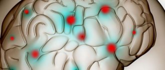
To understand the nature of the disease, it is necessary to become familiar with the anatomical features of the brain. The arachnoid membrane, which is affected by inflammation during arachnoiditis, is located between the soft and dura mater. Moreover, it is not fused with them, but simply fits tightly. Unlike the pia mater, the arachnoid membrane does not penetrate the cerebral convolutions. Small spaces filled with cerebrospinal fluid are formed under it.
All these spaces connect to the fourth ventricle. Through these spaces there is an outflow of cerebrospinal fluid from the cranial cavity. The mechanism of occurrence of arachnoiditis is as follows: due to the influence of various causes and provoking factors, the body activates the production of antibodies to the arachnoid membrane, which then provokes its inflammation. In patients with arachnoiditis, there is clouding and noticeable thickening of the arachnoid membrane, as well as the appearance of cystic expansions and connective tissue adhesions in it.
Pathogenesis
Arachnoiditis causes morphological changes in the form of clouding and thickening of the arachnoid membrane, which may be complicated by fibrinoid deposits. Most often they are diffuse, but in some cases they can be limited, that is, we are talking about more severe local disorders initiated by an extensive process of arachnoiditis. Macroscopic changes in this case are:
- clouding and thickening (hyperplasia of the arachnoid endothelium) of the arachnoid membrane, its fusion with the choroid and dura mater of the brain;
- diffuse infiltration;
- expansion of subarachnoid slit-like formations and cisterns at the base of the brain, development of their hydrops (overflow of cerebrospinal fluid).
The further course of the pathology leads to fibrosis and the formation of adhesions between the choroid and arachnoid membrane, impaired circulation of the cerebrospinal fluid (cerebrospinal fluid) and the formation of one or more arachnoid cysts. In this case, the normal circulation of the liquor fluid is disrupted and, as a result, hydrocephalus , the mechanism of which is based on two development paths:
- occlusive - resulting from a violation of the outflow of fluid from the ventricular system, for example, the closure of the Luschka or Magendie holes by the resulting adhesions or cysts;
- aresorptive – in which the processes of fluid absorption through the structures of the dura mater are disrupted, as a result of a diffuse “adhesive” process.
Classification of arachnoiditis
- Arachnoiditis of the meninges
- Optico-chiasmatic arachnoiditis
- Arachnoiditis of the posterior cranial fossa
- Arachnoiditis of the spinal cord membranes
This type of disease is also called cerebral. Cerebral arachnoiditis is localized in the posterior cranial fossa, on the convex surface of the brain and its base. The clinical picture of this disease is characterized by regular headaches and impaired circulation of the cerebrospinal fluid. In the most severe cases, the disease is accompanied by convulsive seizures, which can even lead to status epilepticus.
Arachnoiditis of the brain is often located in the central gyri and anterior parts of the cerebral hemisphere. Due to the resulting pressure on the sensory and motor centers, the patient may experience sensitivity and movement disorders. If the cerebral cortex is compressed or a cyst forms in it due to arachnoiditis, the patient may experience epileptic seizures.
This type of arachnoiditis is localized mainly in the chiasmal region. Frequent causes of this form of arachnoiditis are tonsillitis, malaria, syphilis, infectious diseases of the paranasal sinuses, and traumatic brain injuries. This type of arachnoiditis is characterized by the formation of adhesions in the area of the intracranial part of the optic nerves and the chiasm. In the most difficult cases, a scar may form around the chiasm.
As a rule, the disease provokes vision problems in the patient. In this case, the degree of decrease in the patient’s vision can vary from its minimal decrease to blindness. In most cases of optochiasmatic arachnoiditis, patients experience optic nerve atrophy. Visual symptoms are often severe, while hypertension symptoms are moderate.
It is the most common type of cerebral arachnoiditis. The severity of the symptoms of the disease depends on the location and nature of the inflammatory process, as well as its combination with hydrocephalus. The formation of cysts and adhesions usually leads to the closure of the openings of the cerebral ventricles, which provokes an increase in intracranial pressure. If intracranial pressure does not increase and is normal, the disease can last a long time.
The acute form of the pathology is characterized by all the symptoms of high intracranial pressure: nausea, dizziness, vomiting, bradycardia, severe headache localized in the back of the head. With a less acute course of the disease, signs of damage to the posterior cranial fossa become most pronounced. Patients may also experience symptoms such as unsteady gait and spontaneous nystagmus.
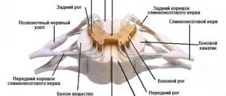
This is a spinal form of arachnoiditis, which occurs mainly due to purulent abscesses and furunculosis. Symptoms of the disease are similar to those of an extramedullary tumor: patients experience motor and sensory disorders, as well as radicular syndrome (limited mobility, parasthesia, trophic changes, pain in the heart, lower back and stomach, neck and limbs).
Spinal arachnoiditis is localized mainly at the level of the lumbar and thoracic segments, as well as on the posterior surface of the spinal cord. Typically, arachnoiditis of the spinal cord membranes is chronic.
Types of arachnoiditis
By nature, arachnoiditis is divided into:
- spicy;
- subacute;
- chronic.
By localization:
- cerebral (brain);
- spinal (spinal cord is affected).
According to morphological and pathogenetic features, they are distinguished:
- cystic (with the formation of cavities);
- adhesive (with the formation of adhesions);
- combined adhesive-cystic.
Cerebral arachnoiditis
The arachnoid membrane of the brain is affected. Cerebral arachnoiditis is a complication of rhinosinusitis - inflammation of the nasal mucosa that spreads to the sinuses. This species is characterized by slow progression; the inflammatory focus is localized in the basal region or on the convex surface. Cerebral arachnoiditis develops up to one and a half to two years.
Spinal arachnoiditis
Spinal arachnoiditis affects the arachnoid membrane of the spinal cord. The lesion is localized on the posterior surface. Cause of development: blunt trauma (bruise). The post-traumatic inflammatory process develops months and even years after the injury.
Adhesive arachnoiditis
Adhesive arachnoiditis is accompanied by purulent inflammation followed by the formation of adhesions between the meninges. These adhesions cause high-intensity pain.
Chronic arachnoiditis
Chronic arachnoiditis develops if the disease is not treated in an acute or subacute course.
Symptoms of arachnoiditis
The first symptoms of the disease appear long after the exposure of the body to the provoking factor that caused its appearance. During this time, autoimmune processes occur in the patient's body.
The duration of this interval is directly related to which factor affected the body. For example, after a patient has had the flu, the first symptoms of arachnoiditis appear after a long period of time - from three to twelve months. After a traumatic brain injury, this period is reduced to 1-2 hours. At first, the patient is concerned about the symptoms characteristic of asthenia: sleep disturbance, weakness, fatigue, irritability. However, over time, more severe focal and cerebral symptoms of arachnoiditis may appear.
Diagnosis F 23.0 Acute polymorphic psychotic disorder without symptoms of schizophrenia
An acute psychotic disorder in which hallucinations, delusions or disturbances of perception are unmistakable but extremely varied, varying from day to day or even hour to hour. In addition, there is often emotional instability with pronounced transient feelings of happiness, ecstasy or anxiety, irritability. Polymorphism and instability characterize the entire clinical picture, and psychotic features do not support the diagnosis of schizophrenia (F20.-). These disorders often begin suddenly, develop quickly (within a few days), and often fade away just as quickly, without reoccurring in the future. If the above symptoms are persistent, the diagnosis should be changed to chronic delusional disorder (F22.-).
Acute delirium without symptoms of schizophrenia or unspecified
Cycloid psychosis without symptoms of schizophrenia or unspecified
General cerebral symptoms of arachnoiditis
The cerebral complex of symptoms of cerebral arachnoiditis is characterized by liquor-hypertension syndrome. Most patients complain of a sharp headache, which is most active in the morning and can be aggravated by coughing, physical activity and straining. The consequences of increased intracranial pressure include such disorders as pain when moving the eyes, vomiting, nausea, and a feeling of strong pressure on the eyes.
Many patients turn to a neurologist with complaints such as decreased hearing, tinnitus, and attacks of dizziness. Therefore, during diagnosis, the doctor should exclude various ear diseases such as labyrinthitis, chronic otitis, cochlear neuritis, adhesive otitis. It is also possible that symptoms characteristic of vegetative-vascular dystonia may appear.
Patients with arachnoiditis occasionally experience liquorodynamic crises - attacks of headache accompanied by vomiting, nausea and dizziness. Rare crises are considered attacks with a frequency of no more than 1-2 times per month, average - 3-4 times, frequent - more than 4 times. Depending on the severity of symptoms during a crisis, its mild, moderate and severe forms are distinguished. The latter can last about two days.
2. Symptoms of the disease
Symptoms of arachnoiditis can manifest in different ways. But often this disease affects the nerves leading to the lower back and legs. The most common symptom of arachnoiditis
is pain, but possible signs of inflammation of the arachnoid membrane may be:
- Tingling, numbness and weakness in the legs;
- Sensation as if insects were crawling on the skin or water running down the legs;
- Severe shooting pain in the legs that feels like an electric shock;
- Muscle cramps, spasms, and uncontrollable leg jerking;
- Bladder, bowel or sexual problems.
As arachnoiditis progresses, symptoms may become more severe or even permanent. Many people with arachnoiditis are unable to work and become disabled because they experience constant pain.
Visit our Neurology page
Focal symptoms of arachnoiditis
Focal signs of the disease occur depending on its location. Convexital arachnoiditis is characterized by mild to moderate disturbances in sensitivity and motor skills of the limbs. More than 35% of patients with this form of arachnoiditis experience epileptic seizures. After the attack ends, the patient experiences a neurological deficit for some time.
Basilar arachnoiditis, which is localized in the optic-chiasmatic region, occurs with serious impairments of attention and memory, as well as a decrease in mental abilities. In addition, patients with this form of pathology complain of a significant decrease in visual acuity and other visual disturbances. In rare cases, opticochiasmatic arachnoiditis is accompanied by inflammation of the pituitary gland, which provokes endocrine-metabolic syndrome, the symptoms of which are similar to those of a pituitary adenoma.
Arachnoiditis of the posterior cranial fossa is characterized by a very severe course. As a rule, patients show signs of facial neuritis and trigeminal neuralgia. Focal manifestations of arachnoiditis also include various cerebellar disorders: cerebellar ataxia, loss of coordination, nystagmus.
Diagnosis of arachnoiditis
Diagnosis of arachnoiditis involves a comprehensive assessment by a neurologist of the characteristics of the course of the disease and its clinical signs. One of the important stages of diagnosis is the collection of anamnesis, during which the neurologist pays attention to the nature and development of neurological symptoms, recent traumatic brain injuries of the patient and infections he has suffered. A study of the neurological status is also carried out, which makes it possible to detect mnestic and psycho-emotional disorders, as well as neurological deficits.
Since arachnoiditis is characterized by visual and auditory disturbances, a neurologist may need to consult an ophthalmologist and otolaryngologist for differential diagnosis. An otolaryngologist checks the degree and type of hearing loss using threshold audiometry. The degree of damage to the auditory analyzer can be determined using the study of auditory evoked potentials, electrocochleography and acoustic impedancemetry.
Instrumental techniques such as skull radiography, electroencephalography and echo-encephalography are not considered sufficiently effective in diagnosing arachnoiditis, since they provide limited information about the presence of the disease in the patient. However, with their help you can detect some symptoms of pathology. For example, cranial radiography detects symptoms of prolonged intracranial hypertension, echo-encephalography detects hydrocephalus, and electroencephalography detects epileptic activity.
More information about the disease can be collected using MRI and CT scans of the brain. Both of these studies are used to identify morphological changes in the brain (atrophic changes, the presence of adhesions and cysts) and the nature of hydrocephalus. These techniques are also used to exclude tumors, hematomas and brain abscess. The doctor obtains accurate information about intracranial pressure by performing a lumbar puncture.
Treatment of arachnoiditis
The main goal of drug treatment for arachnoiditis is to eliminate the source of infection using antibiotics. The use of antihistamines and desensitizing medications (diazolin, histaglobulin, diphenhydramine, suprastin, pipolfen, tavegil, calcium chloride) is indicated. Drug therapy also involves improving metabolism and local circulation, as well as normalizing intracranial pressure.
Patients who experience increased intracranial pressure are advised to take diuretics and decongestants (furosemide, mannitol, glycerin, diacarb). To eliminate convulsive syndrome, antiepileptic medications (carbamazepine, finlepsin, keppra) are used. According to indications, the doctor may prescribe drugs from the following drug groups:
- absorbable (rumalon, lidase, pyrogenal);
- antiallergic (loratadine, tavegil, diazolin);
- neuroprotectors and metabolites (mildronate, nootropil, ginkgo biloba);
- psychotropics (tranquilizers, antidepressants, sedatives).
1.What is arachnoiditis and its causes?
Arachnoiditis
is a pain syndrome caused by inflammation of the arachnoid membrane of one of the membranes surrounding and protecting the nerves of the spinal cord. The main signs of arachnoiditis are severe stinging and burning pain, neurological problems.
Causes of arachnoiditis.
Inflammation of the arachnoid membrane - arachnoiditis - can lead to the formation of scar tissue and failure of the spinal nerves. Arachnoiditis can develop for one of the following reasons:
- Direct damage to the spine.
- Chemical exposure. It is believed that certain chemicals that were previously used for myelograms and injected into the area adjacent to the spinal cord and nerves could cause arachnoiditis. A similar effect is attributed to the substances included in the epidural injection.
- Bacterial or viral infection. Some infections such as viral and fungal meningitis and tuberculosis can affect the spine.
- Chronic compression of the spinal nerves. The cause of such compression may be osteochondrosis or extended spinal stenosis (narrowing of the spinal column).
- Complication after spinal surgery or other invasive procedures in the back area.
A must read! Help with treatment and hospitalization!
Surgical intervention
If drug treatment does not produce the desired results, the patient experiences occlusive hydrocephalus or progressive vision loss, the doctor decides on surgical intervention. During the operation, adhesions are separated and cysts are removed. To reduce the manifestations of hydrocephalus, shunt operations are prescribed.
The prognosis for the patient is often favorable. Only arachnoiditis of the posterior cranial fossa, which is almost always accompanied by occlusive hydrocephalus, can pose a great danger. With frequent relapses of the disease, epileptic seizures and its optico-chiasmatic form, the labor prognosis for the patient may worsen.
Tests and diagnostics
When making a diagnosis, a differential diagnosis must be made with abscesses and neoplasms in the posterior cranial fossa or other parts of the brain. To determine arachnoiditis, it is important to conduct a comprehensive and detailed examination of the patient.
Electroencephalography , angiography , pneumoencephalogram , scintigraphy , survey craniograms , skull x-ray, myelography , CT, MRI are indicative These studies make it possible to identify intracranial hypertension, local changes in biopotentials, expansion of the subarachnoid space, cisterns and ventricles of the brain, cystic formations and focal changes in the brain substance. Only if there is no congestion in the fundus, then a lumbar puncture to detect moderate lymphocytic pleocytosis and slight protein-cell dissociation. In addition, it may be necessary to perform an index and finger-nose test.
Prognosis for arachnoiditis
In most cases, patients with arachnoiditis receive the third disability group. However, if they have severe visual impairment and frequent epileptic seizures, they may be assigned a second disability group. The first disability group includes patients with opticochiasmatic arachnoiditis, which caused complete blindness. Work in transport, at heights, near fire, in noisy rooms, in unfavorable climatic conditions, and with toxic substances is contraindicated for patients with arachnoiditis.