Classification of autoimmune diseases in neurology
The main autoimmune diseases affecting the central nervous system are:
- Multiple sclerosis and some rarer demyelinating diseases. Antibodies are produced against the sheath of nerve processes - myelin. The result is a disruption of signal transmission along nerve fibers. Main symptoms: various visual impairments, weakness and numbness of the arms and/or legs, unsteadiness, urination problems.
- Autoimmune encephalitis. A large group of diseases, the common feature of which is damage exclusively to the brain. Each encephalitis is distinguished by a specific type of antibody and, therefore, by some features of the clinical picture. However, for all autoimmune encephalitis there are a number of common symptoms: mental disorders, memory impairment, seizures.
Autoimmune diseases affecting the peripheral nervous system (nerve roots and nerves):
- Guillain-Barre syndrome. Acutely developing autoimmune disease. Antibodies are produced against the myelin of peripheral nerves. It manifests itself as ascending muscle weakness up to weakness of the respiratory muscles, less often numbness.
- Chronic autoimmune polyneuropathies. A group of diseases that manifest themselves as various combinations of weakness and numbness of the limbs and have a slowly progressive course.
Autoimmune diseases affecting the muscles and neuromuscular system:
- Myasthenia. Antibodies in myasthenia gravis affect the area where nerves and muscles connect—the synapses. As a result of exposure to antibodies, the sensitivity of the muscles to the signal coming from the nerves is reduced. Clinically, the condition is manifested by muscle fatigue - muscle weakness is not felt constantly, but increases gradually during movement.
- Myopathies. Muscle damage can also be autoimmune in nature. These diseases are united by a common symptom - muscle weakness, the severity and localization of which, however, varies depending on the disease and the type of antibodies.
Patients with encephalitis and meningoencephalitis are often encountered in the clinical practice of neurologists, anesthesiologists, resuscitators, infectious disease specialists and, as recent years have shown, psychiatrists. Even an experienced doctor often finds it difficult to suspect encephalitis, much less quickly establish its etiology, given the many infectious and non-infectious causes of this condition and the wide range of diseases that can mimic encephalitis.
General diagnostic criteria for encephalitis or encephalopathy of infectious or autoimmune origin (2013)
Major criterion (mandatory for all patients):
- qualitative and/or quantitative impairment of consciousness, lasting 24 hours or more and not explained by any other reasons.
Minor criteria (it is necessary to identify at least two for a possible and at least three for a probable or confirmed (if laboratory confirmation) diagnosis of encephalitis):
- fever ≥ 38°C within 72 hours before or after hospitalization;
- first-time generalized or focal seizures;
- new focal neurological symptoms;
- cerebrospinal fluid pleocytosis ≥ 5/µl;
- pathological changes in the brain parenchyma during neuroimaging;
- pathological abnormalities on the electroencephalogram, comparable to encephalitis and not associated with other causes.
Differential diagnosis
Diagnostic criteria are common both for encephalitis (of any origin - both infectious and autoimmune) and for encephalopathy. At the first contact with a patient in real conditions, it is difficult to immediately distinguish encephalitis from encephalopathy, and when encephalitis is detected, it is almost impossible to confidently speak about its infectious or autoimmune origin without additional laboratory and instrumental diagnostic methods.
Encephalitis is a diffuse inflammation of the brain substance and is, in fact, a pathomorphological diagnosis, but in practice it is established clinically based on laboratory and instrumental research methods (primarily the results of lumbar puncture and neuroimaging). It can be infectious (primary, caused directly by the invasion of an infectious agent, and secondary, associated with the development of immune reactions induced by an infectious agent), as well as autoimmune (idiopathic or paraneoplastic - associated with various neoplasms).
Encephalopathy, in contrast to encephalitis, is a clinical syndrome of qualitative and/or quantitative impairment of the patient’s consciousness, which is based on various functional disorders in the central nervous system (CNS). At the first examination, it is sometimes difficult to differentiate encephalopathy and encephalitis, however, when a provoking factor is identified and quickly eliminated, encephalopathy resolves in a short time (this is not typical for encephalitis - an organic lesion of the brain). The etiology of encephalopathy is extremely diverse: systemic infections (septic encephalopathy), metabolic disorders (hyponatremia, hypothyroidism, hepatic encephalopathy, uremic encephalopathy), hypoxia, vasculitis, psychoactive substance use, etc.
The etiology of 40–50% of all encephalitis still remains unspecified. The different origins of encephalitis often dictate the need to choose diametrically opposed approaches to treating the patient. An incorrectly chosen tactic can significantly aggravate the rapidly deteriorating condition of a patient with encephalitis and even lead to death in the first few days of the disease, so it is extremely important to differentiate at least the categories of infectious and non-infectious encephalitis.
Prevalence and stages of progression
The results of a large population-based study of the prevalence of various types of encephalitis in the United States, published in 2021, showed that the frequency of autoimmune encephalitis is comparable to the frequency of encephalitis of infectious origin.
In 2007–2011, the California Encephalitis Project drew the attention of doctors to the fact that autoimmune encephalitis with antibodies to glutamate receptors (anti-NMDAR encephalitis) under the age of 30 years is diagnosed 4.5 times more often than the seemingly most common encephalitis in the population , caused by the herpes virus type 1.
There are several different variants of autoimmune encephalitis, but the most common in clinical practice is encephalitis with antibodies to extracellular (cell surface and synaptic) antigens of neurons.
Currently, at least 18 types of autoimmune encephalitis with antibodies to extracellular neuronal antigens have been described, and their spectrum continues to expand. Anti-NMDAR encephalitis is the most common. This type is characterized by an acute or subacute onset, a frequent onset with psychotic symptoms (patients may be mistakenly hospitalized in psychiatric departments), subsequently the appearance of neurological symptoms and a number of rapidly progressing life-threatening complications. Very often in women, anti-NMDAR encephalitis is associated with ovarian teratoma (in most cases, a benign encapsulated tumor containing components of various human organs and tissues, including nervous tissue).
Conventionally, there are three stages of anti-NMDAR encephalitis. In the first (prodromal) stage of the disease, the patient may have fever, mild catarrhal syndrome, headache, myalgia, and sometimes vomiting and diarrhea. This stage lasts from several days to 2–3 weeks and is also characteristic of many infectious lesions of the central nervous system. At the second stage of anti-NMDAR encephalitis, psychotic symptoms dominate (changes in personality and behavior, irritability, anxiety, aggression, hallucinations, delusions, memory impairment, concentration, catatonia, etc.), and sometimes seizures may develop. At the third stage, neurological disorders prevail (dyskinesia, respiratory disorders, progressive decrease in the level of consciousness up to coma, vegetative dysautonomia).
Numerous descriptions of patients with anti-NMDAR encephalitis indicate that psychiatric symptoms and movement disorders are most common, with orofacial dyskinesias, dystonic limb posture, choreoathetoid movements, oculogyric crisis, myoclonus, and opisthotonus dominating. In some patients, neurological manifestations of anti-NMDAR encephalitis can be extremely scanty, if present at all, and in the clinic, even at the height of the disease, psychotic symptoms dominate, which significantly complicates early diagnosis, especially if doctors are not alert enough. Prescribing neuroleptics to a patient, even atypical ones, can lead to unpredictable reactions that mimic the clinical picture of neuroleptic malignant syndrome and should suggest an organic lesion of the nervous system. Convulsions refractory to antiepileptic therapy, autonomic dysautonomia, and long-term short-term memory impairment can also be reported in patients with anti-NMDAR encephalitis, but are much less common compared with limbic encephalitis.
Signs of encephalitis with antibodies to extracellular antigens of neurons:
- can sometimes be associated with certain neoplasms (often benign);
- clinically manifested by symptoms simulating neuroinfections, psychiatric diseases or sudden onset epilepsy;
- development is based on a temporary and most often reversible dysfunction of specific neuronal receptors;
- good response to immunomodulatory therapy even with a long course of the disease with complete recovery or minimal neurological and psychiatric consequences.
Laboratory and instrumental methods
General clinical examination of cerebrospinal fluid (CSF), neuroimaging methods (MRI of the brain) and electroencephalography (EEG) are used as laboratory and instrumental diagnostic methods for anti-NMDAR encephalitis. CSF in 50–90% of patients shows pleocytosis with a predominance of lymphocytes, increased protein concentrations and normal glucose levels. At the same time, the absence of abnormalities in the CSF does not allow autoimmune encephalitis to be excluded. MRI of the brain in 30–50% of cases reveals changes in the form of zones of hyperintense signal on T2/FLAIR in the hippocampus, cerebral or cerebellar cortex, fronto-basal and insular areas, basal ganglia, brain stem, but these changes are not specific to anti-NMDAR -encephalitis and do not correlate with its severity, often persisting even after the patient has recovered. Changes in the EEG can manifest themselves in the form of focal or diffuse slow-wave activity during episodes of dyskinesia, and epiactivity is sometimes recorded. But the most specific pattern for anti-NMDAR encephalitis is the extreme delta brush pattern—rhythmic, sustained delta activity with superimposed beta activity on each delta wave (detected in 30% of patients). [18F]-fludeoxyglucose PET may play a certain role in the diagnosis of anti-NMDAR encephalitis. A study by Kerik-Rotenberg N. et al (2019) analyzed [18F]FDG-PET of patients with anti-NMDAR encephalitis. Compared with healthy volunteers, they showed focal/bilateral hypermetabolism in the temporal lobe, insula, cerebellum and pronounced bilateral hypometabolism in the occipital and parietal lobes.
Unfortunately, the listed laboratory and instrumental diagnostic methods do not always indicate any pathological abnormalities in anti-NMDAR encephalitis; to confirm the diagnosis, it is extremely important to determine specific IgG autoantibodies to the NR1 subunit of the glutamate receptor, including in patients with normal CSF data , neuroimaging and EEG. It is optimal to determine antibodies to glutamate receptors not only in the blood, but also in the CSF, since 12–15% of patients have anti-NMDAR antibodies in the CSF but are absent in the serum.
Debut of psychiatric disease or anti-NMDAR encephalitis?
In every patient with a new episode of psychosis, it is important to first exclude organic damage to the nervous system. In November 2019, the Lancet Psychiatry published an international consensus on approaches to the diagnosis and treatment of psychoses of autoimmune origin. For the early identification of patients with autoimmune encephalitis in psychiatric practice, two groups of criteria have been proposed:
- “yellow flags”, when the presence of at least one sign suggests the inclusion of autoimmune encephalitis in the differential diagnosis, and “red flags” , when the presence of even one of the criteria of this group implies mandatory testing for antineuronal antibodies in patients with psychotic symptoms.
In a study by Shou M. et al. (2018) analyzed 340 patients consecutively admitted to a psychiatric hospital with new-onset acute psychotic symptoms. Antineuronal antibodies were detected in 41 of 340 patients (12.1%) (anti-NMDAR in 21, CASPR2 in 14, GAD65 in 9). At the same time, clinical manifestations in two groups of patients - with autoimmune encephalitis and psychiatric diseases - did not allow a convincing differential diagnosis without the use of additional methods. Another study by Baumgartner A. et al. (2018), retrospectively analyzing 50 patients with laboratory-verified autoimmune encephalitis, found that 40 patients had characteristic clinical signs of encephalitis at the time of hospitalization, but 60% of them initially had an alternative diagnosis (epilepsy, psychiatric disease, transient ischemic attack, dementia , meningitis, cerebellitis, etc.).
Thus, given that up to 80% of patients with autoimmune encephalitis may be inappropriately admitted to psychiatric wards, all patients with a new episode of acute psychosis (lasting up to 3 months) should be screened for anti-NMDAR antibodies. Optimal choice: lumbar puncture and neuroimaging study in all patients with new-onset psychotic symptoms.
Treatment tactics
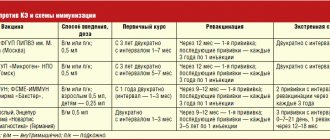
The basis of the treatment of autoimmune encephalitis, including encephalitis with antibodies to glutamate receptors, is the use of immunomodulatory therapy (see table) . The most commonly used first-line treatments are pulse therapy with intravenous methylprednisolone and/or normal intravenous human immunoglobulin, as well as plasmapheresis. Some patients may require second-line therapy (in which case the diagnosis of autoimmune encephalitis is usually already laboratory-verified and the patient does not respond or has a suboptimal clinical response to first-line therapy after day 10), including rituximab, cyclophosphamide, or a combination thereof, as well as alternative agents (tocilizumab, aldesleukin). The addition of plasmapheresis/immunoadsorption to immunosuppressive therapy with methylprednisolone, rituximab or cyclophosphamide can significantly accelerate the recovery of patients with autoimmune encephalitis, especially if it is refractory to first-line drugs.
If the condition of a patient with encephalitis is rapidly deteriorating, immunomodulatory therapy ex juvantibus should be prescribed for life-saving indications if infectious origin is excluded/low probability. Thus, in a description of a series of cases, Sahoo B. et al. (2018) among 9 children with suspected autoimmune encephalitis, only 2 had a laboratory verified diagnosis. Moreover, all children responded to first- or second-line immunomodulatory therapy.
In case of autoimmune encephalitis refractory to treatment, oncological search is indicated. If a tumor is detected, it must be removed, since preservation of the tumor does not allow achieving a pronounced clinical response to conservative therapy, even with the use of modern immunomodulatory drugs. In the case of detection of autoantibodies potentially associated with tumors, it is necessary to repeat the oncological search every three months, since paraneoplastic lesions of the nervous system can debut long before the detection of tumors associated with them.
Therefore, it should be remembered that:
- not all encephalitis is of infectious origin (autoimmune encephalitis should not be overlooked);
- not all autoimmune encephalitis has abnormalities in the CSF, brain MRI and EEG - screening for autoantibodies is mandatory in every patient with suspected autoimmune encephalitis;
- with the acute development of psychotic symptoms, it is first important to exclude organic lesions of the central nervous system , and then diagnose and treat psychiatric diseases;
- Every patient with an autoimmune lesion of the central nervous system is advised to undergo a thorough oncological search - failure to detect a tumor, if present, can significantly reduce the effectiveness of immunomodulatory therapy and lead to suboptimal disease outcomes;
- in cases where it is not possible to laboratory verify autoimmune encephalitis, in a severe patient with encephalitis immunomodulatory therapy should be used empirically according to vital indications (intravenous normal human immunoglobulin is preferable to pulse therapy with methylprednisolone).
Between this light and this light.
A few days after the chickenpox blisters appeared on my body, I felt worse. It felt like the skull was being sawed through with a dull saw, and the body was spinning out of control. First, my arms, legs, and face suddenly began to experience cramps, after which I continued to feel a strange trembling for a long time. And then gradually the muscles stopped obeying me - I could not even raise my hand to reach a bottle of water. And if I managed to reach it, my fingers could not take it, and I powerlessly grabbed the air.
Luckily for me, my parents and I always had a great relationship, and they quickly became worried when I didn't answer two of their calls. But I couldn’t hold the phone in my hand and just prayed that my father and mother would understand: something wrong had happened to me.
The parents had to open the door with their own key. My wife was on a business trip, and I could no longer get out of bed. From somewhere, as if from afar, I heard my mother’s voice, how she called an ambulance, how they laid me on a stretcher, and already completely losing consciousness, I noticed the buzz of the elevator. What happened next, I know only from the words of my parents and my wife, who arrived on the first flight. The most severe condition, when I did not even recognize my relatives, lasted 4 days. During this time, the disease fundamentally changed me.
Write to WhatsApp
COST OF TREATMENT
I lost the ability to feel anything other than primitive desires - to eat, drink, sleep. As doctors noted more and more obvious signs of my physical recovery, my psyche indicated the opposite. My feelings underwent the opposite changes - now I began to feel everything that was possible. I could fall into deep despondency and even cry if my wife did not ask me convincingly enough about my well-being. It seemed false to me, and I began to seriously think that she no longer needed me, and about how I should live further. I often worked myself up to the point that when I saw my wife, I didn’t immediately understand what she was doing here: we divorced a long time ago, and she left me.
I began to observe strange rituals. For example, if my toothbrush ended up with the bristles facing the wall, I couldn't do anything else until I turned it the other way. Or, after walking my wife to work, I had to return to the front door exactly three times and check that I had locked it. Twice was not enough. And four times is too many. And if I miscalculated, then I went crazy from internal tension: should I count the fourth time as the first and check the door two more times, or start counting again. After several such episodes, I got used to carrying a small notebook in the pocket of my dressing gown or shirt, in which I wrote down the sticks. Yes, I counted my ritual trips to the door….
And, of course, the “male failure” hurt his pride. Illness, prolonged bed rest, tons of medications and God knows what else made me impotent. With the remnants of common sense, I understood that my wife was a young, healthy woman; she would not tolerate an impotent man in her bed for long. Or maybe he can’t stand it anymore and is having fun on the side? In a word, my normal life remained there - until encephalitis. But (who would have thought!) my notebook brought me luck.
Having seen it one day, my wife asked what I was writing in it. After examining the “picket fence” that marks the completion of each ritual, she was silent for a long time. And then she asked me to go with her to the doctor.
And so I went.
What are the stages of Rasmussen's encephalitis?
The period of anomaly that occurs between the incubation stage and the disease itself can last for several years. According to average statistical indicators, doctors observe in 70% of patients the formation of a simple type of focal paroxysms of the convulsive direction of motor abilities. In 20% of situations, the disorder begins with epileptic seizures. There is a possibility of progression of secondary generalized seizures. At the initial stage, the number of seizures is minimal, but over time the growth of paroxysms increases significantly. In the debut stage, doctors observe the formation of Todd's palsy.
The active period is marked by the addition of focal neurological manifestations (violation of the speech apparatus, hemianopsia, hemihypesthesia, etc.), mnestic anomalies and muscle cramps of a constant nature. Scientists report that in practice, simple motor failures occur in 80% of cases, complex ones in 20%, secondary generalized ones in 40%, and somatosensory paroxysms in 23% of patients. Only in 10% of situations can hemi-seizures of epileptic origin occur, which affect the muscle area of only one part of the body. The duration of motor impairment in Todd's disease is constantly increasing, and hemiparesis of a permanent nature manifests itself. Over time, it can transform into a persistent characteristic form.
The stabilization moment in 80% of cases occurs after three years from the onset of the development of the abnormal reaction. Doctors note normalization and minimal reduction in the number of seizures in patients. However, patients continue to complain about the progression of sensorimotor, visual and cognitive disorders. A quarter have neuroendocrine disorders, early puberty and signs of obesity.
Dietary supplements inexpensively
I heard about such a disease as encephalitis many times as a child. Mainly from parents and teachers who warned about the dangers of tick bites. On any trip to nature, I was equipped as a polar explorer - my whole body had to be covered with clothes, and after returning home I had to carefully examine myself. What’s interesting is that not a single tick set its sights on me. And encephalitis came from where no one expected it. At the age of 30 I got chickenpox. The doctor warned me about mandatory adherence to bed rest and other conditions under which I would have a better chance of recovering from this illness with minimal losses.
But I didn’t even think that some childhood illness could lock me in the bedroom and put me in bed. And I was right: she put me in the intensive care unit.
Free consultation right now!
Online consultation with a specialist on your issue!
License number: LO-77-01-019036
Diagnostic methods
Drawing up a correct conclusion in the prodromal period is quite difficult due to the absence of focal symptoms. At the active stage of the disease, a doctor in the neurological field during research discovers the presence of central hemiparesis with an increased degree of reflexes and signs of a pyramidal nature, regular contractions of myocolonic origin, malfunctions of the speech apparatus, a high degree of mental insufficiency, a decrease in the level of memory and attention.
Differential diagnosis is carried out in the presence of intracerebral neoplasms, cerebral cysts, encephalitis of other origins and other groups of epileptic seizures in childhood, etc. By undergoing electroencephalography at the active stage of the anomaly, doctors are able to detect disorders in all examined patients. Patients are prescribed to undergo:
- Hemianopsia.
- Ophthalmoscopic scanning.
- Laboratory testing.
- Diagnostics of cerebrospinal substance.
- Computed tomography or magnetic resonance imaging.