G 40.9 – Epilepsy, unspecified – example of writing a call card
Patient, 34 years old
Reason for calling – Unconscious (reason unknown)
Complaints, medical history
At the time of the examination he makes no complaints and does not remember what happened. Located in a public place (shop), sitting on the floor.
According to others, about 10 minutes ago there was an attack of tonic-clonic seizures with loss of consciousness and foam at the mouth. No treatment was undertaken. This is not the first time such conditions have occurred; I previously came to 03 with epileptic seizures.
Anamnesis of life
Epilepsy, is on an outpatient basis. Regularly takes anticonvulsants.
Comfortable blood pressure – does not explain.
Physical examination
Condition: moderate;
Consciousness – stunned, Glasgow scale – 13 points, behavior – inhibited;
Pupils – normal, D = S, reaction to light – lively, no gaze paresis, horizontal nystagmus;
Skin – physiological color, dry, clean;
Heart sounds are clear, rhythmic, no murmurs. The pulse in the peripheral arteries is of satisfactory quality, rhythmic;
Nervous system – no meningeal symptoms, increased muscle tone D = S, no focal neurological symptoms;
The pharynx is calm, the tonsils are normal;
Excursion of the chest - normal, type of breathing - normal, percussion - pulmonary sound, auscultation - vesicular breathing, no shortness of breath;
There is no peripheral edema;
The tongue is clean and moist. The abdomen is soft, painless, participates in the act of breathing, there are no symptoms of peritoneal irritation, the liver is not enlarged, stool is formed, 1 time per day;
Urine output is normal, SSPO is negative.
Main pathology
Consciousness is depressed to the point of stupor, inhibited, inadequate, disoriented in space and time. There are bite marks on the tongue. Traces of involuntary urination. Gradual restoration of consciousness during the examination. No signs of head trauma were found.
| time | 17-30 | 17-50 | 18-10 | Etc. peace |
| NPV | 16 | 16 | 16 | 16 |
| Pulse | 86 | 84 | 76 | 72 |
| Heart rate | 86 | 84 | 76 | 72 |
| HELL | 150/90 | 130/80 | 130/80 | 130/80 |
| Pace. ºС | 36,6 | |||
| SpO2 | 97 | 97 | 97 | 97 |
Glucometry – 7.5 mmol/l
Electrocardiography
17-35 – Sinus rhythm 86 per minute. There are no signs of acute coronary pathology. There is no ECG archive.
Diagnosis by the EMS team
Epilepsy, unknown origin, condition after an attack.
Therapeutic measures
- 17-39 – Sol. MgSO4 25% – 10 ml i.v.
- hospitalization in the neurological department of the City Hospital.
Treatment result
Improvement. Complete restoration of consciousness. During transportation, the convulsive syndrome did not recur.
Status epilepticus
V.A. Karlov Department of Neurology and Neurosurgery MMSI
Status epilepticus (SE) is defined as a condition in which epileptic seizures are so frequent and/or prolonged that a stable and qualitatively different epileptic state is formed. With ES, each subsequent seizure occurs before the patient has completely recovered from the previous attack, i.e., he remains with severe disturbances of consciousness, hemodynamics, breathing, or homeostasis.
According to WHO experts, epilepsy is a disease of various etiologies, caused by excessive neural discharges and characterized by repeated epileptic seizures and other clinical and paraclinical disorders. For their development (in the presence of an epileptic focus), the formation of an epileptic system is necessary, including mechanisms that promote the spread of an epileptic discharge beyond the boundaries of the epileptic focus. This is prevented by the antiepileptic defense system - mechanisms that suppress the spread and generalization of epileptic activity.
In patients with epilepsy, epilepsy should be considered as a manifestation of an extreme degree of insufficiency (failure) of the antiepileptic defense system. In this case, changes in neuroglial relationships occur with the accumulation of K+ in the extracellular fluid, causing repeated depolarization; disturbances of interneuronal interactions with an increase in the intensity and frequency of discharges between them; the occurrence of pathological excitation in neural circles, as happens in the myocardium during its fibrillation.
In addition, as our studies have shown, during SE, secondary pathogenetic mechanisms are formed that support SE in a vicious circle. The most important of them, associated with repeated seizures, are cyclic breathing disorders, apnea (asphyxia), hyperventilation (hypocapnia), which have an epileptogenic effect. This applies to the most dangerous type of epileptic seizure - its convulsive form, although there are other forms corresponding to the variety of epileptic seizures. Below is the classification of ES.
Generalized ES
- ES of convulsive seizures: tonic-clonic, tonic, clonic, myoclonic.
- ES of absence seizures
Partial ES
- ES of simple partial seizures: somatomotor, somatosensory, dysphatic, adversive, sensitive, vegetative.
- ES of complex partial seizures.
Neonatal ES
Next, we will present, first of all, the results of our own research conducted over 30 years (more than 200 observations).
SE can occur both in patients with an established diagnosis of epilepsy and in patients without indications of epileptic seizures in the past (initial SE). Until the 70s, among patients admitted with epilepsy, patients with epilepsy made up the majority; in the 90s, the majority were patients with initial ES. To a certain extent, this indicates an improvement in the quality of epilepsy treatment.
The main causes of epilepsy with an established diagnosis of epilepsy are the following: violations of the regime (sleep deprivation, alcoholism, etc.); a break in taking antiepileptic drugs (AEDs); stopping AEDs too quickly; somatic and infectious diseases; pregnancy; a relative reduction in the dose of AEDs due to a significant increase in body weight (for example, with age-related changes in children); treatment of epilepsy by a psychic.
The specific mechanisms of these influences are different; for example, during pregnancy, the fluid content in the body increases, hormonal relationships change (the level of estrogen, progesterone, etc.), the absorption of AEDs is disrupted, etc.
The most common causes of initial ES are current brain diseases: cerebrovascular, especially acute, cerebrovascular accidents, meningitis, encephalitis and others, as well as traumatic brain injury. The cause may be metabolic disorders of cerebral (in newborns) and extracerebral origin (renal failure, hypoglycemia, hyponatremia, cardiac arrest, hepatogenic encephalopathy, etc.). An iatrogenic factor may be important - an overdose of medications (antidepressants, phenothiazines, tefillin, isoniazid, etc.). ES is often initiated by the sudden withdrawal of sedatives and narcotic drugs in patients who have been taking them for a long time. Finally, in approximately 5% of patients with ES, the latter serves as the debut of epilepsy, and in the future a status course of epilepsy may occur. We have shown that this variant of SE most often occurs in frontal (prefrontal) epileptic foci.
According to the variants of convulsive epileptic seizures, in addition to the status of clonic-tonic seizures, tonic and clonic SE should also be distinguished. Tonic SE occurs more often in children: with Lennox-Gastaut syndrome and intractable epilepsy, often against the background of mental retardation. It can also occur at any age as a variant of prefrontal epilepsy.
Clonic SE is more often focal with or without subsequent generalization of convulsions, which is characteristic primarily of somatomotor (Jacksonian) seizures. It also occurs with epilepsy of infancy, febrile convulsions of early childhood, NHE Gastaut syndrome (hemiconvulsions, hemiplegia, and subsequently epilepsy).
Finally, among the convulsive forms of SE, myoclonic status should be distinguished. However, it must be remembered that pathological myoclonus (there are also physiological myoclonus, for example, myoclonic jerks when falling asleep) can be epileptic and non-epileptic. In accordance with this, a distinction is made between epileptic and non-epileptic status myoclonic. The latter is also called a “myoclonic storm” or storm.
Myoclonic ES occurs in myoclonic forms of epilepsy and in familial progressive myoclonus - Lafar disease, Hunt disease, Unferricht-Lundborg syndrome; for lipoidosis, sialidosis, gangliosidosis (Tay-Sachs, Gaucher, Bielschowsky diseases, etc.).
Non-epileptic myoclonic status is most often postanoxic, for example in acute anoxic encephalopathy due to cardiac arrest, and dysmetabolic - in dysmetabolic encephalopathies, for example renal, hepatic, etc.
Finally, myoclonic SE may be a manifestation of the final stage of SE tonic-clonic seizures, when, due to the depletion of metabolic processes in the brain, only myoclonic phenomena can occur.
ES of convulsive seizures is a serious condition, as it leads to deepening disturbances of consciousness (stunning - stupor - coma), as well as other progressive somatic, metabolic disorders: circulatory - cardiac arrhythmias, arterial hypertension, hypotension, collapse; respiratory – obstruction of the upper respiratory tract, hypoxia, pulmonary edema, pneumonia, aspiration; renal - acute tubular necrosis, myoglobinuria, ischemic kidney; metabolic – acidosis, hypercapnia, hyperkalemia, hypoglycemia, hypoglucocorticemia; vegetative – hyperthermia, vomiting, loss of fluid and electrolytes, hypersecretion (tracheobronchial, salivatory, sweat); hemostatic – obligate disseminated intravascular coagulation syndrome – DIC (described by us).
ES is an urgent condition that requires urgent and adequate actions. Management of patients with ES is based on the following principles: • the earliest possible start of therapy (on site, in an ambulance); • prevention and elimination of disorders of the body's life support systems; • hospitalization in neuroreanimation or general intensive care units; • if possible, use a minimum number (one to two) of anticonvulsants; • their intravenous jet administration (at least in the initial stages); • correct dosage (in mg/kg), monitoring the level of AEDs in the blood; • electroencephalographic control.
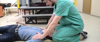
Initial measures - removal of dentures and other foreign objects from the oral cavity, laying the patient on his side, inserting an air duct if necessary, injection of diazepam intravenously - are carried out on the spot, after which the patient is transported to the hospital.
Diazepam is administered slowly intravenously in a glucose solution to adults 10 mg, children 0.05 - 0.1 mg per year of life.
The following activities are carried out in the hospital: • ensuring patency of the upper respiratory tract; • inhalation of a mixture with a high oxygen content; • repeated administration of diazepam if there is no effect from its initial administration; • in case of failure, drip intravenous administration of 100 mg of diazepam in 500 ml of 5% glucose solution is used at a rate of 40 ml/hour or phenobarbital - 20 mg/kg at a rate of no more than 100 mg/min; • in case of failure, anesthesia is carried out (I - II stage of the surgical stage), in the absence of a lasting effect - an extra-long combined anesthesia with the use of muscle relaxants and artificial pulmonary ventilation (ALV). • administer heparin and other drugs to eliminate DIC syndrome; • dexamethasone – for symptomatic ES (often a brain tumor); • monitoring and correction of hemodynamic, metabolic and visceral functions.
Alternative AEDs for ES may be phenytoin (diphenin); an ampoule form of the drug is available abroad; lorazepam (currently the drug of first choice abroad); lidocaine intravenously, infusion of 100 ml in 250 ml of 5% glucose solution; paraldehyde 100 ml in 0.9% sodium chloride solution intravenously over 10 - 15 minutes.
Phenytoin is administered at a rate of 8 mg/kg at a rate of 50 mg/min. It is considered as a first choice drug for symptomatic (stroke, traumatic brain injury, etc.) and idiopathic epilepsy, since, unlike other AEDs, it does not cause depression of consciousness. At the same time, it can only be used under ECG control, as it leads to a slowdown in heart rate. Contraindicated for arrhythmias, prolongation of the P–Q interval.
Phenobarbital remains the first choice for neonatal status ES caused by sudden cessation of barbiturates, in the absence of other AEDs.
In case of prolonged ES using combined anesthesia, the use of phenobarbital is advisable due to its antihypoxic effect on the brain.
Hyperthermia can be a severe disorder in HS. If hyperthermia is present, its nature should be clarified. Hyperthermia can be somatogenic, and treatment of the corresponding somatic disease is required (with ES, most often pneumonia); neurogenic, associated with central thermoregulation disorders.
Physical cooling, hibernation - intravenous administration of diazepam, haloperidol, antihistamines, reopirin as such or in the form of lytic cocktails, as well as muscle relaxants (nosh-pa, etc.) are used in order to increase the heat transfer of vasodilators. In the absence of proper action, as shown in our studies, mechanical ventilation with muscle relaxants has a pronounced effect: body temperature drops to low-grade and even normal within a few hours. In children, ES is especially dangerous. The lability of homeostasis, the increased need for fluid, the threat of developing exicosis, on the one hand, and cerebral edema, on the other, require particularly careful monitoring of homeostasis indicators, in particular water-electrolyte balance and body temperature. Dehydration therapy is indicated - the administration of glucocorticoids (dexamethasone 0.2 - 0.5 mg/kg per day) in combination with increased fluid administration, the amount of which is calculated according to the well-known formula: for body weight less than 10 kg - 100 ml/kg, for weight body weight up to 20 kg – additional 50 ml for each kilogram over 10 kg; for body weight over 20 kg - 20 ml for every kilogram over 20 kg. Due to poor vein development, AEDs are best administered intrarectally to infants. In newborns, the cause of ES can be hyperglycemia, hypocalcemia, hyposulfatemia, hypopyridoxemia. Therefore, in the absence of data confirming the cerebral genesis of ES, newborns, if seizures are not eliminated on the first attempt and their origin is unclear, are sequentially administered intravenously at intervals of 5 minutes: 5 - 10 ml of 20 - 30% glucose solution, 2 - 6 ml 2.5 - 5 % calcium gluconate solution, 2 – 6 ml 2 – 3% magnesium sulfate solution, 10 ml 5% pyridoxine solution.
Analysis of our own observations and literature data shows that the reasons for failure in the treatment of ES are: • late start of treatment (usually due to late admission of the patient); • prescription of an insufficient dose of AED; • use of the drug intramuscularly or too slowly intravenously; • failure to take measures to maintain the maximum concentration of the drug in the blood; • ignoring the causes of ES; • failure to provide maintenance anticonvulsant therapy after seizure control.
The immediate causes of lethal outcomes of ES are respiratory circulatory and metabolic disorders due to seizures (collapse, cardiac arrest, respiratory paralysis, etc.); ischemic kidney; acute hypoxic-ischemic encephalopathy with depletion of metabolic and plastic reserves of the brain; DIC syndrome; acute adrenal insufficiency; iatrogenic factors – inhibitory effect of massive amounts of AEDs and barbiturates; underlying disease (with symptomatic ES).
According to our data, serious risk factors for the fatal outcome of epilepsy in adult patients with epilepsy are: the onset of epilepsy in early childhood, its severe course; age 16 – 18 years with delayed puberty; severe early organic brain damage with severe neurological and intellectual deficits.