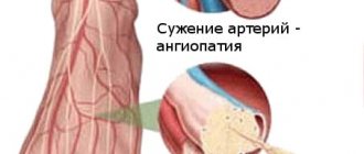
The functioning of the organs and systems of our body is regulated by nerve impulses - signals emanating from the brain. “Outgoing” and “incoming” impulses are transmitted along the nerves, as if through wires. Damage to the nerves disrupts this connection and can cause serious disruptions in the body's functioning. Indeed, along with disruption of nerve transmission in the affected area, cellular nutrition and blood supply deteriorate.
A condition characterized by damage to nerve fibers and accompanied by a violation of the conduction of nerve impulses along the nerve fiber is called neuropathy (neuropathy) .
If one nerve suffers, we are talking about mononeuropathy , if there is multiple symmetrical damage to peripheral nerves (for example, when the process affects both lower and/or upper limbs at once, etc.) - about polyneuropathy . The pathological process can involve both cranial and peripheral nerves.
Lesions of the peripheral nerve trunks, which are based on the infringement of a nerve enlarged as a result of inflammation and swelling in the muscle-bone tunnel, are called tunnel syndrome (there is also a name for compression-ischemic neuropathy ).
There are dozens of tunnel syndromes, the most famous of which is carpal tunnel syndrome.
1
Neuropathy. Diagnosis and treatment.
2 Neuropathy. Diagnosis and treatment.
3 Neuropathy. Diagnosis and treatment.
Our specialists
Bezgina Elena Vladimirovna
The doctor is a neurologist of the highest category. Botulinum therapist. Physiotherapist. Experience: 24 years.
Tarasova Svetlana Vitalievna
Expert No. 1 in the treatment of headaches and migraines. Head of the Center for the Treatment of Pain and Multiple Sclerosis.
Somnologist.
Epileptologist. Botulinum therapist. The doctor is a neurologist of the highest category. Physiotherapist. Doctor of Medical Sciences.
Experience: 23 years.Derevianko Leonid Sergeevich
Head of the Center for Diagnostics and Treatment of Sleep Disorders.
The doctor is a neurologist of the highest category. Vertebrologist. Somnologist. Epileptologist. Botulinum therapist. Physiotherapist. Experience: 23 years.
Symptoms of neuropathy
The variety of types of disease explains the huge number of specific manifestations. And yet we can identify the most characteristic signs of neuropathy :
- swelling of tissues in the affected area;
- disturbance of sensitivity (pain, numbness, coldness, burning of the skin, etc.);
- muscle weakness;
- spasms, convulsions;
- difficulty moving;
- soreness/severe pain in the affected area.
Mononeuropathies of the extremities are never accompanied by general cerebral symptoms (nausea, vomiting, dizziness, etc.); cranial neuropathies can manifest similar symptoms and, as a rule, accompany more serious diseases of the nervous system of the brain.
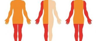
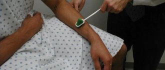
Polyneuropathies are manifested by impaired sensitivity, movement, and autonomic disorders. This is a serious pathology that initially manifests itself in the form of muscle weakness (paresis), and then can lead to paralysis of the lower and upper extremities. The process can also involve the trunk, cranial and facial nerves.
1 ultrasound
2 Electroneuromyography
3 Electroneuromyography
Read also
Lambert-Eaton syndrome
The first description of the disease in the literature appeared in 1953. J. Anderson described a case from practice: a 47-year-old man with lung cancer experienced symptoms similar to myasthenia gravis.
After tumor removal... Read more
Intracranial hypertension on MRI
Intracranial hypertension is an increase in intracranial pressure. Normal intracranial pressure is 15 mm Hg. When blood pressure doubles, a stroke occurs. At a pressure of 50 mm Hg, the patient can...
More details
Brain concussion
A concussion is a mild traumatic brain injury without damage to the bones of the vault and base of the skull. In this condition, there are no pronounced disturbances in the functioning of the brain and there are no changes...
More details
Sudden memory loss/transient global ischemia
Transient, that is, temporary memory loss occurs in elderly patients and people suffering from migraines. At the same time, memory for past and present events disappears. But the person is conscious, accessible...
More details
Myasthenia gravis
MYASTHENIAS – a disease associated with a disruption of the immune system, as a result of which antibodies are produced against the body’s own tissues involved in the transmission of nerve impulses, which...
More details
Facial neuritis - symptoms and treatment
Neuritis (neuropathy) is a disease of the nervous system, manifested in dysfunction of a nerve or a certain group of nerves.
In recent years, the Greek word “pathos”, which means “suffering”, has been used to denote syndromes of damage to the peripheral nervous system, and previously used terms, for example, “neuritis”, have been replaced by “neuropathy”, “sciatica” by “radiculopathy”, etc. d. If several nerves are inflamed, then it is polyneuropathy, if one nerve is inflamed, then it is mononeuritis. When the cause of inflammation of the nerves is, for example, diabetes, we talk about diabetic polyneuropathy, [1] and if there is an infection, then about infectious polyneuropathy (for example, herpes, diphtheria, etc.); [2] if the factor is hereditary, then it is hereditary polyneuropathy; if associated with a nutritional disorder, for example, alcohol abuse, then alcoholic polyneuropathy; if polyneuropathy appears against the background of reduced immunity, then it is idiopathic polyneuropathy, etc. [3]
Many types of peripheral neuropathy are often caused by exposure to toxic chemicals, malnutrition, injury and nerve compression, and also occur as a result of certain medications, such as those used to treat cancer and HIV/AIDS. [4]
As an example, consider such a common type of neuritis as neuritis of the facial nerve, also called Bell's palsy, the incidence of which is 23 people per 100 thousand, in all age groups, regardless of gender. The average age of patients is 40 years.
Most often, facial paralysis occurs as a result of local hypothermia. The source of infection is often chronic processes in the mouth, throat, and ear. In acute otitis media, nerve damage is caused by perineural edema of vascular origin. But more often, facial paralysis is caused by the herpes zoster virus in the area of the external auditory canal and eardrum. [5]
Facial neuritis develops as a result of:
- tumor processes in the cerebellopontine angle and posterior cranial fossa, temporal bone, parotid gland;
- traumatic brain injuries;
- acute, chronic otitis, mastoiditis;
- infections - syphilis, tuberculosis, Lyme disease, HIV infections, malaria, diphtheria, typhus, etc.;
- sarcoidosis, collagenosis, amyloidosis;
- Guillain–Barré syndrome;
- multiple sclerosis and many other diseases.
- Sometimes the development of neuropathy of the facial nerve is observed during pregnancy against the background of nephropathy.[6]
Peroneal nerve neuropathy
Neuropathy of the peroneal nerve, or peroneal neuropathy, occupies a special position among peripheral mononeuropathies, which also include: neuropathy of the tibial nerve, neuropathy of the femoral nerve, neuropathy of the sciatic nerve, etc. Since the peroneal nerve consists of thick nerve fibers that have a larger layer of myelin sheath, then it is more susceptible to damage due to metabolic disorders and anoxia. This point is probably responsible for the fairly wide prevalence of peroneal neuropathy. According to some data, neuropathy of the peroneal nerve is observed in 60% of patients in traumatology departments who have undergone surgery and are treated with splints or plaster casts. Only in 30% of cases, neuropathy in such patients is associated with primary nerve damage.
It should also be noted that often specialists in the field of neurology have to deal with patients who have a certain history of peroneal neuropathy, including the postoperative period or time of immobilization. This complicates treatment, increases its duration and worsens the result, since the earlier therapy is started, the more effective it is.
Anatomy of the peroneal nerve
The peroneal nerve (n. peroneus) arises from the sciatic nerve at the level of the lower 1/3 of the thigh. It consists predominantly of fibers of the LIV-LV and SI-SII spinal nerves. After passing through the popliteal fossa, the peroneal nerve exits to the head of the bone of the same name, where its common trunk is divided into deep and superficial branches. The deep peroneal nerve passes into the anterior part of the leg, descends, passes to the dorsum of the foot and divides into internal and external branches. It innervates the muscles responsible for extension (dorsal flexion) of the foot and toes, pronation (raising the outer edge) of the foot.
The superficial peroneal nerve runs along the anterolateral surface of the leg, where it gives off a motor branch to the peroneal muscles, which are responsible for pronation of the foot with its simultaneous plantar flexion. In the area of the medial 1/3 of the tibia, the superficial branch of n. peroneus passes under the skin and is divided into 2 dorsal cutaneous nerves - the intermediate and medial. The first innervates the skin of the lower 1/3 of the leg, the dorsum of the foot and the III-IV, IV-V interdigital spaces. The second is responsible for the sensitivity of the medial edge of the foot, the back of the first toe and the II-III interdigital space.
Anatomically determined areas of greatest vulnerability of the peroneal nerve are: the place where it passes in the area of the head of the fibula and the place where the nerve exits the foot.
Symptoms of ischemic neuropathy
Most often, this disease affects only one eye, but approximately 1/3 of patients may also experience problems of a bilateral nature. In some cases, the pathology affects the second eye after some time (usually within 2-5 years).
As a rule, the pathology appears suddenly - after a hot bath, physical activity, or even after sleep. Visual acuity drops sharply (sometimes only light perception is preserved, and with total nerve damage, blindness occurs). Vision may deteriorate within minutes. Vision deterioration may be preceded by blurred vision, pain behind the eyeball, or headaches.
The peculiarity of this disease is that during its development, peripheral vision is always impaired. In this case, scotomas, concentric narrowing of the visual fields, and loss in various segments (lower, nasal, temporal halves) may be observed.
The duration of acute ischemia is several weeks. Then the swelling of the optic nerve head gradually subsides, and the hemorrhages resolve. Optic disc atrophy persists, with varying degrees of severity. Visual defects also remain, but may be reduced.
Compression-ischemic neuropathies of the lower extremities
Neurosurgery Tags:
Sergey Ilyalov:
Good afternoon, dear listeners. The program “Microsurgery with Dr. Ilyalov” is on air and I am presenting it, Sergey Ilyalov. The topic of our today's program is “Compression-ischemic neuropathies of the lower extremities.” My guest today is Dr. Artem Grigorievich Fedyakov. And we continue our conversation about compression-ischemic neuropathies, which we started several programs ago. We talked about the upper limbs and, in general, found out that there are a number of anatomical features that contribute to the occurrence of this type of pathology. Artem Grigorievich, what are the features of the functioning of the peripheral nervous system of the lower extremities?
Artem Fedyakov:
Good afternoon. Returning to our previous meetings, when we talked about the pathology of the upper extremities, I would still like to note that humans, as a unique biological species, are affected by some diseases that are not typical for other species, for example, animals and so on. Again, the nerves in the hands are affected because the person has a very functional part of the body. It is anatomically special, it is an opposable finger and so on. As for the legs, man is a unique creature on earth that has the ability to walk upright. And in connection with this, a number of problems arise, anatomically already, as a rule, having the prerequisites for the emergence of various pathologies unique to humans, including pathologies of the peripheral nerves. Today I wanted to talk about the 2 leading pathologies associated with the nerves of the legs - this is damage to the small tibial nerve...
Sergey Ilyalov:
Let us perhaps then immediately clarify about the lesser tibial nerve how it is structured, how it is formed, what it consists of and what contributes to its damage, both chronic traumatic influences and acute ones.
Artem Fedyakov:
The lesser tibial nerve determines a very important function - raising the foot, that is, in medical terms it is called extension of the foot. And it innervates the muscle of the anterior surface of the leg, which provides this function. This nerve is a section of the sciatic nerve, where in the area of the popliteal fossa, this is behind, just above the knee joint, it departs and passes through a narrow place - the fibular canal. This place is located in the upper third of the lower leg.
Sergey Ilyalov:
On the outer surface?
Artem Fedyakov:
This is on the side of the shin, where you can feel such a bone in thin people, this is exactly the head of the fibula. He goes around it and is already moving forward just to the muscles that he innervates.
Sergey Ilyalov:
What features of the passage of the nerve exist, specifically its anatomical course?
Artem Fedyakov:
Firstly, it is a spiral-shaped course, it is almost adjacent to the bone for a long distance.
Sergey Ilyalov:
And it even forms a channel there.
Artem Fedyakov:
Yes, nature has already provided this channel for him. The second anatomical feature is that this nerve lies superficially, in fact it covers the fascia, these are tendon formations, and on top there is only skin.
Sergey Ilyalov:
By and large, this leads to the fact that in this place the small blood supply to the nerve differs from that in more pronounced soft tissues.
Artem Fedyakov:
That's right, yes. It is noted that around this nerve there are already anatomical conditions such that there is not enough structural tissue that both supplies it with blood and holds it. Well, the third anatomical feature is that this nerve is located very close to the large joint. The movement of the leg, in fact, can provoke, that is, constant flexion or some kind of tension in the ligaments of the knee joint, causing a chronic traumatic effect on the nerve as well.
Sergey Ilyalov:
We thus approach the main risk factors that contribute to the occurrence of this neuropathy, this is the forced position of the leg in a bent state, for example, in berry pickers, say, or in parquet layers or some pipe layers, some working professions. Or, on the contrary, in those working professions where there is constant monotonous movement, for example, seamstresses, motorists, and so on. In them, too, this may contribute to permanent chronic injury to the nerve in this place.
Artem Fedyakov:
In addition to the specialties that you named, in our practice we quite often meet athletes, and not even in the active phase of sports, and this is already a consequence, chronic overload in this area. Dancers, sometimes, but there are also just casuistic cases when you ask a person: “Have you noticed what could have such an impact?” It turns out that at the workplace the wall of the table was simply adjacent and he was constantly resting his knee, the side surface of his shin. Then they already note that there were some microtraumas and so on, that after all, these were moments. But it was not acute, it was not some kind of injury that I hit, and all this arose at once, it was chronic traumatization.
Neuropathies of the fibular nerve usually occur in working people.
Sergey Ilyalov:
In the first place we still have chronic traumatization, do I understand correctly? The second is probably an acute traumatic effect, both associated with damage to the knee joint, with bruises of the knee joint, with dislocations, and so on, and with fractures or other traumatic effects in the area of the fibula.
Artem Fedyakov:
That's right, yes. Damage to the lesser tibial nerve is often accompanied by sprains of various ligaments; there are a lot of them in the knee joint. Sometimes it is even enough to damage not the knee joint, but a dislocation of the ankle; it would seem to be the ankle joint, which is lower, but nevertheless, often leads to its pathology.
Sergey Ilyalov:
What is this due to, due to biomechanics?
Artem Fedyakov:
Due to the fact that there is still a stretching of the muscle ligaments in the lower section, and since it is moving, again, its spiral course goes around this axis at a great distance, it seems to be stretched. And this type of damage is called traction, that is, not due to a direct bruise, but due to traction - overextension.
Sergey Ilyalov:
Due to stretching of the nerve trunk.
Artem Fedyakov:
Sometimes there are often cases when this pathology occurs in people who have been squatting for a long time, for example, doing something, repairing, painting or some other moments. A person, after sitting, finishes the evening, goes to bed, and in the morning the signs of this disease are already felt.
Sergey Ilyalov:
Well, in this regard, then we probably move on to the symptoms of damage to the fibular nerve.
Artem Fedyakov:
The main symptom that is difficult to miss is weakness of foot extension.
The main symptom of damage to the fibular nerve is weakness of foot extension.
Sergey Ilyalov:
I apologize, it should probably be noted that the lesser tibial nerve, like most peripheral nerves, has both motor and sensory portions, but in the first place are motor disorders?
Artem Fedyakov:
Exactly, yes, because this is the first thing a person notices when he either receives an injury or, due to chronic acute traumatic actions, develops neuropathy - nerve damage. At first this is weakness, uncertainty of gait. It is very important to note that sometimes at the beginning of this disease a person does not even always notice, or rather, characterizes or interprets this condition as weakness of extension. But he begins to stumble more often, he begins to stumble more often, and just what they say, there is a subluxation in the ankle joint, the foot does not obey.
Sergey Ilyalov:
In fact, the foot begins to sag and the patient is unable to pull the toe of his foot up. Due to this sagging, the phenomenon of stumbling actually occurs.
Artem Fedyakov:
Yes, in medical terminology this gait is called a rooster gait; a person must raise his knee high so that the toe of the foot does not cling to the surface, to the plane along which he is walking. Therefore, you can see the type of walking that is called that. These are the main symptoms. But again, in the early stages, this classic rooster gait is not completely formed, the person just begins to notice that he stumbles more often, gets subluxations of the ankle joint, and so on. Already when this clinical picture develops completely, the foot sags, and he cannot lift it.
Sergey Ilyalov:
Is this the only symptom, or can we pay attention to some other details, sensory disturbances?
Artem Fedyakov:
Of course, they come in second place because people tend to pay less attention to them. But nevertheless, sensitivity in some cases may be the first sign of the development of this pathology. Sensitivity along the front surface of the leg and the back of the foot is impaired. And this numbness, that is, a decrease in sensitivity, reaches the surface of the first finger. It should be noted that with pathology of the fibular nerve, as a rule, pain syndrome very rarely develops. These phenomena - numbness and weakness of extension - are the main ones. And the pain syndrome, which is characteristic of other nerves, which is a red rag when you can immediately understand that something is wrong, is absent here. Therefore, very often this pathology is insidious in that it arises latently, slowly and...
Sergey Ilyalov:
It becomes chronic and patients do not come in for a long time.
Artem Fedyakov:
Chronic, yes, and already when the problem is obvious, when we seek medical help, we are dealing with an advanced situation.
Sergey Ilyalov:
There are situations in medical practice when such types of neuropathy occur acutely and within the walls of a medical institution. Cases have been described where, during, say, long operations, the incorrect position of the leg on the operating table led to the fact that after the operation the patient may experience such neuropathy.
Artem Fedyakov:
Yes, such things, unfortunately, do occur, but this is not to say that this is an exception, but these are the risks of any long-term surgical operation.
Sergey Ilyalov:
Let's start with the fact that, in general, doctors, primarily anesthesiologists, are informed, they know about this and, of course, take preventive measures.
Artem Fedyakov:
Yes, naturally, but sometimes there are cases when the positioning of the patient makes it possible to perform the main stages, more serious, abdominal or chest surgeries, including life-saving ones. Of course, here you sometimes have to risk the possibility of other complications.
Sergey Ilyalov:
If we started talking about iatrogenic medical injuries, what other types can we talk about? Where are the bottlenecks and risk factors specifically from the point of view of providing medical care? For injuries, when applying skeletal traction?
Artem Fedyakov:
You know, there are such moments too. In my practice, there were several patients, if we are already talking about such a thing as iatrogenic medical injuries, when the nerve was damaged after a venectomy. A venectomy is made by making small incisions, literally a centimeter, and the vein is pulled out. And when this happens near or directly in the area of the nerve, it is also damaged. The vein was removed, but after the operation the patient can no longer lift his foot. There were several such patients. There were moments after applying a tourniquet in the upper third of the leg during surgery, again, orthopedists on the bones of the ankles and so on. Because with this tourniquet a very slight tension is enough to press it against the bone structures of the head, and neuropathy develops.
Sergey Ilyalov:
Just analyzing all these possible situations, I want to emphasize once again that there is a very striking anatomical feature here, namely the superficial occurrence of the small tibial nerve in the bone canal, in the bone groove on the fibula, which, in fact, is the very risk factor, and immediate reasons, they can be very diverse.
Artem Fedyakov:
Very often, if we are talking about other types of damage, not iatrogenic, let’s digress a little, this is still direct damage. Sports, when football players get hit in this area. Or, when doing something, chopping wood, a splinter or fragment of an ax may fall in there. Relatively recently, I had a young girl who crossed this nerve with a lawn mower trimmer. It was a big wound, we had to stitch this nerve together because, I repeat once again, its depth is extremely small, a centimeter, maybe a little more, that is, skin, subcutaneous tissue, and now the nerve.
Sergey Ilyalov:
Well, suturing a nerve is already such an extreme option for surgical treatment, and before we get to it, let's talk about diagnosis. It is clear that there are symptoms, and the symptoms for a competent neurologist and neurosurgeon, as a rule, do not cause any particular difficulty in interpretation. However, are any additional diagnostic methods used, in particular, to perhaps identify the level of nerve damage?
Artem Fedyakov:
It is for this nerve, for the tibia, that there is one very interesting feature - the fact is that the clinical picture is very similar to a disc herniation at the level of L4, L5. But in contrast to hernial compression of the fifth root, which also causes a feeling of numbness and weakness in extension of the foot, up to its complete sagging, radicular symptoms are more typical in the lower back; they descend like stripes on the leg, including on the thigh, that is, higher head of the fibula. And hernial compression is still characterized by a pain syndrome, in contrast to isolated neuropathy of the small tibial nerve.
Sergey Ilyalov:
Moreover, there is pain, both in the back and radiating pain.
Artem Fedyakov:
So it goes down into the leg along the innervation zone of this root. As for the lower leg and foot, they match. Therefore, the first point is, of course, it is necessary to exclude hernial compression, because there are forms of hernias in the lower back, when, after all, the pain syndrome is also not so pronounced, and such disorders come first. Therefore, it is often necessary to start with an MRI of the lumbar spine, magnetic resonance imaging, in order to exclude hernial compression.
Sergey Ilyalov:
We have ruled out hernial compression, we have characteristic symptoms for the fibular nerve, what other diagnostic methods are there?
Artem Fedyakov:
The main method that helps us determine the level of the lesion and still differentiate, distinguish whether it is a root or not, and that it is precisely at the level of the head of the fibula, this research method is called electroneuromyography. Experienced specialists give us a quantitative assessment of the disturbance in the conduction of nerve impulses and its level, and also help us distinguish, but not clinically, what the patient tells us or what the doctor observes. Namely, in mathematics, in numbers, in the strength of this response current, where which nerve is affected. In particular, electromyography is the first main method to suspect and make this diagnosis.
Sergey Ilyalov:
Okay, and after we are already convinced of what we are dealing with, where does treatment usually begin if the anatomical integrity of the nerve is not compromised?
Artem Fedyakov:
One more thing, I’m still talking about diagnostics. When the diagnosis is confirmed, if we are talking about tactics, just to determine how to treat it, it is imperative to do an ultrasound examination of the nerve.
Sergey Ilyalov:
What does it give us?
Artem Fedyakov:
Because here it is important to understand whether the nerve is not working because intra-trunk damage has occurred: some pathological processes have arisen inside the trunk and the nerve impulse does not pass through. Or the second option - when the nerve is compressed by external factors, some scars, adhesions, a narrow channel, and so on. Then the tactics will be fundamentally different. And one more thing I want to note is that sometimes there is a situation, the so-called double trap - this is when there is both a hernia and neuropathy of the small tibial nerve. It’s just that the pain syndrome that occurs with a hernia, of course, masks the symptoms associated with neuropathy.
Sergey Ilyalov:
An interesting option, actually. In a nutshell, what tactics can be used in this case, how is differential diagnosis carried out?
Artem Fedyakov:
In this case, the differential diagnosis is made using electroneuromyography. And it is performed first of all, if there are indications, and a disc herniation is detected, of course, surgery for a disc herniation, because this is the upper level, it must be freed. And subsequently the patient is observed for some time, he undergoes conservative treatment and rehabilitation. And if there is no recovery, then sometimes the second stage is an intervention or treatment aimed at eliminating the pathology of the fibular nerve.
Sergey Ilyalov:
Thank you, we'll take a commercial break and get back to you.
Sergey Ilyalov:
We continue our program, and Sergei Ilyalov and my guest Artem Fedyakov are in the studio. Today we are discussing compression-ischemic neuropathies of the lower extremities. Artem Grigorievich, what are the main therapeutic methods used for neuropathy of the small tibial nerve?
Artem Fedyakov:
The first stage of treatment, when, of course, there is no catastrophe, absolute compression, according to ultrasound or compression, compression, conservative treatment is carried out. Usually, once the diagnosis is established, conservative treatment is carried out under the supervision of a neurologist. It includes various medicinal methods of influence, massage, physiotherapy, compulsory physical therapy, this, one might say, is one of the methods, for a month. And then the results are assessed, respectively, clinically, as the patient feels, and the results are confirmed by electromyography. If there is positive dynamics there, there is no obvious reason for surgical intervention, then we have achieved success. If the dynamics are negative, and this is confirmed, or in the case when it is already initially clear that there is a catastrophe in the form of compression and so on, or with severe symptoms, in this case surgical intervention is already used.
With minor damage to the lesser tibial nerve, conservative treatment is possible.
Sergey Ilyalov:
What do such conservative measures contribute to? Do they improve blood circulation, improve nerve conduction?
Artem Fedyakov:
Yes, there are special drugs, just vascular drugs, and drugs of the proserin or neuromidin group, other drugs, metabolic drugs, B vitamins are necessarily used in the treatment of this pathology. There must be a comprehensive intervention: not only medications, but also other non-drug methods of influence. Only in this case can it be called adequate treatment. That is, I repeat, medications, massage, physiotherapy, electrical stimulation, if necessary, and therapeutic exercises - everything must go together for quite a long time. And only after that we can understand the results.
Sergey Ilyalov:
Perhaps we should also mention the use of special orthoses in the event that the foot hangs, in order to prevent the formation of muscle contractures?
Artem Fedyakov:
Necessarily, because this drop foot causes the anterior part of the joint capsule of the ankle joint to gradually stretch. And even if extension function is restored and the foot begins to move, this saggy part of the capsule will still disrupt function. Therefore, an orthosis that holds and supports the foot in case of these disorders is mandatory.
Sergey Ilyalov:
It's clear. And if conservative treatment does not give us the desired result, if electroneuromyography confirms conduction disturbances and impaired muscle response, what type of surgical interventions are most often performed, which are most effective in treating this pathology?
Artem Fedyakov:
Most often, of course, open surgery is performed. An access is made in this area, behind the head of the fibula, and this nerve is freed from all compressing factors.
Sergey Ilyalov:
Is it simply traceable over a certain distance?
Artem Fedyakov:
Yes, and just before it is released, it can be seen during the operation that it is no longer clamped, there are further channels going up and down. In cases where we have only traction injuries without external compression, we have developed a technique in which the first step is to inject a local anesthetic solution into the nerve, usually novocaine, in order to destroy adhesions and pathological processes occurring inside the nerve. This is usually the first stage, and sometimes it is even combined with the first attempt at conservative treatment.
Sergey Ilyalov:
But this most likely does not require a surgical incision? Most likely, this can be done under ultrasound guidance?
Artem Fedyakov:
That's how it's done, yes. The nerve is located superficially, in a small operating room, so to speak, the functional diagnostics doctor shows us the nerve, and through a puncture in the skin we perform this manipulation. Previously, these things were done openly anyway. But since high-resolution ultrasound sensors are now available, this allows us to do minimally invasive interventions.
Sergey Ilyalov:
In your experience, how effective are surgical treatment methods, in particular methods of direct suturing of a nerve when it is anatomically interrupted? How often does the fibular nerve function recover?
Artem Fedyakov:
It should be noted that the lesser tibial motor nerve often does not recover very readily, even when we are talking about its simpler forms of damage. As for stitching, the type of injury is very important, because of which it, in fact, lost its anatomical integrity, why these fragments. One thing is a sharp knife or a piece of glass, and another thing is an ax, or, again, a trimmer from a lawn mower. In the last two cases, we are talking about the fact that the area of injury still receives a large area of bruise, that is, not only what happens...
Sergey Ilyalov:
Irritation of tissues and the nerve itself.
Artem Fedyakov:
And the nerve itself, yes. Therefore, the effectiveness of these interventions is lower here. But on the other hand, it gives a chance for recovery. If, for one reason or another, our operations are not successful, it may be an advanced case, or such extensive lesions, then when all surgical measures have been exhausted, there is an orthopedic technique. It is palliative, when orthopedists transfer a tendon, a fragment from behind, to the front surface in order to somehow restore the function of the foot.
Sergey Ilyalov:
That is, the function of dorsiflexion?
Artem Fedyakov:
Yes.
Sergey Ilyalov:
Artem Grigorievich, in my opinion, the topic is quite extensive and, probably, we cannot cover everything today, however, we tried to tell quite a lot about the lesser tibial nerve. What other types of peripheral neuropathies of the lower extremities have clinical significance, such as epidemiological significance, which are the most common?
Artem Fedyakov:
I would like to dwell on this type of lesion, which is little illuminated for the general public, it is called, it sounds, perhaps, somehow unusual and complex, it is called Morton’s neuroma.
Sergey Ilyalov:
Morton is...?
Artem Fedyakov:
Morton is a surgeon. It is very interesting how this disease was discovered in the first place. The court pedicurist to Queen Victoria of England first wrote about it in the 18th century in his treatise. And in his treatise on the care of feet, corns and calluses, he first described such a condition, calling it the Queen’s favorite disease, a disease that constantly attracts the attention of Her Majesty. The surname of this court pedicurist was Durlacher, such an interesting one. And in medicine, this syndrome was only described 30 years later by Thomas Morton. This is an American general surgeon, he studied appendicitis and so on, he described it for the first time. Although the term neuroma was introduced by a pedicurist. Morton thought that it was just a lesion of the heads of the feet and metatarsal bones, and only in 1940 was it fully clarified what kind of disease this was and the main cause was confirmed.
Sergey Ilyalov:
What is this Morton's neuroma?
Artem Fedyakov:
Morton's neuroma - it can really be described as a callus, only this callus occurs on the plantar nerve. There are also anatomical features in the area between the third and fourth intermetatarsal bones. And between these metatarsal bones the plantar nerve passes. It is believed that the main reason is the thickening of the so-called metatarsal ligament, it is transverse, which, roughly speaking, puts pressure on the nerve, it gradually irritates and thickens. Indeed, like a callus on a nerve that is now called Morton's neuroma.
Morton's neuroma is similar to a callus that occurs on the plantar nerve
Sergey Ilyalov:
Morphologically, what is it like: is it a tumor or is it a modified nerve sheath?
Artem Fedyakov:
This is not a tumor, it is actually a changed nerve that increases in size due to chronic inflammation. And pathological connective tissue appears right in the nerve itself.
Sergey Ilyalov:
Roughly speaking, scars?
Artem Fedyakov:
Yes. The blood supply is disrupted and such a vicious circle arises, which leads to the clinical picture that occurs with this disease.
Sergey Ilyalov:
What is characteristic of this clinical picture?
Artem Fedyakov:
Patients who suffer from this disease feel sharp, shooting, burning pain in this area, in the foot area. They are clearly localized, and most often occur, between the third and fourth spaces. That is, a person walks calmly, and then there is this explosion. This is an acute, burning, shooting, extremely unpleasant pain, like electricity. And this period of acute pain lasts 2-3 minutes, and then, as a rule, 2-3 hours, and for some time this pain becomes so aching, quieter.
Sergey Ilyalov:
And due to what, due to what mechanisms does pain change so much, why does it not have such a constant character?
Artem Fedyakov:
The mechanism of occurrence is precisely subtle pathophysiological moments, there are a lot of them, I will not dwell on them now. There are different theories and so on, which indicates, in fact, that we still do not fully know why this happens. But there are many theories. And these patients are also characterized by a feeling, a feeling of a pebble in this area, a feeling of a pebble. There is such a syndrome with this disease, called Mulder's sign, this pain, the occurrence of pain, is felt by the patient as a certain click, this is called Mulder's sign. And these patients, having walked 100-200 meters in advanced cases, are forced to stop and take off their shoes, knead this place in order to somehow alleviate their condition with such self-massage. Then he puts on his shoes again, again he walks through something, this is such a vivid, of course, clinical picture, again some distance, again he feels this electric shock, like that.
Sergey Ilyalov:
And again, returning to the diagnostic features, on the basis of what, in addition to clinical manifestations, can we clarify this diagnosis, maybe even visualize this neuroma? Is it possible? MRI, CT, ultrasound?
Artem Fedyakov:
There are, of course, visualization methods, but the main predictor, predisposing factor, is the presence of some pathologies in the foot, all kinds of flat feet, arthrosis, arthritis. These are people who are most likely at risk. The main diagnostic methods are ultrasound and MRI, but it is still believed that ultrasound shows the picture better.
Sergey Ilyalov:
And what method of treatment, let’s say, do you use most often? Surgical, maybe conservative methods, again, are ahead? Or do they no longer have such a meaning if the neuroma has developed?
Artem Fedyakov:
If we have a small neuroma and the initial stages of the disease, we also use conservative methods, in particular, the introduction of diprospan into this area under ultrasound. And orthopedists continue to work, select insoles, and eliminate the cause. But when the size is large and already clinically pronounced, we use direct surgery to remove it.
Sergey Ilyalov:
Artem Grigorievich, in fact, in my opinion, the problem of compression-ischemic neuropathies is of enormous importance in clinical practice. On the one hand, due to the fact that a large number of people of various working specialties, various ages and, first of all, perhaps even of working age, suffer from them. And on the other hand, even the elementary symptoms of these neuropathies are not always adequately interpreted at the outpatient level, and so on. Therefore, first of all, I would like to express my gratitude to you for enlightening us in this way about this type of pathology, thank you very much for coming. At the end of our program, I would like to wish all our listeners in the coming year, first of all, health, all-round positive emotions, events, good luck in everything, in all endeavors. Happy New Year everyone and be with us immediately after the new year with our radio broadcasts. All the best.
Artem Fedyakov:
Thank you, good health to everyone!

