In children, young people, mature and elderly people, regardless of their lifestyle, oncologists detect brain cancer. How long do they live with this disease? The prognosis depends on the histological type of tumor and the stage of the pathological process.
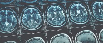
Oncologists at the Yusupov Hospital diagnose brain tumors using modern neuroimaging methods. Early diagnosis can significantly prolong the patient's life. Treatment of patients with brain cancer is carried out using innovative surgical interventions, gentle radiation therapy techniques, and the latest chemotherapy drugs registered in the Russian Federation. They are highly effective and have a minimal range of side effects. As part of scientific drug research conducted at the Oncology Clinic, patients have the opportunity to be treated with new chemotherapy drugs, the safety of which has been proven by previous studies.
Brain tumor – what is it?
A tumor is a voluminous formation, which is a group of atypical cells that are rapidly increasing their population. A tumor in the brain can be either benign or malignant.
All neoplasms of the central nervous system are divided into primary and secondary:
- Primary - those that developed directly in the brain
- Secondary - are metastases of malignant neoplasms of a different location (lungs, kidneys, etc.)
There are a large number of different classifications of tumors of the nervous system. But the histological structure of the neoplasm and its location are of greatest clinical importance.
Based on location, primary tumors are divided into two large groups:
- Brain tumors – account for up to 90% of all central nervous system tumors
- Spinal cord tumors – 10% of central nervous system neoplasms
There are also oncopathologies localized simultaneously in the brain and spinal cord. But their share of the total number of tumors is insignificant - it is a fraction of a percent.
Neoplasms can also be of origin:
- Intracerebral - originate from brain cells
- Extracerebral - comes from blood vessels, nerve sheaths, fragments of embryonic tissue, pituitary gland
Malignant primary brain tumors of intracranial localization have a number of features that distinguish them from cancer of any other location:
- They practically do not metastasize. That is, they do not form daughter tumors in the lymph nodes and other organs.
- The central nervous system is separated from the rest of the body by the blood-brain barrier. Therefore, such tumors usually do not extend beyond the brain.
- Due to the high risk of damage to functionally important areas of the brain, most of these tumors are inoperable.
Due to the above reasons, the classification of tumors according to TNM (tumor size, regional and distant metastases) is practically not used in neuro-oncology. Stages are determined based on the histological type of tumor, and not the extent of the oncological process.
According to WHO, there are 10 histological types of tumors of the central nervous system:
- From neuroepithelial tissue
- From the meninges
- From the nerves
- From hematopoietic tissue
- Germinogenic - from germ cells
- Cysts and tumor formations
- Neoplasms growing into the cranial cavity
- Tumors of the sella turcica (this is a fragment of one of the bones of the skull in which the pituitary gland is located)
- Metastatic tumors
- Unclassified neoplasms
First symptoms
Most brain tumors are asymptomatic for a long time or the existing signs do not allow one to suspect the presence of a space-occupying tumor in the skull. Oncologists at the Yusupov Hospital recommend seeking specialized medical care if you have the following symptoms:
- Frequent and prolonged headaches, often appearing at the site of tumor development, accompanied by nausea and vomiting;
- Loss of coordination, weakness, inability to maintain balance;
- Vision problems and increased photosensitivity;
- The appearance of sudden seizures resembling epileptic ones.
Relatives should arrange a consultation with a neurosurgeon if the patient has strange changes in habitual behavior or unreasonable aggression. If such symptoms occur, it is better not to delay visiting a specialist. If a brain tumor is diagnosed at an early stage of development, when symptoms are mild, patients live much longer. When stage 4 brain cancer is detected, the prognosis for life is disappointing.
The localization of neoplasms is often determined by their biological nature. Neurosurgeons often find malignant gliomas in the cerebral hemispheres of the brain, and benign neoplasms in the brain stem and cerebellum.
Brain tumors are classified according to the degree of maturity of their cells and histological characteristics:
- Mature tumors include astrocytomas, ependymomas, oligodendrogliomas;
- Immature neoplasms include astroblastomas, ganglioblastomas, oligodendroglioblastomas;
- The group of completely immature neoplasms includes medulloblastomas, spongioblastomas, and multiforme tumors.
Brain tumors are divided into different groups according to histogenesis (tissue development):
- Neoplasms of the neuroectodermal or glial series (astrocytomas, neuromas, medulloblastomas, pineoblastomas);
- Meningeal-vascular tumors - develop from the arachnoid endothelium of the meninges and vessel walls (angiomas, meningiomas, chordomas, fibrosarcomas, osteomas);
- Tumors of the chiasmatic-sellar localization - the pituitary gland, growing from the anterior lobe of the adenohypophysis, and craniopharyngiomas;
- Bidermal neoplasms, which consist of elements derived from two germ layers;
- Heterotopic tumors (chondromas, dermoids, epidermoids, lipomas, pyrathemas).
In 1% of cases of brain tumors, systemic neoplasms are identified - multiple meningiomatosis, multiple neurofibromatosis and multiple angioreticulomatosis. Metastases to the brain (unfavorable prognosis) are found in 5% of patients, and neoplasms that grow into the cranial cavity (sarcomas, glomus tumors) - in 1.8% of cases. At the moment, about 90 different tumors of the nervous system are distinguished based on histological and histochemical characteristics. According to the location of the brain tumor, supratentorial tumors are located in the anterior and middle cranial fossa and subtentorial, localized in the posterior cranial fossa.
Gliomas
Gliomas are tumors of neuroepithelial tissue. They make up more than half of all tumors of the central nervous system. This type of brain tumor is most often the result of genetic mutations. These include glioblastomas, astrocytomas, and ependymomas. Some develop for a very long time - years. Others progress rapidly and lead to the death of the patient within a few months.
Gliomas have 4 degrees of malignancy. Typically, grade 1-2 tumors are called benign, grade 3-4 tumors are called malignant. The latter include glioblastoma and anaplastic astrocytoma, the most common neuroepithelial tumors of the central nervous system. They account for up to 80% of all neoplasms of this type.
What does success depend on?
- The operation must be performed by an experienced surgeon based on innovative techniques and detailed diagnostics;
- It is better to perform surgery for anaplastic astrocytoma of the brain in a modern clinic, which will eliminate the formation of infections and side effects;
- If a patient has stage 3 anaplastic astrocytoma Who Grade III is not amenable to surgical treatment, removal by radiosurgery is prescribed. In this case, tumors are destroyed by high doses of radiation, precisely targeting cancer cells with high precision;
- A positive prognosis is influenced by the general condition of the patient and the stage of development of the disease.
Tumors from the meninges
Neoplasms from the meninges account for about 20% of primary CNS tumors. 95% of cases of cancer of this type are meningiomas. The remaining 5% are fibrous histiocytomas, hemangiopericytomas, melanomas, diffuse sarcomatosis and others.
Some risk factors for tumors from the meninges have been established. Here's what can cause a brain tumor:
- Head injuries
- Ionizing radiation (including radiation therapy)
- Eating Nitrites
Genetic defects in chromosome 22 have been identified, due to which a brain tumor can develop. The cause is a mutation at the 22q12.3-qter locus.
According to malignancy, meningiomas are divided into 3 groups:
- Grade 1 - includes 9 histological types, the most common of which are meningoepithelial (60% of cases), transitional or mixed (25%) and fibrous (12%)
- Grade 2 – these are atypical meningiomas, which are marked by rapid cell division and rapid growth
- Grade 3 – anaplastic meningiomas (old name – meningosarcoma)
Rarely, multiple brain tumors occur. The reasons for their occurrence are not known. They make up about 2% of all diagnosed meningiomas. Such neoplasms are characterized by a favorable clinical course. In 90% of cases, a person lives a full life with them, without any symptoms. Only 10% of cases require surgical removal of tumors.
Send a request for treatment
Life expectancy with liver metastases
Secondary cancer foci in the liver arise after the penetration of modified cellular components through lymph or blood. This lesion usually accompanies cancer of the gastrointestinal tract, skin and lungs. Symptoms of liver metastases are as follows:
• weakness; • bad feeling; • loss of ability to work; • abnormally rapid fatigue; • sharp deterioration in appetite; • loss of initial bodily mass; • defects in the functioning of the digestive system.
The danger of this oncopathology is due to the destruction of tissue components of the liver by metastases. How long do you have to live and what does life expectancy depend on in this case? Even with an impressive arsenal of treatment techniques, people with such a lesion live a maximum of 15 months after diagnosis. A slightly longer life expectancy can be achieved with successful cytoreductive operations on the affected organ.
Pituitary tumors
Up to 10% of all intracranial neoplasms are pituitary tumors. They are almost always benign. They usually develop from cells of the adenohypophysis (the anterior lobe of this gland). Such neoplasms are called adenomas, and if they have a diameter of less than 1 cm - microadenomas. What causes a brain tumor of the corresponding localization has not yet been established.
Although some risk factors are known:
- Brain infections
- Traumatic brain injuries
- Toxin poisoning
- Use of oral contraceptives
- Obesity
The causes of a tumor in the brain that grows from pituitary gland cells may be due to gene mutation. Hereditary predisposition is of no small importance. At what age can a brain tumor of adenohypophyseal origin develop? This is one of the “youngest” tumors. It occurs in everyone, including children. The peak incidence occurs in working age - from 30 to 50 years.
Most pituitary adenomas are not accompanied by severe symptoms, so their detection rate is low. Treatment is usually carried out conservatively (normalizing the level of hormones in the blood). When it stops working, surgery is used.
Germ cell tumors
Germ cell tumors in the brain develop from embryonic tissue. These neoplasms include germinoma, choriocarcinoma, yolk sac tumor, and embryonal carcinoma. They are located in the area of the epiphysis. The most common is germinoma. It accounts for up to 0.5% of all brain tumors in representatives of the European race, and up to 3% in Asians. The reasons why this brain tumor occurs more often in the Asian population are not known. It is diagnosed more often in boys. The tumor is malignant - it metastasizes through the cerebrospinal fluid (the fluid that washes the brain).
Metastatic brain tumors
About 20% of the structure of CNS neoplasms are metastatic brain tumors. The causes of these diseases are obvious: the spread of metastases from other parts of the body. It is believed that their actual prevalence is even higher. After all, cancer patients with stage 4 cancer are often not examined too carefully due to the inappropriateness of in-depth diagnostics. Even if there is a suspicion that they have a brain tumor due to cancer of another location, such patients are no longer referred to neurosurgeons.
What can cause such a brain tumor? Here are the most common reasons:
- Lung cancer – 40% of cases of metastatic damage to the central nervous system
- Breast cancer – 10%
- Kidney – 7%
- Stomach or intestines – 6%
- Melanomas - on average 5%
Anaplastic astrocytoma of the brain, grade 2
Treatment at this stage has a positive prognosis. The disease is determined by the following signs:
- The presence of severe malaise, nausea and vomiting;
- Intensive weight loss accompanied by decreased appetite;
- Fatigue;
- Increased fatigue;
- Decreased level of performance;
- Asthenic syndrome.
This stage is not considered too difficult. Treatment of anaplastic astrocytoma of the brain is based on tumor removal. To do this, a number of diagnostic measures are carried out. An ophthalmologist, neurologist, neurosurgeon and oncologist take part in the treatment process. To fully understand the picture, MRI and CT are performed, and these procedures are recommended to be carried out with a contrast agent. A biopsy and puncture is performed, hearing, vision, and vestibular examinations are carried out.
Removal of the astrocytoma provokes an improvement in the general condition. Most often, the process of surgical implementation and drug treatment is carried out. Removal is prescribed by a doctor only if there are no contraindications and if this operation is possible. Since the ability to perform an operation also depends on the location.
Tumors of childhood
Even children can get a brain tumor. Among childhood cancers, brain tumors account for about 20%. This is second only to leukemia. More often they are observed in children of the first year of life. Teratomas are found mainly in children. In older children, benign astrocytomas (more than 30%), primitive neuroectodermal tumors (medulloblastoma, pineoblastoma - 20%) and ependymomas (15% of cases among children over 1 year of age) predominate.
At present, it is not known for certain how brain tumors form in children. It has been established that they develop more often in boys than in girls. The reasons why brain tumors are more common in male children have not been established.
How can you get a brain tumor?
Patients often ask their doctor how they can get a brain tumor. They suggest that knowing the causes will help prevent the disease. Unfortunately, it is not. For many types of cancer, the underlying etiological factors have indeed been established. For example, it is well known that lung cancer occurs mainly due to smoking, cervical cancer is caused by human papillomavirus infection, and liver cancer is caused by viral hepatitis C. But what causes a brain tumor is still not known. Despite numerous clinical studies, scientists have not been able to establish the causes of brain tumors.
However, some risk factors have been identified:
Radiation . There are many ways you can get a brain tumor due to radiation. Most often this is radiation therapy for cancer or occupational hazards (radiologists, nuclear industry workers). In children, in the old days, the cause of brain tumors was radiotherapy used for fungal infections of the scalp.
The connection is not always clearly visible. After all, the first symptoms appear only 10-15 years after irradiation. But a retrospective analysis of medical records shows what can cause a brain tumor. Anamnesis data demonstrate that neoplasms of the central nervous system are more often observed among individuals exposed to radiation exposure.
Does this mean that a brain tumor occurs from radiography, fluorography or computed tomography? After all, these methods are based on the effects of ionizing radiation. No, no such connection has been established. Even decades ago, when the devices were much less advanced and gave tens of times more radiation load, there was no reason to believe that a brain tumor could occur as a result of the diagnostic procedures performed. Today, the devices used are so precise and perfect that the radiation is minimal, without damaging cells or causing mutations.
Heredity . Some genetic abnormalities have been identified that may cause a brain tumor. These include neurofibromatosis type 1, type 2, tuberous sclerosis, Hypeel-Lindau disease, Li-Fraumeni syndrome. There are other genetic causes of brain tumors, but they are much rarer.
Immunity . One of the types of neoplasms of intracranial localization is lymphoma. This brain tumor can occur due to decreased immunity. This is caused by AIDS, treatment after organ transplantation (immunosuppressive therapy), long-term use of immunosuppressants and glucocorticoids for dermatological diseases or systemic connective tissue lesions.
There are also several mythical reasons worth noting. Many people are afraid of them, but these fears are false.
So, here's what a brain tumor can't cause:
- Using a mobile phone – contrary to popular belief, it does not emit ionizing radiation
- Playing soccer (soccer players often head the ball)
- Exposure to electromagnetic fields
- Hair dye
- Stress and bad habits
We also note several controversial reasons why a brain tumor can occur. These risk factors are suspected but not yet definitively proven.
Among them:
- Aspartame (sweetener for diabetics)
- Exposure to vinyl chloride (occupational hazards in plastics production)
- Viral infections
- Exposure to petroleum products
Thus, scientists do not yet know exactly what causes a brain tumor. Therefore, the only thing you can do to prevent this disease is not to expose yourself to massive ionizing radiation and, if possible, maintain a good immune system.
Prognosis in the presence of bone metastases
With this type of cancer, pain discomfort manifests itself quite early, thanks to which the defect is diagnosed almost without problems and treatment begins. About 80% of secondary cancerous lesions of the supporting system appear due to the spread of cancer cells from the prostate and mammary glands. With bone pathology, the clinical picture is as follows:
• there is a development of pain discomfort with a subsequent increase in its intensity; • bone fragility increases (frequent fractures occur); • dense swelling of tissue components is formed due to the accumulation of lymph in the area of localization of the metastatic focus; • signs of cancer intoxication gradually appear.
The described deviation progresses in 2 forms.
• Osteoplastic, in which a large mass of atypical bone tissue is formed. • Osteoplastic, in which bone tissue, on the contrary, decreases.
How long to live with bone metastases? According to statistics, the patient lives 6-10 months. For this lesion, maintenance therapy is carried out, involving the use of chemotherapy and painkillers.
Meningeal brain tumor - prognosis
The survival rate for meningiomas is high. The clinical course of the disease is favorable. Usually these are neoplasms of 1st degree of malignancy. If there are no symptoms of pathology, an observation strategy is used. Surgery may not be necessary because most of these brain tumors have a good prognosis.
The first thing patients are interested in is how long do they live with a brain tumor of meningeal origin? This depends on a number of factors. First of all, it depends on the degree of malignancy, as well as the treatment performed.
If the brain tumor was completely removed during surgery, how long people live depends solely on their age and health status, because meningioma does not recur with a 95% probability. Only 5% of patients experience recurrences within 15 years after surgery.
But sometimes the neoplasm is localized in functionally active structures of the central nervous system. In such cases, complete removal is not possible. However, even if it was partially removed, the risk of recurrence within 15 years is only 50%. In the remaining 50% of cases, the tumor remains the same size - it does not grow. If growth of meningioma is observed, it responds well to treatment with radiation therapy. Even for malignant meningioma, radiation helps control tumor growth for at least 5 years.
The age of the patient is also important. Below you can see data on how long people live with a meningeal brain tumor, depending on age.
The percentage of five-year survival is indicated - that is, the number of people who will live for 5 or more years after diagnosis:
- Up to 45 years – 87%
- Up to 55 years – 77%
- From 55 to 65 years – 71%
In most cases, after 5 years the tumor no longer recurs if it was completely removed during surgery.
Treatment
The goal of treatment is to improve the patient’s quality of life and reduce various disorders of the central nervous system. Most tumors cannot be cured by surgery alone, so combined treatment is used.
The main methods are: surgical removal and radiation therapy involving tissue more than 3 cm. It is the latter method that significantly prolongs the patient’s life. Radiation therapy can be used before and after surgery. In the first case, it reduces the size of the tumor, and in the second, it destroys the remaining tumor cells.
If the localization of the tumor is difficult to reach, making its excision impossible, radiation therapy is used as an independent method. In this case, it does not completely destroy the brain tumor, but stops its growth.
Radiosurgery (gamma knife and cyber knife) is an innovative technique that is a type of radiation therapy. In this case, the radiation beam hits the tumor directly - this is possible under the control of CT or MRI. An important point of this method is its non-invasiveness, which reduces the risk of complications.
Another treatment option is chemoradiotherapy (radiation and chemotherapy at the same time) after surgery. This method is more effective in achieving disease-free survival.
Despite comprehensive treatment, almost all gliomas have a tendency to recur. Only some patients are offered repeated surgery and radiation therapy. Chemotherapy is not very effective in this case.
Recently, in severe cases of relapse, drugs have been used that suppress the formation of new vessels, reduce the vascularization of the tumor and suppress its growth. With their use in glioblastomas, long-term survival of patients in whom standard chemotherapy was ineffective is achieved.
Surgeons at the University of California began treating recurrent cancer with a virus obtained by genetic engineering. It destroys only tumor cells.
Neuroepithelial brain tumor – how long to live?
If you have been diagnosed with a neuroepithelial brain tumor, how long to live cannot be definitely said without establishing its histological type. It is the structure of the neoplasm that determines how long a brain tumor takes to develop.
Based on this parameter, there are 4 degrees of malignancy of neuroepithelial neoplasms of the central nervous system:
- Grade 1 – pilocytic astrocytoma
- Grade 2 – protoplasmic, pleomorphic, hemistotic, fibrillary astrocytoma, xanthoastrocytoma and ependymoma
- Grade 3 – anaplastic astrocytoma
- Grade 4 – glioblastoma
Grade 1 and 2 neuroepithelial brain tumors are considered low grade. They have a more favorable prognosis. Grades 3 and 4 are neoplasms of high malignancy or simply malignant. The worst prognosis is characterized by a grade 4 brain tumor.
How long do people live with glioblastoma ? Unfortunately, this tumor has a worse prognosis. It develops very quickly and is manifested by necrosis (death) of parts of the brain. From the moment the tumor appears to the first symptoms, sometimes not even months, but weeks pass. The average life expectancy for patients under 40 years of age is no more than one and a half years, and for patients over 40 years of age – less than 1 year.
Five-year survival rate in developed countries, depending on age:
- Up to 45 years old – 19%
- From 45 to 55 years – 8%
- From 55 to 65 years – 5%
Patients do not even always undergo surgery, since they die from a grade 4 brain tumor quite quickly. Often only radiation and chemotherapy are used. But these methods do not significantly prolong the patient’s life. Unfortunately, glioblastoma is the most common glioma (neuroectodermal tumor). It makes up about 50% of the structure of these neoplasms.
Anaplastic astrocytoma is another malignant brain tumor. How long do people live with this disease?
Average life expectancy, depending on the patient’s age at the time of diagnosis:
- Up to 40 years – about 3 years
- From 40 to 60 years – on average 2 years
- After 60 years – less than 1 year
Five-year survival rate, depending on age:
- Up to 45 years old – 54%
- From 45 to 55 years – 32%
- From 55 to 65 years old – 14%
The pathology is characterized by infiltrative growth. That is, the tumor penetrates the brain tissue, rather than displacing it. Anaplastic astrocytoma is the second most common neuroectodermal tumor after glioblastoma. In the structure of these neoplasms it makes up about 30%.
The next most common tumor of the central nervous system from neuroectodermal tissue is oligodendroglioma . It accounts for about 5% of all gliomas. The average life expectancy of patients in developing countries, including the CIS, is 6 years. In developed countries (Germany, USA) the indicators are much better.
The five-year survival rate, depending on the age of the patient at the time of diagnosis, is:
- Up to 45 years – 88%
- From 45 to 55 years – 81%
- From 55 to 65 years – 68%
Unfortunately, sometimes oligodendroglioma becomes malignant. In this case, it acquires grade 3 malignancy, and the prognosis worsens. It is not known for certain what causes the brain tumor anaplastic oligodendroglioma. However, the life expectancy of patients is reduced.
Five-year survival rates for this type of neoplasm in developed countries:
- Up to 45 years – 71%
- From 45 to 55 years – 61%
- From 55 to 65 years – 46%
Ependymomas make up about 3% of gliomas. They are more common in children than in adults. Most often these are benign tumors. Anaplastic ependymomas (grade 3 malignancy) are rare. There is no data on what causes this brain tumor in children. Survival rates vary significantly between countries. In the CIS, the five-year survival rate for children over 3 years of age is about 50%, for adults – 70%. In developed countries, the statistics are much better due to better treatment.
Five-year survival rate for patients with ependymoma in countries with well-developed medicine:
- Children's age – 75%
- Patients from 20 to 45 years old – 92%
- From 45 to 55 years – 89%
- From 55 to 65 years – 86%.
How long do they live with a brain tumor from fetal tissue?
Germ cell tumors are malignant. They occur in children. They are characterized by metastases. The most common tumor is germinoma. It is also the most favorable. In the vast majority of patients, even without the use of surgical methods, this malignant brain tumor is cured. How long do they live after treatment? Life expectancy is potentially the same as that of a person who has never suffered from this pathology. To cure the disease, radiation therapy and sometimes chemotherapy (in children under 4 years of age) are sufficient. The younger the age, the better the prognosis. Unfortunately, the prognosis for other types of germ cell tumors is much worse. They are very rare. But patient survival is extremely low. These are inoperable brain tumors. How long do patients live? Only 5% of patients will live more than 2 years after diagnosis. Radiation therapy and chemotherapy are used for treatment, since surgical treatment does not improve the prognosis. Only palliative operations are performed. For example, shunting surgical interventions for blockage of the cerebral aqueduct.
Metastatic brain tumor – how long to live?
In many cases, cancer is detected after metastases have appeared in the central nervous system. How long people live with a metastatic brain tumor depends on the treatment provided. In the natural course of the disease, the average life expectancy of patients is about 3 months. With surgical treatment or radiation therapy, it can reach 2 years or more. Life expectancy also depends on the histological type of cancer, location and number of metastases. In approximately 50% of cases they are multiple, which worsens the prognosis.
Send a request for treatment
Observation after treatment
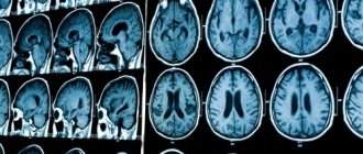
Regardless of the type of brain tumor, the patient will undergo regular MRI examinations in the future.
The frequency of MRI procedures depends on the diagnosis.
- If the tumor is characterized by changes and growth , the patient needs to undergo MRI more often - perhaps once every 2 months.
- If the tumor grows slowly or is a benign formation , scanning is prescribed once every 3-4 months.
In the absence of pathologies, examinations are carried out once every six months, then once a year. In any case, the patient should keep in mind that after diagnosis - regardless of whether he underwent surgery or not - he will undergo regular MRI procedures. Magnetic resonance imaging is the best way to monitor the condition of the tumor.
Where to go if you have brain cancer?
The diagnosis and treatment of brain cancer and astrocytoma are the most successful in Germany and other developed countries, which is confirmed by patient survival statistics. If you want to use the services of German doctors, please contact ]Booking Health[/anchor]. We organize treatment abroad.
Our advantages:
- A team of doctors who understand modern methods of treating various diseases will select the best clinic for the treatment of brain cancer for you.
- Thanks to direct contracts with the best medical institutions in Germany, you can save up to 70% of the cost of the treatment program. In addition, the waiting time for a doctor’s appointment will be reduced.
- The initially agreed cost of treatment is guaranteed not to change. Even if complications arise and additional therapeutic measures are required, all unforeseen expenses will be covered by insurance.
- A full package of services: we will completely organize the entire treatment process in Germany, provide an interpreter, and provide transfer from the airport to the clinic.
- Absolute transparency of financial relationships: you can always find out what services you paid for and in what volume. Unused funds after treatment will be returned.
To take advantage of all the benefits of German medicine, leave a request on our website. After processing it, we will contact you and you will be able to receive comprehensive advice on the treatment of brain cancer abroad.
Diagnostics
Symptoms of the disease vary, and a patient with a larger tumor may experience less severe symptoms than a patient with a small tumor. The first thing the patient should do is visit a local physician, who will prescribe further tests.
An ophthalmologist can identify the disease by the condition of the eyeball.
When a patient consults a doctor with symptoms of the disease, he is prescribed:
- neurological examination;
- positive emission tomography;
- CT;
- MRI;
- angiography and other radioisotope techniques;
- magnetoencephalography;
- surgical intervention;
- stereotactic biopsy;
- venticuloscopy;
- Lumbar puncture.
Ependymoma - causes, symptoms, diagnosis and treatment
Only after carrying out all the necessary studies can the doctor draw conclusions about the patient’s health status, determine the stages and possibilities of treatment.