general information
The shoulder joint is one of the most mobile in the body. The head of the humerus is spherical in shape, and the corresponding surface of the scapula is almost flat. The structure is additionally fixed by a strong articular capsule, numerous tendons and ligaments. As a result, a person can move his arm in any direction.
The cause of pain can be any of the elements of the joint apparatus, as well as nearby nerve bundles and muscles. The problem can also come from neighboring structures: the spinal column, heart, stomach, esophagus. Often, only a doctor can accurately determine the cause of pain in the shoulder joint of the arm and prescribe the correct treatment.
Make an appointment
Treatment of arthrosis of the shoulder joint at ArthroMedCenter
The ArthroMedCenter in Moscow uses the following methods for treating symptoms of arthrosis of the shoulder joint:
- Hivamat therapy. A massage is performed using a special electric field. It represents a slight vibration. Thanks to this procedure, damaged tissues are restored and swelling goes away. This technique copes well with pain symptoms.
- Electrophoresis. Thanks to this procedure, medications penetrate as deeply as possible into the tissues and act more effectively on the affected area.
- SMT therapy. This technique improves blood circulation, triggers recovery processes and relieves severe pain.
With the help of a correctly established diagnosis and competently prescribed treatment, you can get rid of pain in the shoulder joint. To stop the inflammatory process, conservative treatment and physiotherapy are used.
Causes of pain
Pain in the shoulder joint can occur due to various reasons, the most common of which are:
- sedentary lifestyle;
- excess body weight;
- excessive load on the joint apparatus (heavy lifting, vibration, professional sports);
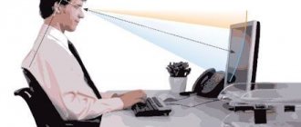
- incorrect posture;
- previous injuries and joint surgeries;
- age over 50 years (tissue regeneration worsens, especially cartilage);
- infectious and autoimmune lesions;
- metabolic disorders;
- disruption of the endocrine glands, as well as sudden hormonal changes (pregnancy, menopause);
- hereditary predisposition to diseases of the musculoskeletal system.
These unfavorable factors cause the development of certain pathologies, which, in turn, cause shoulder pain.
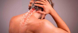
How often does pain in the cervical spine radiate to the shoulder? Who is in the “risk zone”?
According to statistics, this symptom is observed with equal frequency in almost half of all adults living on different continents and representing a wide variety of segments of the population (especially in people with long-term forced “sedentary” work or study). Moreover, it is more often registered in women than in men (with an approximate ratio of 3:2). Sometimes cases where pain in the cervical spine radiates to the shoulder also occurs in adolescents with poor posture and leading a sedentary lifestyle. By the way, it has been noticed that this symptom in most cases recurs again over the next five-year period.
A separate “risk group” for which the occurrence of this symptom is not uncommon consists of athletes who excessively engage in strength exercises with a barbell (especially on their own), contact wrestling, and swimmers.
Possible diseases
Pain in the shoulder joint can be caused by diseases of the musculoskeletal system, nervous and cardiovascular systems, cancer and other processes.
Arthritis
One of the most common causes of shoulder pain. The disease is associated with the development of an active inflammatory process in the shoulder joint, which is accompanied by:
- severe pain syndrome;
- increased temperature and swelling of the affected area;
- redness of the skin over the sore joint.
Pathology occurs against the background of trauma, infectious diseases, hypothermia, high physical activity, allergic and autoimmune reactions. Depending on the duration and severity of symptoms, it can be acute or chronic.
Arthrosis
Arthrosis of the shoulder joint is a chronic degenerative disease in which the gradual destruction of cartilage tissue occurs. As a result, the contacting areas of the bones lose their smoothness. Increased friction during movement causes pain, which gradually increases as the disease progresses. Subsequently, bone growths (osteophytes) form inside the joint cavity, the bones themselves are deformed, and movements are almost completely blocked. In addition to pain, the disease manifests itself as stiffness of the affected joint and a characteristic rough crunch.
Bursitis
Bursitis is an inflammatory disease in which fluid accumulates inside the joint capsule. Most often occurs against the background of trauma. Pathology manifests itself:
- acute pain during movement and less severe at rest;
- increased body temperature (in mild cases, only the area of the affected joint becomes hot);
- pronounced swelling, when pressed on, the movement of fluid inside the limited cavity is clearly felt.
Capsulitis
Chronic inflammation of the capsule surrounding and stabilizing the joint. The second name is “frozen shoulder syndrome”. As a result of a long pathological process, the capsule contracts, and the surrounding ligaments partially atrophy. The pain usually occurs at night, and the disease itself has a wave-like character and either worsens or subsides. After active inflammation subsides, a person feels a pronounced difficulty in movements, especially outward rotation of the arm.
Humeroscapular periatritis
The inflammatory process affects the tissues surrounding the shoulder joint, resulting in severe pain that intensifies with movement. A person cannot raise his arm up and put it behind his back. As the process enters the chronic phase, noticeable stiffness is noted.
Tendinitis
Inflammation of the tendon - tendonitis - occurs against the background of significant physical overload of the shoulder area. Depending on the severity of the pathological process, pain is provoked by physical activity or occurs at rest. The lesion can involve various tendons, which determines the symptoms.
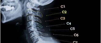
Osteochondrosis of the cervical spine
The disease is associated with flattening of the intervertebral discs, resulting in pinched nerve endings. The intensity of pain depends on the degree of damage, often intensifying when turning and tilting the head. The process may also be accompanied by numbness of the hand and sharp “shots”. Often the pain spreads to other parts of the body.
Ligamentous calcification
The disease is associated with the deposition of calcium salts in the tissues of the ligaments. The exact reasons have not yet been studied. The process causes intense pain during movement and at rest.
Brachial neuritis and brachial plexitis
Damage to the brachial nerve or nerve plexus in this area usually occurs due to injury, hypothermia, or an infectious disease. The pain affects not only the shoulder, but the entire arm, often accompanied by numbness, burning, and the feeling of goosebumps running across the skin. In severe cases, paralysis of individual muscles may develop.
Tumors
Pathological formations in the shoulder area appear quite rarely. As they grow, they compress surrounding tissue. As a result, the patient first experiences discomfort and a sensation of a foreign body in this area, and then pain of varying degrees of intensity.
Diseases of internal organs
Of all the non-musculoskeletal conditions that cause shoulder pain, myocardial infarction is the most dangerous. The pain is usually of a squeezing or burning nature and appears first in the chest area or between the shoulder blades, and then spreads wider, including to the left shoulder, arm, left half of the neck and face. Pathology is usually accompanied by:
- severe weakness;
- sweating;
- dizziness;
- feeling of lack of air;
- fear.
This condition is fatal and requires emergency medical attention.
If the cause of the problem is acute cholecystitis or cholelithiasis, the pain will begin in the solar plexus and radiate to the right shoulder. The attack often occurs against the background of overeating and is accompanied by nausea and vomiting.
Injuries
Bruises, sprains, ruptures and tears of ligaments, joint dislocations, bone fractures inevitably cause acute pain. Depending on the type of lesion, it may be accompanied by:
- limited mobility;
- visual deformation of the joint;
- hematoma formation;
- pathological mobility of bones and crunching of fragments.
The diagnosis is usually not in doubt, since the patient himself clearly associates the occurrence of pain with injury.
Shoulder pain treatment
1. Tendopathy (tendonitis) of tendons
rotator cuff (shoulder rotators)
The unique anatomy of the shoulder joint provides high mobility of the arm in all planes, including 360-degree circular movements. Knowledge of anatomy and functional features will help to understand the cause of diseases that affect the shoulder joint. In its structure, it is simple, consisting of 2 bones and a complex spherical joint. The large, rounded head of the humerus touches the small, flat glenoid cavity of the scapula. The diameter of the head is 3 times the diameter of the glenoid cavity. This discrepancy between shape and size provides a wide range of movements. The strength and stability of the shoulder joint is given by the elastic capsule and the articular lip located in the scapular cavity - cartilage, the curved edges of which extend beyond the boundaries of the bed and cover the head of the humerus.
Stability and mobility are achieved through the tendon-muscular system of the shoulder joint (rotator cuff or shoulder rotators). The main function of the rotator cuff is to center and press the large head of the humerus against the small glenoid cavity of the scapula.
The rotator cuff contains tendons from several muscles.
1. Supraspinatus muscle.
2. Infraspinatus muscle.
3. Teres minor muscle.
4. Subscapularis muscle.
All of them carry out movements in the shoulder joint (flexion, extension, abduction, rotation).
Tendopathy is a degenerative tendon. Tendinitis is inflammation of the tendons. With rotator cuff tendinitis, the ligaments of all the above muscles are affected almost simultaneously or sequentially. Options are possible with different pathological processes occurring in different tendons.
Causes:
There are several groups of people at risk:
· Athletes who constantly strain and injure the shoulder joint (tennis players, swimmers, water polo players, etc.).
· Workers whose professions involve constant tension with their arms raised above their heads.
· People who are forced to wear plaster casts for a long time, limiting movement in the shoulder joint.
· Patients with cervical osteochondrosis.
· Women over 50 years old.
· Persons with diseases of the endocrine system (diabetes mellitus, thyroiditis).
· Patients with poor posture.
Forms of pathology and clinical picture
There are several variants of pathology. They are divided according to the principle of localization and the nature of the processes.
Based on localization, there are general (covers the entire ligamentous apparatus of the rotator cuff) and local tendinitis (separate groups of ligaments are affected).
Depending on the nature of the disease, there are several options.
· Post-traumatic. Classic inflammation of the tendons is associated with their damage (microtears of altered tendons under loads, which subsequently “heal” poorly).
· Classic inflammatory variant. Characterized by common tendon inflammation.
· Calcifying variant. Inflammation occurs
in those tendons that have undergone calcification (deposition of calcium salts). Most often it occurs after prolonged immobilization and in old age.
In form, all the types of inflammation described above can be acute or chronic. It all depends on the duration of the inflammatory process.
The clinical picture of the disease is dominated by pain in the shoulder joint. It is associated with hand movements in any plane. At rest, it lasts much longer than other shoulder pathologies.
When palpating the shoulder, tendon nodules and pain are detected. During the calcification process, every movement is accompanied by crunching and creaking.
Diagnosis and prognosis
Diagnosis of the disease consists of a combination of the clinical picture and data from laboratory and instrumental research methods.
MRI examination is the main diagnostic method that allows us to clarify the nature and extent of the pathology.
The presence of pain during movements in the shoulder joint with simultaneous signs of inflammation (determined by a general blood test) are factors for tendonitis. The characteristics of pain during various movements make it possible to suspect the nature of the damage to certain tendons.
2. Subacromial bursitis.
Subacromial bursitis is a particular manifestation
inflammation of the periarticular bursa located under the acromion (the process of the scapula above the shoulder joint). It occurs most often in young and active people. Most often, it affects male athletes (volleyball players, water polo players, basketball players) and workers whose professions involve a constant forced position of tense arms above the head.
Causes and pathogenesis
Subacromial bursitis in 99% of cases has previous causes:
· injuries;
· loads.
Very rarely (no more than 1% of all patients with bursitis) the disease occurs for no reason.
More often, damage can be the result of injury or prolonged physical activity. The latter lead to disruption of the blood supply to the bag. As a result, they lose their barrier function and their permeability increases.
Clinical picture and diagnosis of the disease
The leading symptom of the pathology is pain when trying to raise the arm above shoulder level. There is no weakness in the muscles surrounding the joint. In the lowered position of the arms, the person does not experience any pain.
In the absence of treatment and as the pathology develops (with severe inflammation), pain can develop when moving the arms to the sides of the body below shoulder level. Only advanced inflammation can lead to redness of the skin over the shoulder joint.
Diagnosis of subacromial bursitis consists of a clinical picture, examination and a number of laboratory and instrumental research methods.
Upon examination, it should be noted that the range of motion in the shoulder joint is limited precisely because of the pain factor. To do this, a so-called Nir test is performed. Why measure the amplitude of active movements in a sore joint? After this, lidocaine is injected into the subacromial bursa (in the absence of allergies, of course). After a few minutes, repeat the amplitude measurement. She meets the standards. Which is proven by the fact that the limitation of range of motion is associated only with the pain factor.
Of the general clinical laboratory tests, a complete blood count is important. It allows you to identify the inflammatory process (increased number of leukocytes and accelerated ESR). It can also be used to judge the effectiveness of treatment.
Among instrumental methods, MRI has an advantage. It allows us to identify not only the expansion of the cavity of the bag and the presence of effusion (inflammatory contents) in it, but also the condition of the tendon-capsular apparatus of the shoulder joint. This will allow us to determine treatment tactics.
3. Biceps tendonitis
(biceps brachii)
The hand is the most important organ of the human body.
Therefore, any pathology is not only a medical, but also a social problem. One of the most common pathologies is tendonitis of the biceps brachii tendon.
Causes
The causes of tendinitis are inflammation of the tendon. It can develop as a result of the following conditions:
shoulder injuries;
· sprain due to forced position of the arms or loads;
· impingement syndrome (incarceration of the tendon between the head of the humerus and the process of the scapula).
Among the patients, athletes predominate - swimmers, tennis players, athletes (shot throwers, discus throwers and javelin throwers). The disease is also common among manual workers, which involves constant straining of the arms when lifting them above shoulder level.
The last reason is most often characteristic of men aged 40 to 50 years. This is primarily due to the fact that this age group still remains highly active. At the same time, the elasticity of the tendons and the amount of synovial fluid in the subacromial bursa are already reduced. In addition, they often (if there has been inflammation of the joint in the past) already have bone growths in the acromion area.
Clinical picture
Tendonitis is characterized by pain along the front of the shoulder. They spread along the biceps muscle up to the elbow bend.
Pain occurs when lifting heavy objects and bending the arm at the elbow joint. They also prevent you from raising your arms above shoulder level. The character is dominated by aching pain, which has a clear connection with movement. The pain goes away with rest.
With instability of the biceps tendons, active movements of the arm are often accompanied by clicking sounds in the shoulder area. They are felt by the patient himself, but can be heard by others.
If left untreated, long-term tendinitis often leads to rupture of the biceps tendon. This is a direct path to disability and surgery. A sure sign of a tendon rupture is a displacement of the biceps towards the elbow. This is very clearly visible when you try to tension it. Many people feel a click just before the rupture.
Diagnostics
Making a diagnosis requires a thorough history taking, examination and a series of instrumental studies. The pathology has very nonspecific symptoms, and therefore requires differentiation from a number of other lesions of the shoulder joint.
The following are widely used as instrumental methods.
· Radiography. It allows you to detect the presence of fractures of the process of the scapula or even the humerus itself. Since they are undoubted signs of the traumatic nature of tendinitis. If the x-ray shows various bone growths, then this is most likely a long-term disease.
· MRI. Non-invasive and harmless method. It allows you to see the condition of the tendons themselves and the underlying tissues.
· Arthroscopy. It is carried out through a small puncture into the joint cavity. The method allows not only to accurately diagnose the nature of the pathology, but also to carry out treatment.
It is important to take into account the fact that without timely contact with a specialist, tendinitis almost always leads to disability.
4. Impingement syndrome of the shoulder joint
Impingement syndrome is a condition in which the rotator cuff tendons and biceps tendon are pinched between the acromion and the humerus (its head).
The rotator cuff is a complex of tendons of several muscles that provide strength and rotation to the shoulder joint.
· Supraspinatus muscle.
· Infraspinatus muscle.
· Round muscle.
· Subscapularis muscle.
When raising the arm above the head, the space between the head of the humerus and the acromion process decreases by almost 2 times. Normally, even with strong contraction, infringement does not occur.
Causes
The syndrome develops in the presence of a number of preceding factors.
1. Constantly raising the arm above the head and straining it.
2. Traumatization of the shoulder joint.
Both processes trigger a chronic inflammatory process in the subacromial space. As a result, the growth of bone growths (osteophytes) is stimulated. Over time, they narrow the subacromial space. In addition, osteophytes disrupt the smoothness of the space between the joint and the process. All this creates conditions for tendon pinching in the subacromial space.
Clinical picture of the syndrome
The syndrome is accompanied by pain in the shoulder area. At first, they look like slight discomfort when raising your arms above head level. Later, real pain syndrome develops. Aching pain. They occur when you raise your arms to the level of the shoulder girdle.
Without treatment, bursitis of the subcaromial bursa quickly joins the syndrome. The pain becomes constant with any movement of the arms. Of course, when it rises to shoulder level and above, they become stronger.
Diagnostics
The presence of pain is a nonspecific sign. But its combination with the results of a number of instrumental methods allows you to accurately determine the diagnosis and treatment tactics.
Therefore, the gold standard for diagnosing the syndrome is a combination of several methods:
· survey;
· collection of data on the development of the disease and the presence of risk factors (age, professional and sports activities);
X-ray and MRI of the joint.
The syndrome is characterized by a connection with movement. Infringement occurs precisely at the moment of raising the arm above the level of the shoulder girdle.
X-rays show a narrowing of the subacromal space and the presence of osteophytic growths.
MRI allows you to visualize structural changes in muscles, ligaments and tendons.
5. Adhesive capsulitis
The disease is a slowly progressive growth of fibrous (a type of connective) tissue in the joint cavity. Traditionally, the pathology refers to the shoulder joint, so its name is omitted in the designation of the disease.
Mostly women suffer from it. They account for about 80% of cases of the disease. With equal frequency, pathology can occur in both the left and right shoulder joints.
Etiology and pathogenesis
For reasons of development, adhesive capsulitis can be of two types.
· Primary. Occurs in a completely healthy joint. The causes of the pathology are not known.
· Secondary. The disease develops against the background of a number of diseases:
- diabetes;
- myocardial infarction;
- stroke;
- surgical interventions on heart vessels;
- thyroid disease;
- malignant processes.
The leading factor in the development of the disease is the formation of collagen threads from which fibrous tissue is gradually formed. It gradually fills the joint cavity, which leads to its reduction. This leads to a decrease in the range of motion of the arm in the shoulder joint.
Clinical picture
The clinical picture of the disease is characterized by clearly defined phases. Signs of pathology are individual for each patient.
1. Initial phase. Lasts from 1 week to 1 year (the reasons for this variation in the primary disease are unknown). Most often, the period does not exceed 3-4 months. During this period, there is a gradual increase in pain in the affected joint. They are not associated with movements in the “sick” hand. Often occur at night (especially after lying for a long time on the side of the affected joint).
2. Stiffness phase. Duration is up to 1 year. But most often the period ranges from 4-6 months. The phase is characterized by a gradual decrease in pain with a simultaneous increase in stiffness in the affected joint. In the final stage, there is almost complete immobilization of the shoulder joint.
3. Resolution phase. The duration of this period is about 1 year. But it strongly depends on the first phase. The longer the pain period lasts, the longer the recovery takes.
The total duration of the disease is from 2.5 to 3 years. In the end, in the absence of treatment, 99 out of 100 patients remain with partial limitation in movements. The recovery period without intervention is extended to 4 years. Accordingly, the risk of lifelong disability increases.
Diagnostics
To diagnose a disease, its symptoms, examination and some diagnostic methods are of key importance.
When interviewed, the patient can quite clearly say (with an accuracy of 2-4 weeks) about periods of pain and stiffness in the affected joint. If a patient first approaches in the last phase of the disease, he most often talks about pain in the joint, which then gave way to stiffness.
Upon examination, you can see a simultaneous decrease in the amplitudes of active and passive movements in the shoulder. Moreover, during the second phase the patient does not experience pain at all when attempting passive movement. They are significantly limited
At the very beginning of the second phase, thinning of the deltoid muscle over the affected joint becomes noticeable. Outwardly, this looks like a smoothing of the shoulder relief.
Among the diagnostic methods, ultrasound and magnetic resonance imaging may be valuable. They are non-invasive and allow detection of thickening of the joint capsule, intra-articular and periarticular changes.
6. Arthrosis of the acromioclavicular joint
Arthrosis of the acromioclavicular joint is a degenerative disease of the joint of the same name. It lasts for a long time. The period from the onset of processes in the joint to the first signs can be measured in years.
Causes
In the risk group, older people who from a young age were involved in traumatic sports (for the shoulder joint) worked for a long time in those professions that required prolonged tension with their arms raised high.
The second most important reason is previous injury to the joint itself or its ligaments. Moreover, degenerative processes develop years after the injury.
To a lesser extent, arthrosis can be caused by injuries to the humerus, which lead to improper redistribution of loads on the arm muscles. Various congenital abnormalities of the spine and chest can also lead to the disease.
Pathogenesis of the disease
It all starts with gradually developing degenerative processes in the acromioclavicular joint. The process looks schematically as follows.
1. Thinning of the articular cartilage of the joint due to wear and tear.
2. Damage to cartilage surfaces. As a result, they lose their smoothness. Friction of articular surfaces
3. The beginning of the development of osteophytes along the edges of the joint and in places of cartilage ruptures.
4. Loss of the joint's functional activity.
Clinical picture
The first two stages of the development of the process proceed almost unnoticed. Rarely, some patients may experience unpleasant (even pain) sensations when pressing on the area of the acromial clavicular joint.
As the process progresses, occasional swelling in the joint area and pain when moving the arms and torso occur. Painful palpation is felt by all patients.
Diagnosis and prognosis
For the diagnosis of pathology, radiography of the acromial clavicular joint area is crucial. It allows you to identify narrowing of the joint space. This is a sure sign of arthrosis.
The prognosis for the development of pathology depends on the time the patient goes to the doctor. Reverse development of the process is impossible. But it is quite possible to slow it down (in some cases even stop it indefinitely). Therefore, the sooner the patient contacts a specialist and the better he follows all his recommendations and prescriptions, the greater his chances of maintaining the mobility of the acromial clavicular joint.
Omarthrosis is a chronic degenerative disease of the shoulder joint. It is characterized by the gradual destruction of cartilaginous plates and articular surfaces of bones. At the same time, osteophytes develop. And the joint itself gradually loses its functional activity.
The disease most often affects people over 50-55 years of age. According to statistics, up to 60 years of age, pathology occurs in 10% of men and 7% of women. After 70 years, both sexes suffer from this disease in approximately the same ratio - 7 out of 10. Among them, joint deformation in the final stage of the disease occurs in almost 60-70% of cases.
Causes
The causes of arthrosis of the shoulder joint are similar to other degenerative pathologies of the joints.
· Excessively strong and prolonged load on the joint. This is typical for painters, plasterers, and weightlifters.
· Congenital anomalies of the joint and humerus. The former lead to early wear of cartilage due to a low safety margin. Abnormalities of the humerus (and the arm in general) cause improper load distribution. As a result, the joint experiences excessive tension even during normal movements. This also leads to early wear.
· Metabolic disorders in the joint cavity. On the one hand, this leads to defects in cartilage tissue due to the low rate of its restoration. On the other hand, metabolic disorders cause the deposition of calcium salts. They themselves lead to damage to the articular cartilage.
There is evidence that arthrosis can be familial. This is based on the pattern that people whose parents suffered from arthrosis are more likely than others to acquire this disease with age.
Not the least of the reasons is previous shoulder injuries. This is especially true for dislocations and sprains. Typically, exposure to grass in youth increases the risk of arthrosis in old age by 50%.
Course of the disease and its clinical picture
Arthrosis occurs in several stages. Each has its own characteristic clinic.
1. First stage or degree. The processes of degeneration of cartilage tissue are minimal. Pain usually occurs in the evening and at night. They are moderate, often aching and constant in nature. The amplitude of habitual movements is preserved. They rarely cause pain. Therefore, patients whose daily activities and occupation do not involve stress on the shoulder joint have virtually no complaints. They attribute evening and night pain to “arm fatigue” or “a reaction to changing weather.” Accordingly, they do not go to the doctor.
2. Second stage. Changes in the joints lead to constant pain. They arise in response to habitual movements. At this stage, most begin to see doctors for the first time. The range of motion in the shoulder joint is reduced due to pain.
3. Third stage. At this stage, pain with the slightest movement is very pronounced. But most of all the patient is worried about stiffness in his hands. It can reach such a state that only their rocking becomes possible. The protrusions of the humerus are visible in the area of the joint.
The last stage is not typical for everyone. Most often, it is reached by those patients who did not seek medical help or ignored medical recommendations.
The disease often develops on both sides. A unilateral process predominates in patients who suffered a shoulder injury in their youth.
Diagnostics
The diagnosis depends on the stage of the disease at which the patient consulted a doctor. To identify pathology at the first stage, it is important not only to question the patient, detail his complaints, but also instrumental studies. Such as radiography and tomography of the joint. The first method allows you to notice a narrowing of the joint space. But it begins to develop at the end of the first stage. Therefore, tomography is considered the ideal method for this period, since it allows you to visualize the condition of the cartilage.
When a patient calls for a second
stage, questioning, examining the patient and performing a simple test are important. The patient's complaints are not specific, but their connection with movement and gradual intensification towards the end of the day speaks in favor of degenerative processes. The test consists of asking the patient to place his hands behind his back and clasp his palms. A healthy person will do this without problems. But this will be difficult for the patient. And when the process is severe, it is simply impossible to become.
Radiography at the second stage will only confirm the diagnosis and clarify the extent of damage to the joint.
The third stage leaves no doubt at the first contact between the doctor and the patient.
8. Dislocation of the acromioclavicular joint
Dislocation of the acromioclavicular joint occurs in people leading an active lifestyle or participating in contact sports. From the point of view of anatomy, under the dislocation of the clavicular
thoracic joint implies a complete or partial rupture of its ligaments.
Causes and clinic
Dislocation develops in the following situations.
· Falling on an outstretched arm.
· A blow to the area of half the chest and the corresponding shoulder. This typically occurs in contact sports. This is hockey, rugby, wrestling, etc.
Clinical manifestations of a dislocation are associated with pain in the shoulder area (sometimes the patient may even indicate the projection of the joint). In this case, the corresponding limb is sharply weakened.
Diagnostics
When examining the victim, a change in the relief of the affected shoulder is visible. When you press on the corresponding collarbone, it “sinks” freely. This is called the "piano key" symptom.
9. Shoulder dislocation
Shoulder dislocation is one of the most common traumatic pathologies of the shoulder joint. In terms of frequency of occurrence, it is second only to a fracture of the humerus.
Causes
Develops as a result of injury. For example, when falling or hitting any surface.
There are primary and habitual (occurring more than once) dislocations. Primary dislocation is associated only with injuries sufficient to cause the joint to sprain, rupture its capsule, and expose the head of the humerus.
Secondary dislocation also occurs with injuries. But for its development there is no longer a need for such a strong traumatic factor. In addition, more than 75% of all habitual dislocations have a congenital predisposition. Most often this is weakness of the ligamentous apparatus or elongation of the connective tissue fibers of the tendon. The latter is typical for individuals with connective tissue dysplasia and for patients with congenital syndromes (Morphan's syndrome is the most common).
Clinical picture and diagnosis
The clinical picture is dominated by two main signs:
· Pain in the shoulder area. It is constant and intensifies when you try to move your hand. This is especially true for primary dislocation. With habitual pathology, pain often occurs when trying to move the arm.
· Restrictions on hand movement. Active movements in the shoulder joint are often completely impossible. Passive movements are greatly limited.
The presence of these signs makes it possible to suspect a joint dislocation with a high degree of probability. Radiography makes it possible to clarify its nature and exclude traumatic bone injuries.
For injuries and other diseases of the shoulder, PRP therapy in Moscow can be effective
Nature of pain
Understanding the nature of pain and the ability to describe it will be useful to everyone, since the quality and speed of diagnosis depends on this. Often, just by the characteristics of the sensations, the doctor can assume the presence of one or another pathology. The pain may be:
- acute: occurs abruptly and, often unexpectedly, usually has a shooting character and has high intensity; often occurs against the background of injury, pinching of the intervertebral nerve;
- aching: the intensity of the sensations is low, often they are of a pulling nature; the symptom often accompanies chronic diseases (arthrosis) and inflammatory processes (arthritis, bursitis, glenohumeral periarthritis, etc.) during the recovery period;
- associated with movement: characteristic of most pathologies of the shoulder joint and surrounding muscles; It is important to note which movements cause increased pain, as this helps to make the correct diagnosis;
- reflected: the epicenter of sensations is not located in the shoulder area, a wave of pain also affects this area; the symptom is characteristic of cardiovascular pathology: angina pectoris, myocardial infarction, as well as cholelithiasis, pleurisy, pancreatitis;
- shooting, pulsating: sharp pain impulses are characteristic of damage to the spinal roots, muscle spasms, etc.;
- constant: sensations do not go away either day or night, while movements can intensify them; characteristic of inflammatory processes.
In addition to describing the nature of the pain, it is important to tell the doctor about the accompanying sensations. Thus, nerve damage is often accompanied by burning and tingling, loss of sensitivity, etc.
Arthrosis of the shoulder joint
Arthrosis is a degenerative pathological process of a chronic type. Accompanied by deformation, destruction and elimination of cartilage tissue. Against the background of such processes, sections of bones begin to rub against each other, come into close contact, and their destruction begins.
With constant friction, painful sensations arise, they become more pronounced as the pathological process develops. In parallel, osteophytes are formed, joints and bones undergo deformation. The motor function of the joint is lost and becomes limited. In the advanced stage of the disease, a person completely loses the ability to move his shoulder.
In the first stages of development, crunching and clicking appears, and an area of compaction and discomfort occurs. After this, discomfort and severe pain occur. To make a correct diagnosis, you must consult a doctor in a timely manner.
Diagnostics
To determine the reason why a person has shoulder pain, the doctor uses the following methods:
- survey: identifying the nature and intensity of pain, the circumstances of its occurrence, the presence of concomitant diseases, etc.;
- examination: the doctor assesses the range of motion in the joint, the color and temperature of the skin, the presence of swelling, limitations in movement, etc.; a neurologist evaluates tissue sensitivity, muscle strength, and quality of reflexes;
- X-ray and CT: pictures and tomograms make it possible to assess the condition of bone tissue and partly cartilage; this is an indispensable method for diagnosing arthrosis, dislocations, cracks and fractures of bones, osteochondrosis of the spine;
- MRI: magnetic resonance imaging allows you to see the condition of not only bones, but also soft tissues, making virtual sections in specified planes;
- Ultrasound: ultrasound examination of the shoulder joint area allows you to see foci of inflammation, accumulation of fluid in the joint capsule, abscesses and neoplasms;
- laboratory diagnostics: a general blood test provides information about the presence of inflammation or allergies; if rheumatoid arthritis is suspected, a test for C-reactive protein, etc. is taken;
- joint puncture: a diagnostic puncture of the joint capsule, through which an analysis of the synovial fluid is taken or arthroscopy is performed (inspection of the joint from the inside using a miniature camera);
- electromyography: assessment of the efficiency of muscle fibers.
If necessary, consultations with narrow specialists and additional examination methods are prescribed: electrocardiogram, ultrasound of the abdominal organs, etc.
Shoulder Pain Treatment
Treatment for shoulder pain depends on its cause. If the problem is related specifically to the musculoskeletal system, painkillers come first:
- non-steroidal anti-inflammatory drugs in the form of injections, tablets and capsules, ointments and creams, patches, rectal suppositories; necessary for quickly stopping the inflammatory process and relieving pain;
- glucocorticosteroids: used in the form of tablets and injections, including for intra-articular administration;
- muscle relaxants: used to relieve muscle spasms in arthrosis, osteochondrosis and other pathologies;
- warming agents in the form of ointments: improve blood circulation in the affected area.
Depending on the cause of the pain, the doctor may prescribe:
- antibiotics (for inflammatory diseases);
- chondroprotectors (for arthrosis);
- B vitamins (for neurological pathologies), etc.
In acute processes and injuries, an important therapeutic factor is hand immobilization. Special bandages and orthoses reduce the range of motion and allow the tissues to fully recover. After active inflammation subsides, a variety of physiotherapy procedures are prescribed:
- magnetic therapy: exposure of the affected area to magnetic waves;
- shock wave therapy: destruction of pathological bone growths by sound waves of a certain length;
- ozone therapy: injection of a special gas mixture into the joint;
- electromyostimulation: exposure of muscles to electrical impulses to cause them to contract;
- ultraphonophoresis and electrophoresis: saturation of tissues with drugs using ultrasound or electric current;
- laser therapy: deep heating of tissues with a laser beam.
These techniques are aimed at improving microcirculation in tissues and stimulating regeneration processes.
Additionally used:
- physical therapy and mechanotherapy;
- massage;
- traction of joints to minimize stress.
If indicated, surgical treatment is used:
- puncture of the joint with removal of pathological contents (fluid, pus, blood);
- restoration of the integrity of bones or ligaments after injury;
- endoprosthetics: replacement of a joint with an artificial one for advanced osteoarthritis;
- removal of tumors.
Make an appointment
Treatment
Help before diagnosis
The hand needs to be rested with a scarf. In case of a fracture, it is necessary to apply a splint from the shoulder girdle to the hand, and give the victim an anesthetic. To reduce swelling, you can apply an ice pack wrapped in a towel or a heating pad with cold water to your shoulder. It is strictly contraindicated to reset fragments on your own; this can lead to additional trauma to soft tissues and nerve damage.
For symptoms of degenerative or inflammatory diseases, gels and ointments with analgesic and anti-inflammatory effects help. Any purulent process is a reason to immediately consult a doctor; self-medication in such cases is ineffective and can cause a worsening of the condition and the spread of infection.
Conservative therapy
Fractures of the humerus in the vast majority of cases are treated surgically; at the initial stage, it is possible to apply skeletal traction to hold the fragments in the correct position and prevent complications. Skeletal traction followed by immobilization with a plaster cast is used as the main method of treatment in cases of combined trauma and the presence of contraindications to surgery.
Conservative techniques in combination with restriction of motor activity are indicated for inflammatory and degenerative lesions of tendons and ligaments, including:
- Orthopedic products
. Usually recommended to reduce the load on the limb during physical activity. - NSAIDs
. They are used in tablets, in the form of local products - gels and ointments. Reduce the severity of pain and have an anti-inflammatory effect. - Blockades with glucocorticoids.
They are performed for persistent pain syndrome that is resistant to other conservative treatment methods. - Physiotherapy
. UHF, electrophoresis, laser therapy and other techniques reduce pain, swelling and inflammation, improve local blood circulation, and activate local metabolic processes.
The listed methods are supplemented with massage, physical therapy, taping, and manual therapy. Patients are referred to sanatorium-resort treatment. For purulent processes, antibiotic therapy and physiotherapy are prescribed.
Surgical interventions
Shoulder surgeries are performed for injuries, purulent lesions, and tumor processes. Depending on the nature of the pathology, the following are performed:
- For injuries
: osteosynthesis of the humerus; suturing the biceps tendon, draining a post-traumatic hematoma. - For purulent diseases
: opening and drainage of the lesion, sequestrectomy for osteomyelitis. - For neoplasms
: tumor removal, marginal or segmental bone resection, shoulder disarticulation, interscapular-thoracic amputation.
In the postoperative period, comprehensive rehabilitation measures are carried out, including exercise therapy, massage, and physiotherapeutic procedures. For neoplasia, patients are prescribed chemotherapy and radiation therapy according to indications.
Possible consequences
The consequences of shoulder pain depend on its cause. Without examination and treatment, damage to the musculoskeletal system and nerves can lead to:
- limited mobility due to arthrosis;
- transition of inflammation to bone tissue (osteomyelitis);
- loss of sensation and motor function;
- severe deformation of a limb, etc.
If a person has a myocardial infarction, cholelithiasis, or pancreatitis, the consequences can be much more serious, including death. It is important to consult a doctor in time, and not use folk remedies.
Diagnosis of pathology
Naturally, when there is discomfort, the question inevitably arises: what to do if your shoulder hurts? Don't wait for the symptoms to go away on their own. It is imperative to consult a doctor, whose specialization will depend on the cause of the disease and the general condition of the patient.
If the pain appears after a fall or bruise, then contact a traumatologist. Osteochondrosis and neuritis are treated by a neurologist, a surgeon and a rheumatologist eliminate the consequences of arthrosis, arthritis and tendonitis.
When painful sensations are accompanied by a general deterioration in well-being, it is recommended to consult a therapist, oncologist, cardiologist or gastroenterologist. Indeed, sometimes pain is reflected when there is a discrepancy between the localization of discomfort and the location of damaged organs or tissues. It is very important to diagnose in time, because shoulder pain can appear due to myocardial infarction, pneumonia, pleurisy, pancreatitis, cholecystitis, and neoplasms in the chest.
For diagnostic purposes, the doctor may prescribe the following types of examinations:
- X-ray of the shoulder or entire upper limb;
- Ultrasound of the shoulder joint;
- MRI of the joint and/or spine;
- ECG.
In addition, you may need to take a general blood test, as well as biochemistry and sexually transmitted infections. The patient is also prescribed fluorography and fibrogastroduodenoscopy.
Prevention
Prevention of shoulder pain is, first of all, prevention of diseases of the musculoskeletal system. It includes:
- dosed physical activity without striving for records;
- elimination or minimization of occupational hazards;
- wearing the right shoes;
- posture correction;
- sleeping on an orthopedic mattress and orthopedic pillow;
- normalization of body weight;
- proper nutrition with a minimum of harmful foods (alcohol, carbonated drinks, excessively fatty or spicy foods, etc.).
At the first signs of trouble, you must urgently contact a specialist for diagnosis and selection of treatment.
Treatment at the Energy of Health clinic
If your shoulder starts to hurt, the doctors at the Health Energy clinic will always come to the rescue. We offer a full range of examinations to identify the disease, as well as modern comprehensive treatment:
- individual drug regimens;
- drug blockades for quick pain relief;
- physiotherapeutic procedures;
- physiotherapy;
- professional massage;
- therapeutic punctures of the joint capsule with the administration of analgesics or an analogue of synovial fluid;
- PRP therapy.
Advantages of the clinic
“Health Energy” cares about the comfort of each patient. At our orthopedic center, we diagnose and treat specific diseases and problems, and also offer comprehensive screening programs to identify hidden pathologies. Comfortable rooms, experienced staff, appointments by appointment and affordable prices for all services - all this makes taking care of your health truly enjoyable.
Shoulder pain is a symptom that cannot be tolerated. Make an appointment at Health Energy and get rid of your problem!
Which doctor should you consult if pain in the cervical spine radiates to the shoulder?
Any (acute or chronic) manifestation of this symptom may indicate the presence of serious pathological processes in the spine, somatic and oncological diseases and requires urgent and accurate diagnosis. Therefore, any painful manifestations in the neck and shoulder girdle are a serious reason to visit specialists such as:
- neurologist;
- orthopedist-traumatologist;
- rheumatologist;
- vertebrologist
Are you or your loved ones concerned that pain in the cervical spine radiates to the shoulder, interfering with normal movements and disrupting the usual way of life? Specialized medical specialists will determine the exact cause of the ailment and offer you the most effective pain relief regimen!