Cervical spine radicular syndrome (cervical radiculopathy) is a clinical description of pain and/or neurological symptoms caused by various pathologies in which the roots of the cervical spine are compressed.
Cervical radiculopathy is much less common than lumbar radiculopathy. The annual incidence is approximately 85 cases per 100,000 population. In the younger population, radicular syndrome (radiculopathy) of the cervical spine is the result of a herniated disc or acute trauma causing local impact on the nerve root. Disc herniation accounts for 20-25% of cases of cervical radiculopathy. In older patients, cervical radiculopathy is often the result of narrowing of the intervertebral joints due to the formation of osteophytes, decreased disc height, and degenerative changes in the uncovertebral joints. Treatment of radicular syndrome of the cervical spine can be either conservative or surgical, depending on the clinical picture and the genesis of compression.
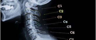
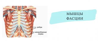
The spinal roots (C1 - C8) emerge from the cervical spine and then branch, innervating the muscles of the upper extremities (shoulders, arms, hands), which allows them to function. They also carry sensory fibers to the skin, which provides sensitivity to the skin in the area of innervation.
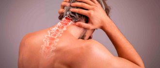
When the roots of the cervical spine are irritated, due to inflammation or compression, pain appears in the neck with radiation to the arms, sensory disturbances, and muscle weakness in the area of innervation of the damaged root.
Symptoms of radicular syndrome in the cervical spine can develop suddenly or gradually, and periods of exacerbation are followed by remission.
Causes of cervical radiculopathy
Any condition that somehow compresses or irritates a nerve root in the cervical spine can cause cervical radiculopathy.
The most common reasons are:
- Disc herniation. If the internal material of the intervertebral protrudes and irritates the nearby root in the cervical spine, then the development of radicular syndrome (cervical radiculopathy) is possible. If a young person (20 or 30 years old) has cervical radiculopathy, the most likely cause is a herniated disc.
- Cervical spinal stenosis. As part of the degenerative process of the cervical spine, changes in the spinal joints can result in decreased space in the spinal canal. Spinal stenosis is a common cause of cervical radicular syndrome symptoms in people over 60 years of age.
- Osteochondrosis (degenerative disc disease). As discs in the cervical spine degenerate, the discs become flatter and stiffer and do not support the spine. In some people, this degenerative process can lead to inflammation or damage to a nearby nerve root. Cervical degenerative disc disease is a common cause of radiculopathy in people over 50 years of age.
- Cervical radiculopathy may be caused by other conditions that put pressure on the nerve roots or cause damage to the cervical nerve roots, such as tumors, fractures, infections or sarcoidosis, synovial cyst, synovial chondromatosis of the facet joints, giant cell arteritis of the radicular vessels.
- Factors associated with an increased risk of developing cervical spine syndrome include: heavy manual labor requiring lifting more than 10 kg, smoking, and long periods of driving or working with vibrating equipment.
Why does the disease occur?
Possible causes of occipital neuralgia:
- Acute injury to the back of the neck and head.
- Constant tension in the neck muscles. For example, office workers who spend most of the day sitting at a computer.
- Protrusions and herniations of cervical intervertebral discs.
- Osteochondroma (benign tumor) of the vertebrae.
- Infectious diseases and inflammatory processes.
- Diabetes.
- Gout.
- Multiple sclerosis.
- A rare cause is metastasis of malignant tumors to the spine.
Sometimes the doctor is never able to fully understand why the disease occurred.
We will call you back
Message sent!
expect a call, we will contact you shortly
Symptoms
Symptoms of cervical spine radicular syndrome typically include pain, weakness, or numbness in areas of the affected nerve root. The pain may be felt in only one area, such as the shoulder, or spread throughout the arm and fingers.
The type of pain may also vary. Some patients describe a dull, constant pain. However, other patients describe the pain as sharp (stabbing) or intense burning.
Patients may experience tingling in their fingers, which may also be accompanied by numbness. Feelings of numbness or weakness in the hand can also affect the ability to grasp or lift objects and perform other daily tasks such as writing or putting on clothes.
Certain neck movements, such as neck extension back, neck tilt, or rotation, can increase pain. Some patients note that the pain is relieved when they place their hand behind their head; movement can relieve pressure on the nerve root, which in turn reduces symptoms.
Types of cervical radiculopathy
Symptoms of radicular syndrome in the cervical spine depend on which root is subject to cervical compression. For example, C6 radiculopathy occurs when the nerve root that extends above the C6 vertebra is damaged.
While specific symptoms in any given patient can vary widely, there are characteristic symptoms for each level of root involvement:
- C5 radiculopathy - may cause pain and/or weakness in the shoulders and arms. A characteristic symptom is discomfort around the shoulder blades; numbness or tingling is rare.
- C6 radiculopathy (one of the most common) causes pain and/or weakness along the entire length of the arm, including the biceps, wrist, thumb, and index finger.
- C7 radiculopathy (the most common) causes pain and/or weakness from the neck to the hand and may involve the triceps and middle finger.
- C8 radiculopathy causes pain from the neck to the arm. Patients may experience arm weakness, and pain and numbness may spread along the inside of the hand, ring finger, and little finger.
- When several roots are simultaneously affected, a combination of symptoms is possible
- Symptoms may worsen with certain activities, such as sitting with the neck tilted for long periods of time (working on a computer), and improve with rest.
- But in some cases, symptoms may become persistent and not improve when the neck is in a supported resting position.
To select adequate treatment tactics for radicular syndrome in the cervical spine, it is necessary to correctly identify the cause of the symptoms. For example, cervical radiculopathy and carpal tunnel syndrome may have similar symptoms, such as arm pain and numbness, so it is necessary to accurately determine the genesis of the symptoms, which will allow targeting the actual source of the problem.
Diagnosis and treatment of pain in the neck and upper extremities
Professor O.S. Levin
RMAPO
Neck pain is one of the most common manifestations in clinical practice [3,9,17]. Neck pain affects almost half of the adult population , with persistent neck pain affecting approximately 23% of women and 17% of men [15]. The cervical spine is the most mobile part of the spinal column, which makes it particularly vulnerable to injury and degenerative changes. However, degenerative-dystrophic changes in the spine are far from the only cause of neck pain, which thus requires careful differential diagnosis [7,17,21]. Pain in the neck and arm is usually divided into vertebrogenic (caused by spinal pathology) and non-vertebrogenic, and the causes of both types of pain are very numerous (Table 1). Each variant of vertebrogenic pathology can lead to the development of 4 main clinical syndromes: • local pain syndrome (cervicalgia), often accompanied by local vertebral syndrome in the form of pain, deformity, limited mobility or instability of one or more adjacent segments of the spine; • reflex (irritative) syndromes include referred pain (for example, cervicobrachialgia, cervicocranialgia), muscular-tonic syndromes, neurodystrophic manifestations, repercussion autonomic disorders with a wide range of secondary manifestations: enthesopathies, periatropathies, myofascial syndrome, tunnel syndromes, etc. d.; • radicular syndrome (radiculopathy), caused by irritation or compression of the spinal roots; • syndromes of compression (ischemia) of the spinal cord (due to herniated intervertebral discs, spinal canal stenosis, etc.).
From a pragmatic point of view, back pain, regardless of its origin, is useful to subdivide according to its course and location (Table 2). Back pain can be nociceptive, neuropathic or psychogenic in origin. Nociceptive pain is associated with the activation of pain receptors - nociceptors, and generally corresponds to the degree of tissue damage and the duration of action of damaging factors. It is felt at the site of origin (local pain) or at a distance (referred pain). Neuropathic pain is associated with dysfunction or damage to the nervous system. Neuropathic back pain is usually associated with root or dorsal root ganglion involvement [1,12].
Acute neck pain in most cases is a consequence of protrusion or herniation of the intervertebral disc, less often - trauma or myofascial syndrome. Chronic (persistent or constantly recurring) pain, which has a particularly important medical and social significance, most often occurs against the background of degenerative-dystrophic changes, although the latter is not always its direct cause. Degenerative changes, as a rule, affect intervertebral discs, joints, ligaments and other tissues that form the spinal motion segment (SMS). An important role in the development of degenerative-dystrophic changes is played by heredity, age, physical activity on the elements of the PDS, and injuries. At different stages of the degenerative cascade, clinical manifestations of spinal osteochondrosis can be predominantly associated with intervertebral disc herniation, in others - with arthrosis of the intervertebral (facet) joints (spondyloarthrosis), the formation of osteophytes and other changes [6].
Herniated intervertebral discs Characteristic clinical features of pain syndrome with a herniated cervical intervertebral disc: - acute onset after physical activity, awkward movement or injury; increased pain in the neck and arm with increased pressure in the epidural space (with coughing, sneezing, straining, compression of the jugular veins);
Table 2. Classification of back pain
| With the flow | By localization |
| Acute - less than 6 weeks Subacute 6-12 weeks Chronic more than 12 weeks | Cervicalgia - neck pain Cervicobrachialgia - neck pain spreading to the arm Cervicocranialgia - neck pain spreading to the head area Cercicothoracalgia - neck pain spreading to the interscapular area |
Table 1. The main causes of pain in the neck and upper extremities Vertebrogenic pain Herniated intervertebral disc Cervical spondylosis Painful dysfunction of the SMS Instability of the SDS Rheumatoid arthritis Seronegative spondyloarthropathy Trauma Tumors Spondylitis Osteoporosis Non-vertebrogenic pain Brachial plexopathy Peripheral nerve lesions Reflex sympathetic dystrophy ia Myofascial syndrome Fibromyalgia Damage to joints and periarticular tissues Damage bones (osteomyelitis, osteoporosis) Damage to blood vessels (thromboembolism, vasculitis, Raynaud's syndrome) Viscerogenic pain (diseases of the pleura, heart) Psychogenic pain
- increased pain in the neck and arm when bending the neck, tilting the head towards the pain with an axial load on it, rotating the head towards the pain with throwing it back; - forced position of the head with a slight tilt forward and the side opposite to the localization of pain, a sharp limitation of the mobility of the cervical spine and tension of the neck muscles; - reduction of pain when traction of the head or placing a hand behind the head (due to the expansion of the intervertebral foramen).
Depending on the level of the lesion, pain can radiate to the medial parts of the scapula, interscapular region, back of the head, shoulder girdle and arm. With radiculopathy, pain, sensory disturbances (paresthesia, dysesthesia), loss of reflexes, and sometimes muscle weakness are detected in the area of the corresponding root (Table 3). The cause of radiculopathy is most often lateral disc herniations C6-C7 and C5~C6, much less often - C7-T1 and C4-C5. Root damage may be associated with its mechanical compression or tension, compression of its arteries and veins with the development of ischemia and edema, inflammation, demyelination, axonal degeneration, fibrosis, but it is difficult to clinically differentiate these types of root damage. An important role in symptom formation may be played by the irritation of the spinal ganglion cells and the sympathetic fibers that follow the root [11].
The cause of spinal cord compression is usually a median hernia, which is most often localized at the C5-C6 or C3-C4 levels. Dysfunction of the spinal cord can be associated with its direct mechanical compression (static or dynamic - during movement) or compression of the vessels supplying the spinal cord. A factor that increases the likelihood of compression is congenital or acquired spinal canal stenosis. At the level of the cervical spine, spinal canal stenosis is diagnosed if the ratio of the anteroposterior diameter of the spinal canal and the vertebral body is less than 0.8. Compression of the spinal cord is manifested by weakness and impaired sensitivity in the arms and legs, hyperreflexia and spasticity in the legs, changes in gait, pathological foot reflexes, pelvic disorders, Lhermitte’s symptom (the sensation of current passing through the spine and legs when bending it) [9,12].
Cervical spondylosis Spondylosis is understood as a set of degenerative changes, which include the formation of osteophytes, degenerative changes and hypertrophy of intervertebral joints (spondyloarthrosis), hypertrophy of the ligamentous apparatus, sclerotic changes in uncovertebral “joints”, inflammatory damage to periarticular tissues, etc. These changes can be a source of pain, and the narrowing of the intervertebral foramina and spinal canal they cause can lead to radiculopathy or myelopathy. In contrast to disc herniation, spondylosis often affects the upper cervical (C2-C4) roots, pain usually increases with extension rather than with flexion, and the prognosis is less favorable [12,17].
Damage to the facet joints can be not only a consequence of increased load on these joints (including disc herniation) or trauma, but also a manifestation of systemic arthropathy (polyosteoarthrosis, rheumatoid arthritis, seronegative spondyloarthropathy). It manifests itself as acute unilateral or bilateral neck pain, often regressing within 7-10 days. When the upper cervical joints are affected, the pain radiates to the back of the head and forehead, when the middle cervical joints are affected - to the area of the shoulder girdle and shoulder, the lower cervical joints - to the scapula and interscapular area. Torticollis may develop as a result of protective muscle spasm (8).
Exacerbations of pain syndrome with spondyloarthrosis occur with varying frequency. They are often caused by minor trauma, unsuccessful movement, hypothermia, or prolonged stay in an uncomfortable position (including during sleep). In some patients, the pain becomes permanent over time. Upon examination, limited mobility of the cervical spine in the corresponding SMS is revealed, especially during extension - while flexion and rotation are preserved; pain on palpation of the facet joints is usually on both sides (approximately 1-2 cm from the midline). Pain is provoked by straightening the neck or bending towards the more affected joint. The diagnosis is confirmed by a decrease in pain after joint blockade. Osteophytes, whose sanogenetic function is to stabilize the SDS, sometimes compress the vertebral artery and esophagus, causing vertebrobasilar insufficiency and swallowing disorders (dysphagia).
| Table 3. Signs of damage to the cervical and upper thoracic roots | |||||
| Signs / roots | Localization of pain | Decreased sensitivity | Decreased reflex | Paresis | Possible location disc herniation |
| C5 | Outer surface of the shoulder, medial part of the scapula | Upper outer shoulder (above the deltoid) | Biceps reflex | Abduction and external rotation of the shoulder, partially flexion of the forearm | C4-C5 |
| C6 | Lateral surface of the forearm and hand, 1st and 2nd fingers |
| Biceps reflex | Flexion and internal rotation of the forearm, partially extension of the hand | C5-C6 |
| C7 | The back of the shoulder and forearm up to 2-3 fingers | 2nd and 3rd fingers, back of the hand and forearm | Triceps reflex | Shoulder extension, wrist and finger extension, partial wrist flexion | C6-C7 |
| C8 | Inner surface of the forearm, hand up to fingers IV-V | IV-V fingers, inner surface of the hand and forearm | No | Flexion and extension of fingers | S7-T1 T T1-T2 |
| T1 | Inner surface of the shoulder and forearm, axillary area | Inner surface of the shoulder and upper forearm, axilla | No | 4 Spreading fingers | |
As a result of narrowing of the spinal canal and intervertebral foramina due to the proliferation of osteophytes, hypertrophy of the articular processes and the yellow ligament, compression of the anterior spinal artery, as well as the vertebral or medullary arteries feeding it, is possible. Compression of the anterior spinal artery at the cervical level can cause damage to the cervical spinal cord. When the vertebral artery is compressed, the following may also be observed: posterior cervical sympathetic syndrome (see below), repeated episodes of vertebrobasilar insufficiency, episodes of drop attacks.
Painful dysfunction of the spinal motion segment Disc degeneration can lead not only to a hernia, but also to uniform moderate protrusion of the disc, a decrease in its height and a change in the relative position of the main elements of the spinal motion segment. This can cause instability or pathological fixation of the segment, leading to chronic pain syndrome.
Myofascial syndrome Myofascial pain is localized primarily in the proximal limbs, muscles of the shoulder girdle (trapezius, levator scapulae, multifidus, erector spinae), suboccipital muscles, facial and masticatory muscles. Referred pain is noted in the head, shoulder, and eye area. The pain syndrome may resemble radicular pain (“pseudoradicular syndrome”), but is sometimes observed simultaneously with radicular pain, which causes difficulties in diagnosis. Spasm of the scalene or pectoralis minor muscles can cause compression of the brachial plexus (myogenic thoracic outlet syndrome).
Cervicocranialgia Cervicocranialgia is characterized by the presence of pain in the cervical region, spreading to the occipital, sometimes frontotemporal region. The pain can be bilateral or unilateral, and the side of the pain usually does not change. The pain often intensifies when moving the head, staying in an uncomfortable position for a long time, palpation of the cervical-occipital muscles, and is accompanied by limited mobility of the cervical spine. Cervicocranialgia may be associated with pathology of the cervical osteoarticular or muscular structures innervated by the roots of the superior cervical spinal nerves (C2, C3, less often - irritation of the sympathetic plexus of the vertebral arteries (with the development of posterior cervical sympathetic syndrome). Constant dull pain in the cervico-occipital region usually occurs caused by myofascial syndrome or pathology of the facet joints [4].
Posterior cervical sympathetic syndrome is a condition associated with irritation or compression of the sympathetic plexus of the vertebral artery and manifested by a combination of neck pain, unilateral migraine-like headache with signs of autonomic dysfunction on the same side (pupil dilation, facial hyperhidrosis, less often - ptosis or miosis), transient dizziness, ringing in the ears, blurred vision, neck pain, dysesthesia in the scalp, which often develop against the background of anxiety-depressive syndrome [12].
In some patients, it is caused by neurodystrophic syndrome of the nuchal ligament (in this case, maximum pain and induration are detected at the site of attachment of the nuchal ligament to the occipital bone). In addition, the cause of cervicocranialgia can be neuralgia of the occipital nerve, damage to the upper cervical roots or the cervical part of the nuclear apparatus of the trigeminal nerve due to cervical spondylosis, as well as the consequences of a whiplash injury to the neck. craniovertebral anomalies or tumors. Neuralgia of the occipital nerve is manifested by short-term paroxysms in the zone of innervation of the medially located greater or laterally located lesser occipital nerves, accompanied by pain upon percussion along the course of the nerve and impaired sensitivity in the zone of its innervation [2,7].
Cervicocranialgia may be accompanied by cervicogenic dizziness, provoked or aggravated by movements in the cervical spine (especially when turning to the sides and throwing back the head) and caused by pathological proprioceptive impulses from the neck muscles, ligaments, joints, less often - irritation or compression by bone osteophytes of the vertebral arteries passing through holes in the transverse processes of the cervical vertebrae. In the latter case, dizziness occurs in the context of other manifestations of vertebrobasilar insufficiency and is more often observed in elderly patients with stenosing atherosclerosis of the main arteries of the head [12].
Psychogenic back pain Chronic back pain is rarely purely psychogenic in nature. More often it occurs against the background of another psychogenic disorder (anxiety-phobic, hypochondriacal syndromes, depression, hysteria, etc.) and potentiates the manifestations of vertebrogenic pathology, contributing to the chronicity of the pain syndrome. The main diagnostic signs of psychogenic back pain can be: discrepancy between the pain zone and traditional topography; the course of the pain syndrome, determined by fluctuations in the psychological state of the patient; unusual localization of neurological symptoms (for example, weakness or numbness is felt not in the characteristic zones of innervation, but in the entire limb): pain of a superficial nature (painful skin); discrepancy between the severity of pain and the absence of limitation of spinal mobility, the appearance or intensification of pain with axial load on the spine (with pressure on the vertex area) [6,20].
Other causes of neck pain Neck pain, especially long-term or increasing pain, should always be the basis for a thorough somatic and oncological search. It is important first of all to exclude infectious diseases (nonspecific or tuberculous spondylitis, epidural abscess, discitis) and tumors. They are characterized by persistent, increasing pain of a non-mechanical nature (intensified rather than relieved at rest and at night), accompanied by systemic manifestations (loss of body weight, increased fatigue, fever, increased ESR, leukocytosis, anemia), severe local pain of the spinous processes . Characteristically, there are signs of compression of the roots and spinal cord. Most cervical spine tumors are metastases from lung, breast, or prostate cancer. Especially often, the metastatic process also affects the bodies of the C7 and T1 vertebrae. X-rays at an early stage may not reveal changes. Radioisotope scintigraphy and MRI are more sensitive.
Idiopathic diffuse skeletal hyperostosis (Forestier disease) affects mainly men after 50 years of age. The clinical picture includes pain, stiffness, and tenderness of the spine to palpation. Cervical spine involvement is often accompanied by dysphagia. X-rays reveal numerous osteophytes and defoliation of the anterior longitudinal ligament. Often the disease is asymptomatic and is accidentally detected during radiography.
Tendinitis of the longus colli tendon causes progressive pain along the anterior neck and dysphagia that worsens with head movement. Upon examination, severe pain is revealed on palpation of the upper cervical vertebrae from the front.
Styloid process syndrome (Eagle syndrome) is characterized by elongation of the styloid process and is manifested by pain along the anterior surface of the neck, radiating to the ear, as well as persistent sore throat. Patients often undergo tonsillectomy by mistake. The diagnosis is made by radiography. Treatment includes analgesics, physical therapy, and sometimes surgery. Inflammatory or neoplastic diseases of the thyroid gland can cause pain along the anterior surface of the neck, radiating to the ear, lower jaw, and back of the head.
Diagnosis When examining a patient with pain in the neck and arm, first of all it is necessary to exclude serious diseases that require emergency intervention: tumor, osteomyelitis, epidural abscess, meningitis, retropharyngeal abscess, fracture or subluxation in the cervical spine, subarachnoid hemorrhage, thrombosis or carotid or vertebral dissection arteries [4,17].
Having ruled out the possibility of a so-called “serious pathology” based on the anamnesis, one should differentiate between radiculopathy and reflex pain syndrome, as well as disc herniation and cervical spondylosis. When trying to identify the source of pain, you need to palpate the muscles, identifying painful and trigger points, spinous processes and facet joints.
Pain in the arm, occurring separately from pain in the neck (brachialgia), combined with sensory disturbances, paresis, amyotrophy and/or vegetative-trophic disorders, may be associated not with damage to the spine, but with involvement of the brachial plexus, compression neuropathies, and reflex sympathetic dystrophy. Pain in the arm, not accompanied by neurological symptoms, is more often caused by damage to soft tissues (arthrosis, enthesopathies, glenohumeral periarthropathy, myofascial syndrome, etc.), vessels of the upper extremities, as well as somatic diseases that cause referred pain (for example, angina pectoris).
Particularly significant diagnostic difficulties are caused by the diagnosis of glenohumeral periarthropathy, mainly associated with the pathology of soft periarticular tissues. There are several variants of glenohumeral periarthropathy: 1) with rotator cuff tendinitis, the pain is diffuse or limited to the lateral surface of the shoulder; Abduction of the shoulder is the most painful, but other movements, such as raising the shoulder, can also be painful. On palpation, pain is detected in the subacromial region; 2) when inflammation spreads from the cuff to the subacromial bursa, subacromial bursitis occurs; 3) biceps tendinitis is manifested by pain in the shoulder and tenderness of the muscle tendon, which is palpated during external rotation of the shoulder along its anterior surface. Flexion of the forearm and supination against resistance are also painful; 4) arthritis of the acromioclavicular joint is manifested by diffuse pain, which intensifies when raising the arm, as well as pain in the joint area; 5) adhesive capsulitis - the final stage of any pathology of the glenohumeral tissues, as well as neurological diseases that limit movement in the shoulder joint. It manifests itself as diffuse pain in the shoulder, limitation of both active and passive movements in the shoulder joint [4,8]. Local pain in the elbow area, as a rule, does not have a vertebrogenic nature and is more often caused by myofascial syndrome, tunnel neuropathy (for example, damage to the superficial branch of the radial nerve), epicondylitis, arthrosis of the elbow joint, or olecranon bursitis [12].
Additional research methods can help in correct diagnosis. However, their results should always be assessed with caution, analyzed in the context of clinical data. In patients with chronic neck pain radiography of the cervical spine most often reveals a decrease in the height of the intervertebral discs, sclerosis of the end plates, hypertrophy of the articular processes, osteophytes, and uneven narrowing of the spinal canal. Oblique films may reveal narrowing of the intervertebral foramina. However, the main purpose of cervical x-rays, which are performed in most patients, is to rule out causes of pain such as tumor, spondylitis, or osteoporosis. Detection of radiological signs of osteochondrosis is not of great clinical importance, since they can be found in the vast majority of mature and elderly people. On the other hand, young individuals with a herniated disc may not have any radiographic changes. To identify spinal instability, functional radiography , taking pictures in extreme flexion and extension. A disc herniation can be verified using CT, MRI or myelog raffia CT and MRI are especially important when signs of spinal cord compression appear . The diagnostic value of myelography has sharply decreased in recent years, but nevertheless it is sometimes performed (as part of preoperative diagnosis). Functional diagnostic methods include ENMG, a method for studying evoked potentials [17].
Treatment In the absence of “serious pathology,” the prognosis is generally favorable - in most cases, complete recovery occurs, which sometimes (especially with radiculopathy) lasts for several weeks or months. Treatment should be aimed at accelerating the regression of symptoms, preventing chronic pain syndrome and further exacerbations.
In the acute period, with severe pain, short-term immobilization of the neck using a soft collar is indicated. It is possible to temporarily wear a cervical collar for 1-2 weeks (primarily at night). Individual selection of the collar is important, since if the collar is too wide, excessive extension of the neck may occur. As with lumbosacral pain, chronic pain can be prevented by returning to normal levels of daily activity as quickly as possible. In turn, this requires quick and adequate relief of pain. To control pain, analgesics and nonsteroidal anti-inflammatory drugs (NSAIDs) are usually used [1,5].
In recent years, NSAIDs have been especially widely used to relieve pain in patients with vertebrogenic syndromes. The main mechanism of their action is associated with inhibition of cyclooxygenases (COX) - enzymes that ensure the biotransformation of arachinodonic acid into prostaglandins. Two isoforms of COX have been identified: COX™ 1 and COX-2. COX-1 plays an important role in the functioning of the gastric mucosa, kidneys, platelets, and vascular endothelium. Its suppression significantly mediates the development of side effects of NSAIDs. COX-2 is induced directly in the area of inflammation and is involved in the development of the inflammatory process and pain syndrome. Inhibition of COX-2 underlies the analgesic and anti-inflammatory effect of NSAIDs. In addition, blockade of COX-2 in the central nervous system appears to be able to further block the transmission of pain impulses at the level of the spinal cord [1,9,16]. Based on their effect on various COX isoenzymes, NSAIDs are usually divided into three main groups: non-selective COX inhibitors - suppress the activity of both COX isoforms approximately equally (diclofenac, indomethacin, ibuprofen, ketoprofen, lornoxicam); agents predominantly acting on COX-2 (nimesulide, meloxicam); • selective COX-2 inhibitors (celecoxib) [1.19]. When using selective COX-2 inhibitors, the risk of developing complications, primarily from the gastrointestinal tract, is lower than with traditional NSAIDs that act on both COX-1 and COX-2. Selective COX-2 inhibitors should be considered as the drugs of choice if traditional NSAIDs are poorly tolerated, if there is a history of gastric or duodenal ulcers, or if long-term use of NSAIDs is necessary. For moderate pain, NSAIDs are used orally or externally, in the form of gels and ointments. For severe pain, NSAIDs are advisable to administer parenterally. It should be noted that patients are individually sensitive to NSAIDs; therefore, if the optimal therapeutic doses of one of the drugs are ineffective, another drug can be tried within 1-2 weeks. Currently, there is a wide arsenal of NSAIDs, the effectiveness of which has not been sufficiently studied (Table 4). An important factor influencing the effectiveness of treatment is the patient’s individual sensitivity to a particular drug. In this regard, if the optimal therapeutic doses of one of the drugs are ineffective, another drug can be tried within 1 week. The choice of drug for a particular patient is determined primarily based on the range of its side effects.
The most common side effects include: gastrointestinal damage, impaired renal and liver function, fluid retention, increased blood pressure. The frequency of side effects depends on the pharmacokinetic and pharmacodynamic characteristics of the action. The use of coxiotes (for example, celecoxib), which selectively act on COX-2, significantly reduces the risk of side effects from the gastrointestinal tract, but at the same time may increase the risk of cardiovascular complications, especially with long-term use [15,19]
Among the drugs with an optimal balance of effectiveness and safety is nimesulide, which in therapeutic doses has a relatively selective effect on type 2 COX. The drug has been used in various European countries for more than 20 years and has proven itself to be a relatively safe remedy. A number of comparative studies have shown that nimesulide is less likely to cause gastrointestinal complications than traditional NSAIDs such as diclofenac, ibuprofen or naproxen, without being inferior to them in effectiveness [13,18,20]. In addition, it has been shown that due to the lower incidence of gastrointestinal complications and, accordingly, lower costs of their treatment, the use of nimesulide is more beneficial from a pharmacoeconomic point of view than the use of standard NSAIDs [14].
However, long-term use of NSAIDs should be avoided in any case, especially in the elderly. In patients with a high risk of erosive and ulcerative lesions of the stomach and duodenum (elderly people with a history of peptic ulcer disease, suffering from diseases of the cardiovascular system, taking corticosteroids and anticoagulants) in combination with NSAIDs to protect the gastrointestinal tract, it is advisable to prescribe proton pump inhibitors (omeprazole 20 mg/day, lansoprazole 30 mg/day) or a synthetic analogue of prostaglandins miso-prostol (100-200 mg 3-4 times a day). It should be remembered that with parenteral or rectal use of NSAIDs, dyspepsia occurs less frequently than when taking tablet forms, but the risk of developing ulcers and erosions is not significantly reduced. If dyspepsia or ulcers appear, discontinuation of NSAIDs and administration of a proton pump inhibitor are indicated (H2 receptor inhibitors and misoprostol are less effective). Non-absorbable antacids can be used to relieve symptoms of dyspepsia, but they do not prevent or treat GI damage caused by NSAIDs.
When there is severe tension in the paravertebral muscles, the obligate component of therapy is muscle relaxants (tizanidine 6-12 mg/day, clonazepam 1-4 mg/day, diazepam 5-15 mg/day, etc.). To reduce local muscle spasm, it is advisable to use therapeutic blockades with a local anesthetic (novocaine, lidocaine, etc.) [10,17]. To increase the long-term effect of the therapeutic blockade, a corticosteroid (hydrocortisone, depo-medrol) can be added to the local anesthetic. For radiculopathy, it is possible to use drugs to treat neuropathic pain, primarily antidepressants (amitriptyline, melipramine, paroxetine, duloxetine, etc.) and/or anticonvulsants (carbamazepine, gabapentin). If intense pain persists, epidural blockades may be performed.
Subsequently, gradual mobilization of the neck, post-isometric relaxation, therapeutic exercises, traction, massage, physiotherapeutic procedures (CMT phonophoresis with hydrocortisone, alternating magnetic field, etc.) are indicated. With facet syndrome, pain can often be relieved with repeated blocks with a local anesthetic and corticosteroid. There is conflicting evidence about the effectiveness of oral corticosteroids, although a short course of corticosteroids is sometimes recommended in severe cases. For myofascial syndrome, injections of anesthetics and corticosteroids into trigger points, applications with dimexide, passive stretching, massage, and therapeutic exercises are performed. At the same time, NSAIDs, muscle relaxants and tricyclic antidepressants are prescribed [9,12].
In the subacute and chronic phases, physical methods are especially important, primarily therapeutic exercises, massage, manual therapy, and balneotherapy. When determining the intensity of the load, it is necessary to take into account the severity of pain. The use of a special orthopedic pillow is important.
In case of chronic pain syndrome, a comprehensive psychophysiological approach is required, taking into account the importance of both peripheral and psychological factors in the origin of pain. Chondroprotectors are used empirically, but their effectiveness remains unproven.
Surgical intervention is indicated for symptoms of spinal cord compression (pelvic disorders, spastic paresis, sensory disturbances), with the appearance and increase of paresis in the area of innervation of the spinal root, as well as with pronounced pain syndrome (with clear signs of radiculopathy and ineffectiveness for several months the entire arsenal of conservative treatment). In case of periarthropathies or enthesopathies complicating the course of vertebrogenic cervicobrachialgia, special attention should be paid to therapeutic exercises aimed at increasing the mobility of the corresponding joint, possibly local administration of corticosteroids (4.6,12).
Literature 1. Ananyeva L, P., Podchufarova E.V. Modern painkillers in the pharmacy. M.: MCFR, 2005. -158 p. 2. Bogacheva L.A., Snetkova E.P. Dorsalgia: classification, mechanisms of pathogenesis, principles of management.//Neurological journal, 1996.-N2. -P.8-12. 3. Veselovsky V.P. Practical vertebroneurology and manual therapy. Riga, 1991.-S. 30-145. 4. Voznesenskaya T.G. Pain in the back and limbs.//Pain syndromes in neurological practice. Ed. A.M. Veina, M.: Medpress, 1999. -2I7-283. 5. Lebedeva R.N., Nikoda V.V. Pharmacotherapy of acute pain, M,: AIR art, 1998.-184p. 6. Levin O.S. Diagnosis and treatment of neurological manifestations of osteochondrosis of the hypochondrium. //Concilium, 2004, -N6. — P.547-554. /, Levin O.S.. Makarov G.V. Neurological complications of whiplash injury. // Neurological Journal, 2002. - No. 3. -P.38-46. 7. Levit K., Zahse J.Yu Yanda V. Manual medicine. M.: Medicine, 1993. - 511 p. 8. Popelyansky Ya.Yu., Shtulman D.R. Pain in the neck, back and limbs.//Diseases of the nervous system. Ed. N.N.Yakhno, D.R.Shtulman. M.: Medicine, 2001.-P.293-316. 9. Fisher Yu, Local treatment of pain, M.: Medpress-inform, 2005. - 160 p. 10. Shtok V.N., Levin O.S. (ed). Handbook for formulating the diagnosis of diseases of the nervous system, M.: MFA, 2006. -495 p. 11. Shtulman D.R., Levin O.S. Neurology. Handbook of a practicing physician. M.: Medpress-inform, 2005. - P. 70-90. 12. Dreiser RL, Riemenfeld D. Nimesulide in the treatment of osteoarthritis: dou ble-blind studies in comparison with piroxicam, ketoprofen and plaeetx>,//Drugs, 1993. -V.46. (SI)-P.191-195. 13. Fekiman M., McMhon AT Do COX-2 inhibitors provide benefits similar to those of traditional nonsteroidal anti-inflammatory drugs with less gastrointestinal toxicity? // Ann, Intern. Med, 2000. - V.132. -P. 134-143. 14. Guez M., Hildingsson C., Nilsson M. et al. The prevalence of neck pain.//Acta Orthop.Scand., 2002. -V.73. -P.455- 459. 15. Henry D., Lim LL, Garcia Rodriguez LA. et al. Variability in risk of gastrointestinal complications with individual non-steradal anti-inflammatory drugs: a results of a col laborative meta-anarysts.//BMJ, 1996, -V.312. -P. 1563-1566. 16. Maigne R. Diagnosis and treatment of pain of vertebral origin. Baltomore. Wiiams&Wilkins, 1996. -550 P. 17. Porto A., Almeda H., Cunha MJ et al. Double-bind study evaluating by endoscopy the toterabilily of nimesulide and diclofenx on the gastric mucosa in osteoarthritic patJents.//Eur J. Rheumatol. Intlamm., 1994. -V.I4. -P.39-50. 18. Shah AA, Thjodteifsson V, Murray FE et al. Selective inhibition of COX-2 in humans is associated with less gastrointestinal injury: comparison of nimesulide and naproxen.//Gul., 2001. -V.48. -P.339-346. 19. Singla AK, Chawla M., Singh A. Nimesulide: some pharmaceutical and pharmacological aspects.//J.Pharm. Pharmacol., 2000. -V.52. — P.467-486. Waddel G. The back pain revolution.//Edinburg. Churchill Livingstone, 1998. -438P.
Source - Russian Medical Journal
Diagnostics
If you have symptoms such as neck pain or related symptoms such as tingling, weakness, or numbness in your shoulder, arm, and/or hand, your doctor will likely start with the following:
- Patient history. The doctor collects detailed information about the presence of any previous or current diseases or conditions, accidents or injuries, family history and lifestyle. This allows you to get a better idea of what may need to be examined further.
- Physical examination. Based on examination and palpation, the doctor determines the presence of abnormalities, painful areas, as well as the range of motion and strength of the neck muscles.
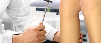
- The Spurling test allows the physician to determine whether compression of the cervical spine may provoke or (temporarily) worsen a patient's radicular symptoms. This test is usually performed by having the patient tilt their head to the side where the symptoms began, and then the doctor's hand applies gentle pressure to the top of the head. This process leads to a narrowing of the foraminal openings from which the nerve roots emerge and this leads to a reproduction of the radicular symptoms that the patient experienced. If the Sperling test reproduces radicular symptoms, then cervical radiculopathy is likely present.
- In patients who already have signs of cervical myelopathy (spinal cord compression) or who have radicular symptoms following an episode of trauma (and therefore may have fractures), the Spurling test is not recommended.
Symptoms
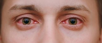
Inflammation of the occipital nerve is manifested by a characteristic throbbing pain in the occipital region of the head, ears, temples, and lower jaw. It gets worse with head movements and may be accompanied by nausea. Often on the affected side there is pain in the eye area.
When palpating the affected area, increased sensitivity is noted, and severe pain is observed at the site of nerve projection. The patient then takes a forced position with the head tilted towards the affected nerve. The skin may acquire a reddish or pale tint. Tearing may occur. Bilateral lesions may simulate migraine.
By deciding to delay a visit to a specialist a little, a person risks driving himself into a more difficult situation. The sooner competent treatment is started, the lower the risk of disruption of the innervation of the area of the diseased nerve!
Instrumental diagnostic methods
- X-ray
of the cervical spine is usually the first method for diagnosing radicular syndrome and can detect the presence of injuries, osteophytes, and narrowing of the space between the vertebrae. Radiography is considered the best initial investigation in all patients with chronic neck pain. - CT (MSCT)
- CT scanning provides good visualization of bone morphology and can be a useful diagnostic tool for the evaluation of acute fractures. The accuracy of diagnosing disc herniations in the cervical spine using CT imaging ranges from 72-91%.
CT scans with myelography have an accuracy approaching 96% in diagnosing cervical disc herniation. In addition, the use of contrast material makes it possible to visualize the subarachnoid space and assess the condition of the spinal cord and nerve roots.
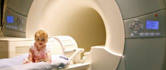
- MRI
- MRI has become the method of choice for imaging the cervical spine and can detect a significant proportion of soft tissue pathologies, such as disc herniation. MRI can detect ligament tears or disc herniation that cannot be detected by other imaging modalities. MRI can clearly visualize the entire spinal cord, nerve roots, and spinal column. MRI has been found to be quite useful in assessing the amount of cerebrospinal fluid (CSF) surrounding the spinal cord when evaluating patients with spinal canal stenosis:
- EMG (ENMG)
- Electrodiagnostic research methods are important for identifying physiological disorders of the nerve root and excluding other neurological causes of the patient's symptoms. EMG (ENMG) studies have been shown to be useful in the diagnosis of radiculopathy and correlate well with the results of myelography and surgical treatment.
Causes of occipital neuralgia
Treatment of occipital neuralgia is prescribed depending on the reasons that caused this pathology, and they can be very different:
- mechanical damage to the spine resulting from trauma;
- hypothermia;
- prolonged muscle tension due to a sedentary lifestyle;
- consequences of osteochondrosis;
- severe stress;
- diseases of the spine of various nature;
- diseases of infectious etiology;
- inflammation of blood vessels;
- gout;
- diabetes.
Treatment
Conservative treatment of radicular syndrome of the cervical spine may include the following treatment methods:
Rest or change in activity
. Wearing a cervical collar during acute pain. Cervical radiculopathy often resolves on its own, especially if symptoms are mild. Limiting strenuous activities such as playing sports or lifting heavy objects or improving your posture while sitting or driving may sometimes be sufficient treatment.
Exercise therapy.
Exercise and stretching may help relieve symptoms. A physical therapy doctor can develop an individual plan for a specific patient. Exercise therapy is the most effective method of treating radicular syndrome in both the short and long term. Exercises aimed at opening the intervertebral foramen are the best choice for reducing the effects of compression on the root. Exercises such as contralateral rotation and lateral flexion are the simplest forms of exercise that are effective in reducing the symptoms of radicular syndrome and increasing range of motion in the neck. Exercises can also be performed to strengthen the muscles, which will improve neck stability and reduce the risk of developing nerve root irritation in the future, if the root compression is not due to reasons for which exercise therapy does not have a therapeutic effect. During the initial stages of treatment, muscle strengthening should be limited to isometric exercises in the involved upper extremity. Once acute symptoms have resolved, progressive isotonic strengthening can begin. Initially, resistance exercises should be done with light weights and frequent repetitions (15-20 repetitions). It is necessary to engage in exercise therapy for a long time, periodically adjusting the volume and intensity of exercise with a physical therapy doctor.
Medicines
. To reduce pain symptoms, it is possible to use various anti-inflammatory drugs (diclofenac, movalis, ibuprofen) and muscle relaxants.
If medications in this group do not have an effect, then opioids may be added for a short period of time.
Cervical epidural steroid injections
used in patients refractory to other treatment methods. When performed correctly by experienced doctors under X-ray control, in most cases of radicular syndrome in the cervical spine, a fairly good effect can be achieved.
Manual therapy
. Manipulations during manual therapy can remove blocks and improve the mobility of motor segments and thus reduce symptoms.
Traction therapy
. Skeletal traction is often used in the treatment of radicular syndrome in the cervical spine. Tractions are performed on specialized traction tables with a controlled load. Traction allows you to slightly reduce root compression by increasing the distance between the vertebrae. •
Acupuncture
, along with other methods, is used in the treatment of radicular syndrome in the cervical spine. This treatment method improves conductivity in nerve fibers, reduces pain and restores sensitivity.
Physiotherapy
. Modern methods of physiotherapy, such as cryotherapy or Khivamat, as well as traditional methods (electrophoresis, phonophoresis) are widely used both in the acute stage of radicular syndrome and in a complex of rehabilitation techniques.
Prevention
Inflammation of the trigeminal nerve is not only important to treat promptly. It is important to prevent relapses, and here prevention plays a huge role:
- Avoid any drafts . It is important and necessary to ventilate the room, but do not get exposed to drafts of air, do not work, do not sleep under the air conditioner.
- Avoid hypothermia of the face and head . Don't ignore hats and scarves during the cold season.
- Protect your head and face from injury.
- Visit your dentist regularly . Avoid periodontitis (inflammation of the tissue around the roots of the teeth). If you treat caries, pulpitis, and periodontitis in a timely manner with modern dentistry, you can avoid it.
- Be careful about herpes . This is not just a “cold on the lips”, but a disease that can provoke the occurrence of ternary neuralgia.
- Fight psycho-emotional stress . Meditate.
- Take up exercise therapy . Focus on exercises that are aimed at working muscles (improving their tone) and increasing heart rate (cardio training).
Remember! Relapses are cyclical. Moreover, exacerbations are more common in the autumn-spring period. Therefore, at this time, pay the most attention to prevention.
Surgery
If conservative treatments do not provide pain relief, or if neurological symptoms such as numbness and arm weakness continue to worsen, then surgery may be considered.
The following surgical techniques are most often used in the treatment of cervical radiculopathy:
Anterior cervical discectomy and fixation
. This surgery is performed through a small incision in the front of the neck to remove the herniated disc, and then fixes that motion segment of the cervical spine to ensure spinal stability. This is the most common operation for root decompression.
Replacement of an intervertebral disc with an artificial disc
. This technique allows you to replace the fixation of the vertebrae. A potential advantage of this technique is that it aims to maintain mobility at this level of the cervical spine rather than fusion of two vertebrae.
Surgical treatment of radicular syndrome in the cervical spine can effectively reduce symptoms and restore conduction through nerve fibers. According to statistics, the efficiency rate ranges from 80% to 90%. As with any surgery, there are some risks, but most often the benefits of surgery outweigh the risks.