Hemianopsia – simple things to say. Features of medical terminology
The medical term "hemianopsia" comes from the Latin "Hemianopsia", where "Hemi" is a prefix meaning half of something, and "anopsia" is the absence or defect of vision, which literally means "half blindness".
Hemianopsia is a violation of the perception of a certain area of the visual field or the complete loss of half of it in one or two eyes. Unlike small, barely noticeable areas of prolapse (scotomas), hemianopsias are usually noticed by patients on their own, or are detected randomly during routine examinations.
The presence of hemianopsia indicates anatomical changes in the visual pathway and certain areas of the brain in the form of their compression or circulatory disorders and is an important diagnostic criterion for the most accurate determination of the affected area even before specific examination methods are carried out.
Hemianopsia can be detected using a special device - a perimeter; as a rule, neurologists, neurosurgeons, and ophthalmologists are sent for this study if they suspect anatomical changes in the brain.
Forecast
After treatment, vision can be completely restored, or it can be completely lost. It depends on many factors and primarily on the cause of the disease.
Here are the main ones:
- chosen treatment tactics;
- severity of the disease;
- duration of visual impairment;
- individual characteristics of the patient’s body;
- stage of the disease at which treatment began;
- patient's age;
- presence of complications.
For example, after a stroke, restoration of lost visual fields is possible in about six months.
Mechanisms of visual impairment
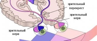
The nerve impulse that, under the influence of light, appears on the retina - the nervous tissue of our eye - is transmitted along a chain of neurons as part of the optic nerve, the visual pathway, to specific areas of the brain to process the received information and transform it into a whole picture.
Since a person has two eyes, we should have two pictures, but thanks to the peculiarities of the anatomy and physiology of the nervous system, the two images merge into one.
Photo: https://pixabay.com/photos/eye-iris-look-focus-green-1132531/
One of the reasons for this transformation is that the nerve fibers from one eye at a certain moment are divided into two groups, one of which continues to transmit information to the brain on its side, and the second goes to the opposite side, connecting with the first group of the other eye. This crossing of fibers is called chiasma.
When the nerve pathway is compressed or its blood circulation is disrupted, the affected area ceases to function, and the transmission of nerve impulses from the eye to the brain is interrupted, and the eye seems to go blind in patches. When examining visual fields, the doctor can make an assumption about where the dysfunctional area is located and can suggest where to look for the pathological process.
In the future, to determine the cause of the development of visual field impairment, an MRI or CT scan of the brain is done, sometimes with contrast.
- If the pathological process is located before the chiasm, blindness or partial loss of vision occurs in only one eye.
- If the chiasm is compressed, then the temporal halves of vision of both eyes fall out - the person perfectly sees what is happening in front of him, but does not notice what is on the side of him until he turns his head in the right direction.
- If the lesion is localized after the chiasm, then loss of the temporal half of vision occurs on one side and the nasal half on the other, or vice versa.
The size of the lost field depends on the force of compression of a particular part of the visual pathway or on the degree of disruption of its blood circulation.
Symptoms
Hemianopia is manifested by narrowing of peripheral vision or the appearance of blind spots in the central part of visual perception. This symptomatology causes the patient difficulty in moving and performing minor work.
The clinical picture depends entirely on the type of disease. Pain may occur due to eye strain. There is no redness.
The heteronymous form differs from the homonymous one in that with the first there is a clear boundary between the visible area and the one that is outside the field of view.
Causes of hemianopsia
The main role in the development of hemianopsia belongs to damage to the visual pathway in any part of it, be it the retina, optic nerve or occipital cortex.
Photo: https://pixabay.com/vectors/eye-diagram-eyeball-body-pupil-39998/
Most often, damage occurs due to compression or poor circulation, and the immediate cause may be:
- Benign and malignant brain formations.
- Abscesses of brain tissue.
- Intracranial hypertension.
- Intracranial hemorrhage.
- Aneurysms of the cerebral arteries.
- Skull injuries.
- Ischemic infarction.
- Congenital brain anomalies: hydrocephalus, microcephaly.
- Meningitis, arachnoiditis.
- "Empty sella" syndrome.
Brain tumors that can lead to hemianopsia are usually localized in the area of the sella turcica, since it is in its projection that the chiasma is located - the zone of intersection of nerve fibers coming from the two eyes. These can be pituitary tumors, gliomas, meningiomas.
Useful video
Irina Sergeevna Mironova’s story about the causes and features of the development of the pathological condition:
Author's rating
Author of the article
Alexandrova O.M.
Articles written
2100
about the author
Was the article helpful?
Rate the material on a five-point scale!
( 1 ratings, average: 2.00 out of 5)
If you have any questions or want to share your opinion or experience, write a comment below.
Classification
Hemianopsia is classified as:
- Homonymous hemianopsia (symmetrical) – when there is a loss of the temporal half of vision in one eye and the nasal half of vision in the other, that is, both right halves of vision or both left halves are lost. Damage to the optic pathway is localized anywhere after the chiasm.
- Heteronymous hemianopsia can be of two types:
- Bitemporal hemianopsia - loss of the temporal halves of vision in both eyes - from a small area to complete loss. Occurs when the chiasm is damaged.
- Binasal hemianopsia is accompanied by loss of the nasal halves of the visual field of both eyes. Occurs when both retinas or both optic nerves are damaged, usually against the background of sclerosis of the internal carotid arteries.
- Double homonymous - with damage to the occipital lobe, when only a small area of the central visual field remains.
- Quadrant hemianopsia:
- Upper quadrant, in which the upper parts of the visual fields fall out.
- Inferoquadrant – accompanied by loss of the lower halves of the visual fields of both eyes.
Depending on the degree of damage to the visual pathway, hemianopia can be:
- partial;
- full.
Diseases starting with "G":
- Hiatal hernia
- Glossalgia
- Hygroma
- Skin fungus
- Glomerulonephritis
- Hepatitis E
- Overactive Bladder
- Hemophilia
- Gynecomastia
- Worms (helminthiasis)
- Hidradenitis
- Atrophic gastritis
- Glossitis
- Hypertrichosis
- Hypertension (hypertension)
- Hypercalcemia
- Gonorrhea
- Erosive gastritis
- Preeclampsia
- Hepatitis A
- Gastritis
- Gonarthrosis
- Herpes
- Hemoblastosis
- Superficial gastritis
- Hypertrophy of the labia minora
- Hypogonadism
- Alcoholic hepatitis
- Hepatitis D
- Gangrene
- Hypothyroidism
- Endometrial hyperplasia
- Hypersomnia
- Hemianopsia
- Hypertensive crisis
- Hepatitis B
- Hypotrophy
- Hepatitis C
- Hyperparathyroidism
- Hyperthermia
- Hypocalcemia
- Hirsutism
- Hyperthyroidism
- Sinusitis
- Hydrocephalus
- Hemochromatosis
- Flu
- Glaucoma
- Hemangioma
- Gingivitis
The main complaints of patients with hemianopsia
Complaints appear, as a rule, already with a significant loss of part of the visual field, and sometimes some changes in behavior are noticed by relatives, and not by the patient himself. For example, a person may see a specific small object, but not notice the large furniture surrounding it, may not recognize the faces of acquaintances or see half of them.
Photo: https://www.flickr.com/photos/nationaleyeinstitute/7544022300/
Patients often even deny the presence of the disease, since orientation in space is partially preserved, and visual acuity can remain at a high level for a long time. Patients often bump into corners, people, if they have to drive a car, they often feel like they are being cut off, and other road users appear as if “out of nowhere.”
Patients may experience problems in daily life, seeing only half the picture on TV, noticing half a portion of food, or reading slowly. On the street, their lives are at risk because they may not notice traffic approaching from the side.
Tips for improving your quality of life
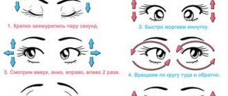
This disease is practically not amenable to conservative treatment; there are only recommendations that can improve the patient’s quality of life. For example, you can make reading easier by deliberately increasing the size of small eye movements to one movement along the line itself. In this case, you should read vertically, and keep the text at a right angle.
With right-sided hemianopsia, you should read from top to bottom so that each subsequent line is in the left preserved visual field.
A number of computer techniques have also been developed that can improve the visual functions of patients. With the help of these training courses, it is possible to partially compensate for lost vision, which will make it easier for the patient to navigate in space.
Once the diagnosis and cause of the disease are accurately established, the doctor will be able to tell you where to treat bitemporal hemianopsia.
Diagnosis of the condition
Since there are no specific symptoms of hemianopia, the main method for detecting visual field impairment is perimetry. There are three main types of this research.
- Approximate is the most primitive control method that allows you to identify large defects in the peripheral visual field. When performing this test, the patient sits opposite the doctor, look straight at him, and the doctor, moving the pencil alternately from four sides - top, bottom, right and left - asks the question - does the patient always see this pencil. The identified defect will be more accurately described when performing another type of perimetry.
- Kinetic perimetry. In this type of examination, a small object moves along an arc, and the patient, looking at the center of this arc, must notice this object as soon as it appears in his field of vision. This method requires specially trained personnel and is especially necessary in cases where it is necessary to monitor the thoroughness of the study (for example, in children). An example is Goldman perimetry.
- Static perimetry is the most accurate way to study visual fields. The patient is presented with a stimulus at a specific point in the visual field, and its intensity and size are changed until the patient notices it. Examples of such perimeters are Humphrey and Octopus devices.
Photo: https://www.flickr.com/photos/goldberg/9536427936/
When visual field loss is detected, it is necessary to determine its immediate cause, and for this, computed tomography (CT), magnetic resonance imaging (MRI) is performed, and referrals are made for consultation see a neurologist and ophthalmologist.
Control studies
It is important to differentiate this disease from such pathologies as decreased perception of certain colors, that is, hemichromatopsia, and deterioration of vision in certain areas - hemihypoxia. These diseases may precede the onset of hemianopsia or develop independently.
To establish the exact number of visual fields that are lost, perimetry is performed.
Additionally, control tests are prescribed, which can only be carried out by doctors with normal vision. To do this, the patient and the doctor stand opposite each other at a distance of 10 centimeters. Their eyes must be at the same level. Both examination participants cover one eye with a bandage or hand. The patient must look carefully into the doctor's open eye.
The doctor begins to move the finger from the periphery to the central region alternately in all directions. When the doctor's finger appears in the patient's field of vision, he must report it. If the doctor himself does not suffer from pathology, and his indicators coincide with the parameters of the person being examined, then the patient has normal vision. Identifying this disease in this way will not be difficult.
Approaches to the treatment of hemianopsia
Treatment of hemianopsia is aimed at eliminating the immediate cause of the development of this pathology. In some situations, neurosurgical intervention is necessary on a planned or emergency basis; for ischemic strokes, thrombolysis is required in the first 6 hours and further conservative treatment. In the presence of cancer, the tactics are chosen by a council of doctors; depending on the stage and scale of the process, radiation therapy, chemotherapy or surgery are offered.
In addition, the role of rehabilitation is important in the process of getting used to the changed living conditions - the patient is taught to constantly move his gaze towards the lost area of vision.
Sometimes it is possible to use special prisms and mirrors that allow you to notice what is happening on the sides of the patient without having to turn your head in that direction.
Prevention
It is impossible to prevent the development of pathology. However, the patient can improve the quality of vision if he follows the recommendations of doctors:
- when reading books, you should not turn your head to the sides;
- when reading books, keep your head straight, and try to cover as much of the text as possible with your eyes;
- you need to read the text not horizontally, but vertically;
- When reading, the book should be held at an angle of 90 degrees.
To expand the visual range, special ophthalmological programs developed by leading doctors are used.
In order not to provoke the development of pathology, you need to protect your head from blows and injuries. It is unacceptable to overstrain the visual organs so as not to injure the optic nerve and blood vessels. Avoid smoking tobacco and staying in environmentally unclean areas. Sometimes intoxication of the body leads to eye pathology, so follow safety precautions when working with chemicals.
Preventative examinations once a year are important for every person after 30 years of age. Left-sided or right-sided hemianopia most often develops in women aged 30-50 years. Pathology at the initial stage can only be noticed at a doctor’s appointment, so do not neglect regular professional examinations. This disease may not show itself in any way, but subsequently vision will begin to decline sharply. Along with decreased vision, the quality of life will deteriorate, even to the point of disability.