In order to form a three-dimensional image in the central structures of the brain, it is necessary to merge two images that were received from both eyes into a single whole. This is called binocular vision.
Binocular vision appears through the process of merging images that were obtained from the surface of the retina of both eyeballs. This helps to perceive objects in volume, that is, it provides depth to the image.
And due to the operation of the binocular vision mechanism, you can fully perceive the world around you and determine the distance to objects and between them. The so-called stereoscopic vision is an achievement of evolutionary transformations. With monocular vision, it is possible to obtain information only about the width and height of an object, its shape, but flat vision does not make it possible to assess the relative position of objects relative to each other.
In addition to the listed advantages, binocular vision is responsible for expanding the visual fields, as a result of which you can perceive objects more clearly. This, in turn, is accompanied by an increase in visual acuity. Only if you have good binocular vision is it possible to work in several specialties, in particular, as a driver, surgeon, pilot, etc.
Mechanism of binocular vision
The fusion reflex is a fundamental mechanism of binocular vision. In this case, the images that were formed in the retinal plane merge into a single picture with stereoscopic characteristics. This fusion occurs at the level of the cerebral cortex.
For the image to become unified, it is necessary to match the images obtained from the right and left retinas. This takes into account the size and shape of the image, as well as the area where it is projected onto corresponding identical areas of the retina. Each point in the retinal plane has its own corresponding point on the opposite side. Asymmetrical areas are called non-identical points, or disparate. When image points fall into these disparate points of the retinal plane, binocular vision becomes impossible. Instead of merging the image, doubling occurs.
In newly born children, coordinated movement of the eyeballs is not possible, and therefore binocular vision is absent. At about one and a half months, babies develop the ability to fix their gaze with two eyes, and at 3-4 months we can already talk about stable binocular fixation. The fusion reflex is formed only by 5-6 months, and full binocular vision - only by 12 years. In this regard, strabismus is more often present in patients of preschool age.
To form normal binocular vision, several conditions must be met:
- Ability to fusion (bifoveal fusion).
- Coherence in the work of all muscle fibers responsible for eye movement. They should ensure a parallel position of the eyeballs while the patient looks into the distance. When the gaze moves to a closer position, a proportional convergence of the visual axes occurs. This process is called convergence. Also, the extraocular muscles must respond to the associated movements of the eyeballs directed towards the object being studied.
- The visual acuity of each eye should not be less than 0.3-0.4, which is sufficient to form a clear image.
- The eyes should be located in the same horizontal and frontal plane. If, as a result of injury, tumor, inflammation or surgery, a change in symmetry occurs, then the merging of images becomes impossible.
- Images projected onto the retinal plane must be of this size (iseikonia). When the size of objects on the retina is different, we are talking about anisometropia, which occurs when the refraction of the two eyeballs is different. In order for binocular vision to be present, the degree of anisometropia should not exceed 2-3 diopters. This must be taken into account when choosing, despite good visual acuity, binocular vision will be absent.
- Also a necessary condition for binocularity is the transparency of the media of the eye (lens, vitreous body, cornea), the absence of pathological changes in the perceptive apparatus (retina) and the conduction system (optic nerve, optic tract, chiasm, cerebral cortex, subcortical centers).
Subcortical visual centers and optic radiance
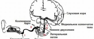
In the lateral geniculate bodies , which are subcortical visual centers, the bulk of the axons of retinal ganglion cells end and nerve impulses are switched to the next visual neurons, called subcortical or central. Each of the subcortical visual centers receives nerve impulses coming from the homolateral halves of the retinas of both eyes. In addition, information also comes to the lateral geniculate body from the visual cortex (feedback). It is also assumed that there are associative connections between the subcortical visual centers and the reticular formation of the brain stem, which contributes to the stimulation of attention and general activity (arousal).
The lateral geniculate body consists of six layers . Each of them has several (usually six) layers of nerve cells. It has been established that in the six-layer subcortical visual center the arrangement of neurons maintains a certain orderliness and topographic-anatomical relationships characteristic of the retina. The alternating layers of the lateral geniculate body receive visual impulses only from the homolateral (right or left) halves of the retina corresponding to the side of the location of the geniculate body of one or the other eye. There is no absolute sequence in the alternation of layers.
Thus, in the left geniculate body, the projections of the right halves of the retinas are located in the following order (from the superficial layer to the deep): left eye, right, left, right, right, left. There is no explanation yet for the incomplete sequence of alternating projections of the homogeneous halves of the retinas of the right and left eyes. The listed projections of the halves of the retinas in the layers of the lateral geniculate body are located exactly one below the other.
The experiment proved that the cells of the lateral geniculate body respond to visual impulses reaching them in approximately the same way as retinal ganglion cells respond to visual impulses coming to them from photoreceptors. At the same time, the central visual neurons of the geniculate bodies and the corresponding retinal ganglion cells, which can be called peripheral visual neurons, have a similar structure of receptive fields with the on- and off-centers of visual neurons and give similar bioelectric responses, depending on the intensity and color of light impulses.
It has also been established that neighboring retinal ganglion cells and central visual neurons of the subcortical visual center are located among themselves in an identical sequence. It is assumed that some neurons of the lateral geniculate body have short axons that provide local interneuronal synaptic connections, which suggests their interaction leading to a possible preliminary analysis and synthesis of visual information entering the subcortical centers. However, there is currently no consensus on the role of the external geniculate bodies in the processing of visual information. D. Hubel in 1990 suggested that, apparently, no significant transformations of visual impulses coming from the retina occur in it. At the same time, J.G. Nicolet, A.R. Martin, B.J. Wallas and P.A. Fuchs (2003) recognize that geniculate neurons are involved “in providing the first steps in the analysis of visual scenes: identifying lines and shapes based on retinal input...”
The axons of the neurons of the lateral geniculate body, emerging from the six layers of the lateral geniculate body, unite into a single bundle and participate in the formation of the posterior limb of the internal capsule, and then form the next part of the visual pathways, the optic radiance, which has a significant extent.
Visual radiance
The axons of optic neurons located in the lateral geniculate body are part of the white matter of the cerebral hemispheres. At the same time, they first form a compact bundle that participates in the formation of the posterior leg of the internal capsule, more precisely its sublenticular part (pars sublenticularis), and subsequently form optic radiation (radiatio optici), or Graziole bundles . After passing through the so-called isthmus of the temporal lobe of the brain, the optic radiance expands and takes the form of a wide ribbon. This peculiarity of the organization of this part of the optic radiance leads to the fact that its damage is often partial, due to its considerable width and the non-compact arrangement of the nerve fibers included in its composition. In this regard, total damage to the optic radiance occurs only with a fairly common pathological process.
The nerve fibers that make up the optic radiation are involved in the formation of white matter in the temporal, parietal and occipital lobes. In the temporal lobe, near the outer wall of the inferior horn of the lateral ventricle, most of the fibers of the inferior part of the optic radiation first pass forward to the pole of the temporal lobe. Then these fibers, forming Meyer's loop , turn back and pass through the white matter of the temporal and occipital lobes.
As a result, they reach the cortex of the lingual gyrus (gyrus linqualis), which forms the lower “lip” of the calcarine groove (sulcus calcarinus), located on the medial surface of the occipital lobe.
The upper part of the visual radiance is straighter and therefore shorter than the lower one. It passes through the white matter of the parietal and occipital lobes of the hemisphere and ends by coming into contact with cortical cells located in the upper lip of the calcarine sulcus, formed by a gyrus known as the cuneus. The cortex of the medial surface of the occipital lobe, surrounding the calcarine sulcus and extending into its depth, constitutes the primary projection visual field , occupying cytoarchitectonic area 17, according to Brodmann.
It should be recalled that the visual pathways conduct visual impulses throughout their entire length, being located in a strict retinotopic order and at the same time maintaining the topographic-anatomical relationships characteristic of the retina.
How to check?
There are several diagnostic tests available to test binocular vision:
- Sokolov's experiment (hole in the palm) involves placing a tube (this can be a piece of paper rolled into a tube) to the patient's eye. The subject must look through it into the distance. From the side of the open eye, a palm is placed at the end of the tube. If there are no problems with binocular vision, then due to the superimposition of images the patient perceives the image as a hole in the center of the palm (occurs due to the superposition of images).
- The slip test (Kalf experiment) requires two pencils (or two knitting needles) to perform. The patient must hold the knitting needle (pencil) in a horizontal plane at arm's length, and the end of it must touch the second knitting needle (pencil) located in the vertical plane. If the patient has normal binocular vision, then this task will not cause difficulties. If binocular vision is absent, then the subject constantly misses. For an accurate diagnosis, the study should be performed with one eye closed. In this case, the patient must miss.
- The pencil reading test is carried out as follows: a pencil is placed at a distance of several centimeters from the tip of the nose, which will cover some of the letters. If binocular vision is preserved, then there will be no problems with reading due to the superimposition effect. The subject will be able to read without changing head position. Otherwise, you will have difficulty reading closed letters.
- For a more detailed diagnosis, you can use a four-point color test. At the same time, the visual fields of the right and left eyes are separated, which becomes possible thanks to the use of color filters. Four objects are also used (two green, one white and one red). Glasses with different glasses (red and green) are placed on the patient's eyes. If the patient has binocular vision, then he is able to distinguish between red and green objects. What is colorless will be colored red-green as it will be perceived by both eyes. If there is a pronounced dominant eye, the colorless object will be colored in the color of the corresponding glass (green or red). In the case of simultaneous vision, if the higher structures perceive data from both eyes, the patient will see five circles. In the case of monocular vision, the patient will be able to recognize only those objects that match the color of the glass on the perceiving eye, as well as a colorless object (it will turn into the corresponding color).
Lecture 4 Cortical centers of the cerebral cortex.
The concept of cortical center was proposed by I.P. Pavlov. Cortical center
has no clear boundaries and consists of nuclear and scattered parts. Cortical centers are located in the lobes of the brain.
I. Frontal lobe of the brain.
1. Motor zone
located in the precentral gyrus.
In the upper third there are neurons that innervate the leg, in the middle - the arm, in the lower third - the face, tongue, larynx and pharynx. Irritation of this area with a weak electric current leads to contraction of a specific muscle group. When the motor area of the brain is impaired, paresis
(weakening of movements) and
paralysis
(complete absence of movements) occur.
2. Center of combined head and eye rotation
located in the middle frontal gyrus. It is a bilateral center and performs a combined rotation of the head and eyes in the opposite direction. If the center in the right hemisphere is damaged, the head and eyes look to the right side, i.e. towards the damage. A patient with such damage cannot turn his head and eyes in the direction opposite to the damaged part.
3. Motor speech center
(
Broca's center
) - located in the posterior lower part of the third frontal gyrus of the left hemisphere (in right-handed people). If damaged, motor speech (the ability to speak) is impaired.
4. Writing Center
(
graphic center
) is located in the posterior parts of the middle frontal gyrus.
If damaged, the ability to write is impaired ( agraphia
).
II. Parietal lobe.
1. Center for General Sensitivities
– located in the parietal lobe behind the central sulcus (Roland’s sulcus). It is a two-sided center. The sensitive centers of the legs are located in the upper section, the arms in the middle section, and the heads in the lower section. If the center is damaged, sensory disturbances in the corresponding organs occur.
2. Center for the perception of complex types of sensitivity
– bilateral center located in the superior parietal lobule. Responsible for the perception of weight, two-dimensionality, etc.
3. Body diagram center
– located inside the parietal sulcus. Lesions of the center lead to violations of the correct ideas about the size, shape, and location of parts of one’s body.
4. Praxia Center
– located above the marginal gyrus of both hemispheres.
Ensures the execution of complex, targeted movements in a specific sequence. When the center is damaged, disturbances in purposeful movements and actions occur while its constituent elementary movements ( apraxia
) are preserved. There are three types of apraxia:
- ideatorial
(apraxia of design) – a disorder in the sequence of movements when performing a task;
- motor
(apraxia of execution) - a disorder of acting on orders or imitation;
- constructive
– the impossibility of constructing a whole movement from its individual parts.
5. Stereognosia Center
– located behind the middle part of the postcentral gyrus. If damaged, the ability to recognize objects by touch is impaired.
6. Lexicon Center
– located in the angular gyrus of the left hemisphere in right-handed people.
Responsible for the ability to recognize printed characters and read. alexia
occurs - a reading disorder.
7. Account center
(
calculia
) - located above the angular gyrus.
When the center is damaged, the ability to count is impaired - acalculia
.
8. Center for Semantic Aphasia
– located in the convergence area of the parietal, occipital and temporal lobes. If damaged, the ability to understand complex verbal, grammatical, temporal, and semantic structures is impaired.
III. Temporal lobe.
1. Sensory Speech Center
(
Wernicke's center
) - located in the posterior part of the superior temporal gyrus of the left hemisphere in right-handed people.
Responsible for understanding spoken language. Damage to the center leads to sensory aphasia
- a violation of the understanding of oral speech.
2. Amnestic Aphasia Center.
If damaged, the ability to name objects whose purpose is well known to the patient is impaired.
3. Hearing Center
– bilateral center, located in the superior temporal gyrus and partially in the transverse temporal gyrus. If damaged, hearing is impaired in both ears, but to a greater extent in the opposite ear, i.e. If there is damage in the left hemisphere, the hearing of the right ear suffers more.
4. Center of taste and smell
– located in the hippocampus region. If damaged, the ability to perceive taste and smell is impaired.
IV. Occipital lobe.
1. Center of vision
– located in the area of the calcarine sulcus and sphenoid gyrus. When damaged, visual impairment and loss of visual fields occur.
2. Center for Visual Gnosia
– located on the superolateral surface of the occipital lobe. If damaged, the ability to recognize objects is impaired.
Lecture 6 Cerebellum
Cerebellum
is a department of the central nervous system. Takes part in coordination of movements, regulation of balance, accuracy, proportionality and correctness of movements, regulates muscle tone, and is the most important center of the autonomic (?) nervous system.
Located in the posterior cranial fossa behind the brain stem. Consists of the cerebellar hemispheres
and
the cerebellar vermis
. The cerebellar vermis is responsible for static coordination, the cerebellar hemispheres are responsible for dynamic coordination of movements. Somatotopically, the muscles of the trunk are represented in the cerebellar vermis, and the muscles of the limbs are represented in the hemispheres.
The surface of the cerebellum is covered by a layer of gray matter that makes up the cerebellar cortex. The cerebellar cortex is covered with thin grooves and convolutions. There are several lobes of the cerebellum.
White matter forms the cerebellar peduncles
, in which the conducting paths are located.
The cerebellar peduncles are divided into inferior
,
middle
and
superior
. The lower legs are connected to the medulla oblongata, the middle ones to the pons, and the upper legs to the midbrain.
The white matter of the cerebellum contains nuclei
gray matter:
1. Tent core;
2. Globular nucleus;
3. Dentate nucleus;
4. Corky nucleus.
Visual system
The lecture on the physiology of vision was given by Vyacheslav Albertovich Dubynin, Doctor of Biological Sciences, leading researcher at the Department of Human and Animal Physiology, Faculty of Biology, Moscow State University named after M.V. Lomonosov.
Vision is the process of obtaining information about the outside world by capturing electromagnetic waves of a certain length. In humans, the range of visible light is 400-750 nm. The shortest wavelengths we see are subjectively perceived as violet; the longest ones are like red ones. Vision is the most significant of our sensory systems.
The eye is the organ of vision
The eyeball is located in the orbit of the skull and has three membranes. This:
- sclera: outer (tunica albuginea); formed by dense connective tissue, performs a protective function; its transparent front part is called the cornea;
- choroid: occupies a middle position; contains blood vessels and pigment cells that supply the eye; its visible part is the iris;
- retina: inner layer; Here are the photoreceptors, as well as the neurons whose axons form the optic nerve.
A little more about the iris
. The pigment cells included in its composition determine the color of the eyes (depending on the amount and distribution of melanin). In the center of the iris there is a hole - the pupil, surrounded by smooth muscle cells. By constricting the pupil, the amount of light entering the retina is regulated. In the dark, the pupil is maximally dilated, which is caused by signals from the sympathetic nervous system.
Inner core
The eye consists of the vitreous body and the lens. The lens is an elastic transparent lens consisting of living cells located immediately behind the pupil. It is surrounded by a special ciliary muscle, which, when contracted, can change the shape of the lens from flatter to more convex.
As a result, accommodation occurs - a reaction that allows us to clearly see objects located at different distances (“focusing”). Looking at close objects requires a convex lens (tension of the ciliary muscle). If you need to clearly see the horizon, the ciliary muscle relaxes and the lens becomes flat. The parasympathetic center of the midbrain regulates the tone of the ciliary muscle.
There are three main disorders of the accommodation system: myopia, farsightedness and senile farsightedness. In all these cases, the refraction of light in the lens and the size of the eyeball do not match. With myopia, the focus of the image is in front of the retina; for farsightedness - behind it; With senile farsightedness, the elasticity of the lens decreases and it cannot take on a sufficiently convex shape. Correcting myopia requires concave lenses, and farsightedness requires convex lenses.
Vitreous body
- the transparent contents of the eyeball, located between the lens and the retina. It is formed by a transparent jelly-like intercellular substance and is devoid of blood vessels (like the lens).
The normal functioning of the eye and the entire visual system is ensured by the lacrimal glands and eyelids (moisturizing the surface of the cornea), as well as the extraocular muscles (6 for each eye).
Retina
The outermost position in the retina is occupied by photoreceptors; deeper there are several layers of neurons that take part in the operational processing of visual information.
Humans have two types of photoreceptors – rods and cones. There are relatively more rods at the periphery of the retina, and cones closer to the middle (opposite the pupil). In the very center of the retina there is an area consisting of the most densely located cones - the macula macula. This is the area of greatest visual acuity. When examining an object in detail, we look directly at it, and the image, refracted in the lens, is projected onto the macula.
The photoreceptor consists of an outer part, a nuclear region and a zone in contact with retinal neurons. The reaction of the photoreceptor to light is possible due to the presence inside its outer part of light-sensitive pigments - substances that are destroyed under the influence of electromagnetic waves of a certain length. The breakdown products of pigments cause a receptor reaction and affect the generation of impulses by retinal neurons.
Cones contain pigments called iodopsins (otherwise known as conopsins). There are three types: red-, green- and blue-sensitive. Each specific cone contains one of the iodopsins, and together they provide color vision.
The rod pigment rhodopsin is sensitive to the entire visible range. In this regard, the reaction of rods to orange light is no different from the reaction, say, to green (black and white vision). The advantage of the sticks is their high photosensitivity. At dusk, when cones cannot function, rods remain the only source of visual information. In bright light, the rods provide, first of all, a clear identification of the boundaries of objects and a reaction to movement.
A genetic disorder that affects color vision is called color blindness. In most cases, it is registered in men (7% versus 0.5% in women) and is associated with the absence of one of the iodopsins. As a result, a colorblind person sees two of the three primary colors, and perceives the remaining one as gray (due only to the rods). Impairment of rhodopsin (and twilight vision) occurs with vitamin A deficiency.
In general, photoreceptors “describe” the image we see as a collection of red, green, blue and gray dots. After additional processing by retinal neurons, this information travels through the optic nerve to the brain. The functioning of the retina can be likened to the work of a scanner or camera matrix, which also describe the image in the form of a collection of pixel points. The difference is that in technical devices all pixels are the same size; in the case of the retina, the "pixels" are minimal in the macula area and much larger in the periphery. This allows us to reduce the overall amount of information and use the “computing resource” of our brain more economically.
Visual centers of the brain
The optic nerve, which contains about 1 million axons, leaves the orbit and goes to the central nervous system. Before entering the diencephalon, its fibers form a decussation - the optic chiasm.
Only half of all fibers cross, the rest go to the visual centers on their side of the brain. After the chiasma, the optic tract axons go to one of the following areas:
- The suprachiasmatic nuclei of the hypothalamus, which use information about light intensity to regulate the internal rhythms of the body - primarily circadian rhythms.
- The superior colliculi of the quadrigeminal (midbrain), which control eye movements, give the command to change the diameter of the pupil and the shape of the lens, and also organize an orienting reflex when new signals appear.
- Nerve centers in the back of the thalamus (the thalamus is the upper half of the diencephalon, where visual information is prepared for transmission to the cerebral cortex. The essence of this preparation is to contrast the image - emphasizing the boundaries between light and dark areas.
- Visual cortex.
The visual cortex occupies the most occipital region of the surface of the cerebral hemispheres. Here, sequential recognition of increasingly complex visual images and the integration of different streams of visual information are realized.
In the very posterior occipital area (primary visual cortex; Fig. 35-5, bottom), recognition of straight line segments occurs. Different neurons respond to lines of different (relative to the horizon) orientation - horizontal, vertical, at an angle of 30º, etc.
If we move forward a little, we find ourselves in the area of the secondary visual cortex. Here's what happens:
- recognition of geometric figures (as the sum of several lines);
- combining black-and-white and color signal streams (information from rods is used to determine the boundaries of objects, information from cones is used to “fill” them with color);
- comparison of information from the right and left eyes (falls into one hemisphere thanks to the chiasmus) and due to this, calculation of the distance to objects and their volume.
Recognition of the most complex and generalized features of images is associated with a zone located on the border of the occipital, parietal and temporal cortices. It is sometimes called the tertiary visual cortex. In monkeys, neurons have been found here that selectively respond to the “face” of another specific monkey. In humans, this area is also associated with the recognition of familiar faces and, in addition, with the visual component of speech - discrimination and reading texts.
Strokes and injuries to the primary visual cortex lead to loss of areas in a person’s visual field; strokes of the secondary and tertiary cortex lead to impaired perception and recognition of visual images. Damage to the primary cortex is almost not compensated (here neurons have an innately assigned function); damage to the secondary and tertiary zones is compensated well, since the properties of the corresponding neurons are the result of learning.
Human vision
What is human vision, or simply about complex things.
Vision is a unique gift given to us from birth.
The value of which you begin to understand only when you begin to lose it. First of all, it must be said that vision is one of the five senses. Thanks to it, we receive up to 98% of information about the world around us. Visual impairment leads to loss of information and decreased quality of life. We use this word often. Since childhood, we have heard: “You need to protect your vision,” and when going through a commission we always hear: “What kind of vision do you have?” And as the years pass, we ourselves often begin to say: “My vision has begun to fail, I have a visual impairment.” But what is human vision, what should it actually be like normally, and what does it depend on? Vision is an extremely complex functional mechanism that begins with the passage of a stream of light from objects through our eyes, with the conversion of the energy of light quanta into a nerve impulse that carries information to the brain.
More precisely, to his occipital lobe. The visual process ends with the formation of an image of an object in our brain. Visual impairment can occur at any stage of visual perception. Therefore, if your vision decreases, it is very important to contact an experienced specialist in order to accurately diagnose and solve the problem. The first and very important stage of the work of the organ of vision is the passage of the light flux through the transparent optical structures of the eye: the cornea, the anterior chamber filled with intraocular fluid, the lens of the eye, the vitreous body. They should all remain transparent and undamaged. Only in this case can light reach the retina of the eye, where the mysterious process of converting light energy into a nerve impulse occurs, in which all the smallest details of the object that emits or reflects the light flux are transmitted. Of course, in order to fully transmit information further, the retina, optic nerve and visual pathways of the brain must be in ideal condition. Dystrophic processes in the retina, tumors and head trauma significantly reduce a person’s vision. And the last stage on which vision depends is the cerebral cortex. It also determines the quality of vision, and, in principle, the ability to see. The human brain begins to learn to see from the moment of birth. But if there is a disturbance in the path of the light flux, then the brain does not learn to perceive visual images and this causes a decrease in vision. This condition is called amblyopia. It also applies to visual impairment. The severity of amblyopia depends on the degree of damage to anatomical structures in the path of light flow. The complexity of the organ of vision leads to the fact that vision has several functions, or rather five: 1. Central vision 2. Visual fields 3. Light perception 4. Color perception 5. Binocular vision. It is the totality of all these functions that defines the concept - human VISION. Violation of at least one of the functions leads to visual impairment and decreased quality of life. You can familiarize yourself with each function of the organ of vision in more detail on our website, understand what the concept of “normal vision” includes, and what deviations there are. All this will allow you to maintain excellent vision for many years. If you want to check your vision in Krasnoyarsk, then our specialists will completely check your vision in 45 minutes and examine all the functions of your eyes. They will make an accurate diagnosis and determine the prognosis for the future.
27.06.2020
