MRI of brain structures Magnetic resonance imaging is considered one of the most modern and accurate diagnostic methods. MRI can clearly visualize organs such as the brain. The method is prescribed for headaches and other pathologies and allows you to identify tumors, vascular pathologies and other life-threatening conditions. Unlike X-rays and computed tomography, MRI of the brain is safe for adults and children. The procedure is allowed as many times as necessary to monitor the dynamics of the disease. Let's figure it out together: is MRI really not harmful?
Is MRI safe?
The main advantage of MRI, in addition to its high informativeness for diagnosis, is the absence of ionizing radiation .
The MRI method is based on the electromagnetic characteristics of hydrogen atoms, which quantitatively predominate over other particles in human tissue. A constant high-power magnetic field is maintained inside the tomograph; radio signals with a frequency close to the vibration frequency of hydrogen pass through it. Due to resonance, the radio wave is amplified, which is recorded in a special matrix and converted by a computer into an image.
Since different tissues of the human body contain different amounts of hydrogen, the outgoing signals from different organs and tissues differ significantly, which makes it possible to obtain fairly accurate images.
There is no medical evidence that this procedure is harmful to health or causes any complications. Among the millions of people who have undergone magnetic resonance imaging, there have been no cases of ill health or harm to the body after the examination.
The only inconvenience for the patient when conducting magnetic resonance imaging is the duration of the study. An MRI scan can take anywhere from 15 minutes to 1 hour. During this period, the patient should lie still . The examination itself is an absolutely painless procedure ; the effect of magnetic waves does not cause any discomfort to the patient.
What is the essence of the MRI method?
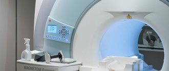
— Magnetic resonance imaging is one of the most complex medical devices. To put it simply, under the plastic shell of the tomograph there is a large magnet in the form of a cylinder, which creates a very powerful magnetic field in the room. As a result, with the help of additional parts of the device, it is possible to receive a signal from hydrogen protons in our body.
Hydrogen in the human body is found in various molecules, and they have different concentrations in different tissues. We apply energy to hydrogen protons and monitor how quickly the protons restore their original properties and emit energy back.
The device captures this energy and produces diagnostic images. Since the properties of protons differ depending on the molecules and tissues in which they are located, these tissues in the images differ in color, and, very importantly, by changing the program settings, we ourselves can influence this. For example, make the liquid bright and the fatty tissue dark. This is one of the main advantages of the method - high soft tissue contrast.
How often can an MRI be done?
MRI is prescribed for various pathologies of the substance and blood vessels of the brain, paranasal sinuses, diseases of the spine and spinal cord, joints, abdominal and pelvic organs. This study is carried out as necessary. As a rule, an initial MRI allows you to clarify the diagnosis and prescribe treatment. A repeat MRI examination is prescribed to clarify the condition of an organ or system after surgery, to monitor the treatment process, and for a more refined diagnosis using a contrast agent.
Since electromagnetic waves do not cause radiation exposure to the human body, unlike X-ray examinations, MRI can be done as often as required for diagnosis and effective treatment. Thanks to the constant improvement of computer technology, the MRI procedure has become absolutely safe for the public, and at the same time the most informative for the doctor.
Is it possible to do an MRI if you have claustrophobia?
- If a person is truly claustrophobic, he simply will not be able to withstand the study inside the apparatus for 10-30 minutes. He will press the bulb in the first minute and ask you to remove it from the machine.
In this case, you should either turn to other methods (ultrasound, radiography, CT) or undergo an MRI on a low-field machine. There will be a magnet at the top and bottom, and open space on the sides. But these devices are much less powerful compared to 1.5 Tesla, and the study will be much less informative.
Contraindications for MRI
In some cases, MRI can cause some harm to health, and therefore doctors do not prescribe this research method to the patient. Common reasons for refusing magnetic tomography include:
- The first trimester of pregnancy (absolute contraindication), the second and third trimesters - for health reasons, strictly individually;
- The presence in the patient’s body of various metal implants for medical purposes (pacemakers, hemostatic clips applied to the vessels of the brain, wires in the bones, orthopedic structures, artificial joints, etc.);
- Fear of closed spaces (claustrophobia);
In addition, there are a number of absolute and relative contraindications to MRI.
What are the contraindications for MRI?
— The strength of the tomograph’s magnetic field is more than 10 thousand times stronger than the Earth’s magnetic field. Therefore, when signing up for a study, pay special attention to whether you have in your body:
— pacemaker, neurostimulator, implanted hearing aid and other devices;
— clips on vessels. If there was an arterial aneurysm and it was operated on with a metal clip, MRI is strictly contraindicated;
— metal fragments, especially in the orbital area. There is a risk that the fragments may dislodge and injure surrounding tissue.
In the cases described above, MRI is not performed in principle, since this procedure is very dangerous for the patient.
As for joint prostheses, it is important to know what metals and alloys they are made of. Each prosthesis has its own name and passport; if it does not indicate whether it is possible to undergo an MRI, there is an excellent online database where you can look. We, professionals, also use this site.
If you don’t know the name of the prosthesis, check with the place where it was installed and take the corresponding extract. They may ask for it in the MRI room.
All modern stents (metal meshes installed in the lumen of blood vessels to widen them) are made of non-magnetic metals. They are usually an alloy of nickel and titanium and are not a contraindication for MRI.
MRI for a child
For young children, MRI examinations are performed according to strict clinical indications in specialized clinics, usually using anesthesia. If it becomes necessary to have an MRI performed on an older child, parents should explain to him that the examination does not cause pain. The only inconvenience can be the loud sound of the tomograph (earplugs are required) and the duration of the examination procedure, during which it is necessary to lie still.
If diagnosing a disease in a child is possible without magnetic resonance imaging, then pediatricians try not to prescribe the study due to the inconvenience that is difficult for the baby to bear. If the study is still necessary, and the child is unable to remain motionless, sedatives and anesthetics are used. An MRI of a child under anesthesia is possible strictly after consultation with an anesthesiologist .
Is it possible to do an MRI on the part of the body and head where there is a tattoo?
— It’s possible, but no one can guarantee that the research will be described reliably, since the picture may be distorted. After all, the higher the magnetic field, the greater the image distortion may be due to the presence of metal in the tattoo on the examined area. You also need to monitor your condition during the study and, if something happens, call a laboratory assistant in time.
Many people watched the series “House M.D.,” where in one of the episodes the machine “sucked” a tattoo out of a patient’s skin using a strong magnetic field. None of my colleagues, of course, have encountered this in their practice, because the metal content in tattoo ink is not that high.
It is worth making a reservation: the devices differ in many characteristics. The main one is the magnetic field strength, measured in Tesla. In our country there are low-field (0.3−0.4 Tesla), high-field (1.5 Tesla) and ultra-high-field (3 Tesla) machines.
Metal in the magnetic field of a tomograph has three properties that are unpleasant for us: it is attracted, heats up, and distorts the image. And although when undergoing an MRI at 1.5 Tesla you will not see the “sucking” of the tattoo from the skin, theoretically in isolated cases in the field of 1.5-3 Tesla you can feel a slight heating of the skin in the tattoo area. You should not be afraid of this, because throughout the entire study the patient has a special signaling device in his hands - a pear. When you click on it, a laboratory assistant will immediately come to the patient, who will ask what happened and, if necessary, complete the procedure.
How does research affect the body?
MRI is performed using special equipment (tomograph). The device emits a constant and/or alternating magnetic field. It is especially important how water molecules in tissues react to the latter. Hydrogen atoms inside these compounds are arranged in a specific way and change the polarity of the substance. When radio waves pass through, the orientation of the molecules undergoes a transformation with the release of energy. The tomograph's receiving coils capture state conversion, which a computer program converts into images. Body tissues are magnetized to varying degrees, depending on the amount of fluid they contain. This feature ensures high accuracy and detail of scanning results.
MRI does not carry radiation, does not stimulate the formation of free radicals in the body, and does not change the function or structure of cells. There were no cases of harmful effects of the procedure recorded.
Main features of children undergoing MRI of the brain
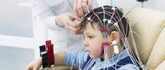
A child can also be a patient for examination on a tomograph, if there are grounds. For example, if the birth was difficult and fetal hypoxia occurred or the woman had an infectious disease, the baby may be prescribed an MRI of the brain.
A child with suspected autism may be referred for examination, since the cause of the disease may be a dysfunction of a certain part of the brain.
Diagnosis in children is often carried out by putting the baby into medicated sleep, since children are too mobile, and for some it is impossible to explain the reasons why it is absolutely forbidden to move for an hour. The most commonly used medication is Propofol.
It is best for a conscious child to undergo diagnostics in an open-type tomograph. Only the baby's head is placed under the scanner; the body is on the medical table. During the procedure, parents can be nearby, talk to the child, and also hold his hand.
Features of the influence of MRI frequency on the brain
When we have decided that the method is completely harmless, we need to answer the question - how often can we undergo examinations to diagnose the brain.
Magnetic resonance imaging may be performed as often as required by your neurologist. Another question is what diseases may require re-examination of your body.
There are several examples of diseases for which just one procedure will be sufficient. These include:
- encephalopathy;
- external non-obstructive hydrocephalus;
- leukoaraiosis;
- chronic cerebral ischemia and other structural changes.
The reason for a one-time examination is that in this situation there are practically no dynamics to be traced. The doctor decides on the need for a re-examination if there are any new symptoms that were not previously observed. The standard frequency is once every two years.
It is also worth paying attention to the fact that in case of hydrocephalus in the case of surgery or shunting, MRI can be performed annually.
Stages of undergoing an MRI of the head
The procedure for examining the brain is practically no different from MRI of other organs and parts of the body. During an appointment with a radiologist, the patient goes through the following stages:
- Before entering the medical office, you must remove all jewelry. It is advisable to change clothes, as they may contain metal parts. Some clinics provide a disposable medical shirt or you can take “home” clothes with you.
- Electronic equipment must also be left outside the room for examination. Signals emitted by a phone or tablet can distort the images transmitted by the tomograph, and the equipment, in turn, can break down.
- The patient lies down on a table placed in the tomograph. His body is first secured with medical straps, and a medical “helmet” is put on his head. A special cushion is placed under the neck. The doctor is in the next room and observes the patient through glass.
- You need to be prepared for the fact that the tomography machine is quite noisy. However, you can use earplugs.
- During diagnosis, it is important for the person being examined to remain completely still, since any movement can distort the image, which will result in an erroneous diagnosis. The MRI takes about an hour. The patient can communicate with the doctor through a special microphone located inside the tomograph.
- Upon completion of the MRI, the radiologist will provide a conclusion written on the basis of the study protocol, which records the sizes of certain parts of the organ under study, indicating their shape and structure.
Head MRI results
Upon completion of the diagnosis, the radiologist prepares a report. The document must contain information about:
- how the structures of the organ being studied are developed;
- blood flow speed;
- what development do cavities with cerebrospinal fluid have;
- are there pathological changes in the soft tissues of the head;
- what state are the hemispheres, cortex and subcortex in;
- what size are the examined brain structures;
- the presence or absence of pathology of the bone structures of the skull;
- How evenly the vascular network is filled with contrast agent.
Brief information can be obtained within an hour. If a detailed report is required, its preparation may take several days. In addition to the conclusion, printed images with elements of pathology or confirmation of the normal state of the organ are issued, as well as an electronic recording of the MRI on a CD. The patient must present the results to the doctor who referred for the examination. Only the attending physician can prescribe the correct treatment regimen.
Operating principle of MRI
MRI is a hardware diagnostic method that is based on the effect of a magnetic field on the human body. Magnetic radiation is absolutely harmless and safe, and therefore there is only one answer to the question “Is MRI harmful” - no! However, MRI, of course, can have a negative impact if contraindications are ignored.
MRI in our center
Power 1.5 Tesla
High image quality
Study for patients up to 250 kg
Burn to disc for free
Why are they sent for an MRI of the brain?
MRI of the brain is necessary to determine the following diseases and pathologies:
- establishing the level of activity of the cerebral cortex;
- assessing the degree of harm caused by skull trauma;
- Parkinson's disease;
- detection of benign or malignant tumors;
- vegetative-vascular dystonia and other vascular diseases;
- schizophrenia and other mental disorders that affected the structure of the organ under study;
- presence of tissue necrosis;
- damaged soft tissues;
- Alzheimer's disease;
- ischemic heart attack;
- violations in the integrity of the myelin sheath of the cortex;
- carcinoma;
- cyst;
- detection of inflammatory and infectious processes;
- autism;
- cerebral hemorrhage;
- manifestations of the gerontological type;
- epilepsy;
- edema;
- cerebral atrophy of the subcortex or cortex;
- multiple sclerosis and the degree of its development.
Before being referred for diagnostics, it is advisable to undergo tests. Test results may indicate an area of the head that needs more targeted examination. For example, if a patient has a high level of prolactin, this indicates problems in the functioning of the cerebellum.
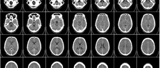
On an MRI of the brain, the doctor examines the condition of the cerebellum, the sections responsible for memory and thinking, the pituitary gland, the ventricles of the brain, the occipital lobes responsible for vision, and assesses the density of gray and white matter.
Can MRI be done for pregnant women?
— Previously, MRI was not performed on patients in the first trimester of pregnancy just in case. Simply because a teratogenic effect on the fetus has not been proven. But in 2021, a large scientific work was published by Canadian researchers who tracked more than 15 thousand pregnant women and found no signs of harmful effects of MRI either on pregnant women or on subsequently born children.
In general, there are not many situations when pregnant women really need an MRI. We are talking here about very serious diseases, such as stroke, fetal developmental anomalies (but most of these issues are resolved by ultrasound), fetal cerebral ischemia (here MRI has no alternative) and tumors in pregnant women themselves.
