Benefits of MRI
Magnetic tomography allows you to obtain a three-dimensional image of the areas under study in three projections. During the procedure, the device takes many slices, the thickness of which can be set individually and is usually 2-4 mm.
Images taken using a tomograph
Obtaining a large number of sections allows you to examine the entire organ and detect even the slightest abnormalities and pathologies.
What types of tomographs are there?
Modern magnetic tomographs are available in various variations with a wide variety of characteristics.
All tomographic devices are divided into:
- open;
- closed.
Despite the fact that conducting a study in an open tomograph is usually considered more comfortable for the patient, closed devices have greater power and detail. If the patient does not have a strong fear of closed spaces and does not have weight restrictions, it is recommended to conduct the study in a closed-type apparatus.
Tomographs are also divided according to the strength of the magnetic field radiation, the unit of measurement of which is called Tesla. Magnetic tomographs can be:
- low-floor – power up to 1.0 T;
- high-field – radiation intensity above 1.0 T.
Low-field tomographs do not provide a clear and detailed picture. A study using a high-field tomograph will allow you to examine the diagnosed area with the highest accuracy.
Modern high-field tomograph
The DiMagnit clinic has a closed-type tomograph from Philips, whose power is 1.5 Tesla. Using the device, it is possible to obtain images of the highest quality and detail.
Should you be afraid of the procedure?
Some patients are worried before the test. But their fears are in vain - magnetic resonance imaging is absolutely painless, and the effect of magnetic radiation on the body is safe.
Unlike other types of radiation diagnostics, MRI does not use ionizing radiation. The magnetic field does not have a carcinogenic or mutagenic effect on the cells of the body. Magnetic resonance scanning can be performed as often as needed.
The difference between MRI and CT and ultrasound
Magnetic resonance diagnostics has a number of advantages compared to ultrasound and computed tomography.
Ultrasound examination allows you to obtain a two-dimensional image of the area under study, but does not allow you to see a three-dimensional image of soft structures.
Computed tomography can be compared to MRI in terms of image clarity, but has a number of serious contraindications. CT is more often used to visualize hollow organs and bone structures, while MRI is much more effective at visualizing soft tissue.
What does an MRI show?
Magnetic resonance imaging is successfully used to diagnose diseases:
- thyroid gland;
- liver;
- gallbladder and ducts;
- pancreas;
- kidney;
- spleen;
- joints;
- spinal cord;
- vessels of the head, neck, abdominal region;
- pelvic organs;
- soft tissues;
- etc.
All of the above anatomical structures are perfectly visualized on MRI images. The diagnostic results make it possible to accurately identify abnormalities in the functioning of the organs being examined.
What can a doctor find?
The pictures clearly show all the structures of the body that contain moisture, and therefore hydrogen, which reacts to the magnetic field. This also applies to the following deviations from the norm:
- abscesses (purulent processes);
- dilation of the renal pelvis;
- hernias and contusions;
- degenerative changes in tissues and organs;
- brain tumors and other ailments.
Often an MRI examination becomes the only way to understand what is happening to a person. This is typical for symptoms related to several diseases simultaneously. It also happens that there are complaints, but the reasons cannot be determined, as well as the source of the malaise.
In what cases is MRI prescribed?
The wide capabilities of magnetic resonance diagnostics make its use indispensable in the following cases:
- the need for a primary diagnosis;
- conducting a comprehensive survey;
- preparation for surgery;
- monitoring the effectiveness of the therapy and treatment methods used.
In each individual case, the choice of diagnostic technique is made by the attending physician. Magnetic resonance imaging is more often used than other methods to detect diseases and injuries of soft tissues.
The MRI technique is indispensable for diagnosis:
- Neoplasms.
Magnetic resonance scanning can clearly identify the boundaries and size of the tumor and the extent of its invasion into soft tissue. No other radiation diagnostic technique is capable of providing such a clear and detailed picture of diseases.
MRI also makes it possible to determine the nature of the tumor with a high degree of probability. Malignant neoplasms have unclear boundaries and grow into surrounding tissues. Benign neoplasms, as a rule, are clearly differentiated from healthy tissues.
- Brain diseases.
The greater accuracy of magnetic resonance diagnostics makes it possible to visualize such small anatomical structures as the pituitary gland and the sella turcica. Also, MRI with contrast of the brain shows high efficiency for diagnosing demyelinating diseases (multiple sclerosis, Parkinson's disease, etc.), as it allows one to clearly see the structure of altered nerve tissues.
Brain images obtained with contrast-enhanced MRI of the brain are particularly clear because magnetic waves are difficult to image solid anatomical structures and there are no artifacts from the skull in the brain images.
- Diseases of the intervertebral discs.
MRI images of the spine
Magnetic resonance imaging is the only diagnostic method that allows you to see intervertebral discs. Even modern diagnostic methods, such as computed tomography, allow you to see only the space between the vertebrae, while MRI gives a complete picture of the condition of the discs, the possible presence of hernias and protrusions.
The use of MRI is not limited only to the above diseases, but is used when it is necessary to identify and monitor a wide range of pathologies, congenital developmental anomalies, consequences of injuries and previous surgical interventions.
MRI diagnostician
This is a doctor who diagnoses various pathologies using MRI. To engage in MRI diagnostics, you must graduate from medical school and specialize as a radiologist. Then the specialist undergoes special courses lasting 30 days or 6 months. But even this period is not enough to carry out MRI diagnostics. Therefore, after a six-month course, a young doctor works for 2 years or more under the supervision of experienced specialists.
An MRI diagnostician only conducts research and establishes a specific diagnosis.
This doctor does not treat diseases diagnosed using MRI.
He must know the method of performing diagnostics, the operating modes of the tomograph, and make a correct analysis of the results obtained, processed by a computer. The specialist must know the anatomy of internal organs, tissue structures in normal condition and in various diseases.
An expert radiologist is at risk of harm from x-ray radiation, so the radiologist's office is equipped with protection. The doctor is obliged to always develop in his specialization, attend special seminars, conferences, participate in research programs, improve his qualification level, study remotely, and read specialized literature.
Video review about the training of MRI doctors:
to contents ^
When to Apply Contrast
Magnetic resonance diagnostics can provide a very high degree of clarity of the resulting images. In most cases, the use of contrast is not required.
But when it comes to diagnosing tumors and small anatomical structures, a contrast agent can still be used.
The staining agents are made from the rare earth metal gadolinium and are injected intravenously into the patient during an MRI.
MRI contrast agents are much better tolerated than CT contrast agents. This makes the use of the dye safe even for patients with kidney pathology and does not require a preliminary test for creatinine, which is necessary for CT diagnostics with contrast.
MRI with contrast is used in the following cases:
- suspicion of a neoplasm;
- the need for differential diagnosis of a malignant tumor;
- study of the pituitary gland;
- the need to diagnose demyelinating diseases.
The use of contrast allows one to obtain a comprehensive picture of the disease, its course and the effectiveness of the therapy used.
Contraindications for MRI
Despite the fact that magnetic resonance diagnostics is a safe technique, the study has a number of absolute contraindications, in the presence of which diagnostics are prohibited:
- presence of a pacemaker, neurostimulator, insulin pump;
- vascular clips on the arteries of the brain;
- the patient’s inability to maintain a stationary position for various reasons;
- early childhood up to 5 years;
- the patient’s weight is more than 130 kg and body girth is more than 150 cm;
- first trimester of pregnancy;
There are also a number of conditions in which MRI examination is carried out with caution:
- severe pain syndrome, in which it is difficult for patients to remain in a motionless position for a long time;
- fear of confined spaces;
- psychical deviations;
- second and third trimester of pregnancy.
The presence of various prostheses and implants in the patient's body may be a contraindication to MRI if they are made of metals sensitive to magnetic radiation. Modern medical devices are most often made of titanium and other materials that are inert to the effects of magnetic fields. Their presence in the body does not interfere with MRI.
Who refers you for diagnostics, how to get it for free
MRI is a method that allows an accurate diagnosis to be made. But it has a significant disadvantage - high cost. A medical policy allows you to undergo a free examination, since MRI is included in the free list of medical insurance services in the Russian Federation. The patient will receive a referral from the attending physician. MRI is performed without payment in public medical institutions.
But not every policy will allow you to undergo such an examination for free. The high cost of the method is the main reason why insurance companies do not include MRI as a free service.
Important! Several MRI procedures are available at the public clinic, for which you do not have to pay; patients are carefully selected for medical reasons.
To get a free MRI, you need to ask your insurance company for a list of procedures provided free of charge. The patient calls the hotline of such a company and finds out the information of interest. He must have with him a referral from a doctor with a passport, insurance policy, and individual account insurance number.
If it is not possible to undergo an MRI without paying, the patient undergoes a CT scan, X-ray examination or ultrasound examination free of charge. If a patient wants to be examined for an MRI, he signs up for a waiting list, which by law lasts no more than a month. There is also an “urgent” queue, where the patient waits for 2 or 3 days. This queue will include patients with serious illnesses suffering from:
- Oncology.
- Stroke.
- Heart attack.
- Vascular diseases in the brain.
- Heart, kidney, liver and other pathologies.
Patients who have undergone surgery will also be included in the “fast” queue.
The patient is referred for an MRI by the attending physician or local doctor. Positron emission tomographic examination will provide higher accuracy than MRI.
If the patient has no contraindications, he goes into the room where the scan is performed. The doctor is in another room equipped with computer equipment. Through the glass, the doctor observes the patient in the tomograph and communicates with the patient using a microphone.
Before the procedure, the patient empties the bladder and bowels, if necessary, since the examination requires not moving. If a person wants to relieve himself at the time of diagnosis, then immobility will not be achieved and the picture will lose clarity.
The examination itself lasts 10 or 20 minutes; when contrast is administered, the duration of the procedure will be from 30 to 40 minutes. Additionally, it takes time to decipher the results and get a conclusion. It describes the state of the tissue structures being studied. If there are no pathological changes, normal tissue parameters are described. When a pathology is identified, the location where it is localized, the structure, the volume of pathological changes and other characteristics are indicated.
to contents ^
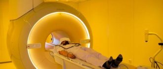
How is an MRI performed?
Before the procedure, the radiologist questions the patient about the presence of contraindications to the study. The patient is asked to remove any metal accessories, including clothing with metal fittings, and lie down on a couch, which is then placed in the CT scanner tube.
During the diagnosis, it is strictly forbidden to move, as this may affect the clarity of the resulting images.
Scanning procedure
High-field tomographs produce a fairly high level of noise, which can cause some discomfort to patients. The medical department will provide you with headphones that will play pleasant music, drowning out the sounds of the operating device.
The tomograph scans the patient’s body in different projections and instantly transmits the images to the computer screen. The interpretation of the results by the research physician begins even before the procedure is completed.
Who will order an MRI of the brain?
If a patient has frequent and severe headaches, he is referred for examination by a neurologist, family doctor, or therapist. In case of TBI (traumatic brain injury), a referral for the procedure is issued by a traumatologist or surgeon. If the patient is diagnosed with severe neurological disorders, a referral is issued by a neurologist.
To determine the functioning of the pituitary gland, an endocrinologist will refer the patient to an MRI. If necessary, a document for examination is issued by a phlebologist, ophthalmologist, neurosurgeon and other specialists.
to contents ^
Getting Results
Scan results are available immediately after the end of the study. Many of the images obtained are carefully examined by a radiologist, and a detailed report is drawn up describing both the normal anatomy of the area under study and possible abnormalities and pathologies.
15-30 minutes after the procedure, the patient is given a written report and a computer disk with the resulting images.
Magnetic resonance imaging is a modern, safe type of radiation diagnostics that allows you to obtain accurate and quick results and thoroughly examine the area under study. MRI helps to identify many diseases and abnormalities even at the initial stages of their development.
| MRI of the pancreas in Rostov-on-Don |
| MRI of the abdomen with contrast |
| MRI of the spine, what does it show? |
| MRI of ureters |
| MRI of the abdominal cavity, which organs are checked? |
| What does an MRI of the knee show? |
Which doctor interprets MRI?
The interpretation of the MRI is done not only by the person who conducted the examination itself, but also by the attending physician who referred the patient for examination. This procedure is quite labor-intensive and requires high qualifications. In addition to anatomical knowledge, the specialist must understand various pathologies and the tomograph’s reaction to them. Thus, a particular disease is determined not only by the echogenicity of the tissue, size, deformation, but also by the shade in the image. Therefore, the interpretation of the MRI obtained by the radiologist is supplemented by the conclusions of the attending physician. Now you understand that you should entrust the interpretation of MRI, especially of the brain, to a competent specialist. Our clinic employs highly qualified doctors with extensive practical experience. Not only can they interpret an MRI correctly and make a diagnosis, but they also provide extensive consultations. The correctness of the diagnosis and the course of treatment depend on which doctor interprets the MRI.