Main symptoms of the disease:
- headache,
- insomnia,
- mental disorders,
- noise in ears,
- loss of consciousness,
- lack of coordination
- convulsions,
- loss of skin sensitivity,
- nausea,
- vomit.
The causes of cysts include bruises and hematomas, congenital disorders, parasitic infections, meningitis, dystrophic transformations, problems with blood circulation in the brain. It is impossible to perform an operation to remove a brain cyst without finding out the true cause of the disease. Even the slightest mistake in making a diagnosis will lead to further inflammation of the meninges and the emergence of new lesions.
Small cysts that are not accompanied by any symptoms are most often discovered during the diagnosis of other diseases. In some cases, they do not require surgical intervention, especially if they do not increase in size over a long period of time. It is enough to monitor your blood pressure, avoid hypothermia, avoid viral infections, and do not abuse smoking and alcoholic beverages.
Indications for the study
The presence of a retrocerebellar cyst can be suspected by some characteristic symptoms:
- Severe headaches that do not go away even after taking painkillers;
- Unreasonable loss of consciousness or fainting state;
- Sensation of squeezing and pulsation in the head;
- Deterioration of hearing or vision;
- Arterial hypertension;
- Numbness of the limbs;
- Impaired coordination of movements;
- Epileptic seizures.
Magnetic resonance imaging is required if surgical intervention in the brain area is necessary.
Note that the clinical picture and severity of symptoms depend on the exact location and size of the retrocerebellar cyst.
Cyst in the brain and nose
If there is a brain cyst, exudate accumulates in the area of the meninges. With a small tumor volume, there are often no adverse symptoms. In the presence of a large cyst, compression of the brain structures is observed, and increased intracranial pressure is often observed.
Depending on the location, the following types of neoplasms are distinguished:
- retrocerebral cyst. It often occurs after a previous stroke. A cyst forms in place of dying tissue. In the presence of a neoplasm, the likelihood of neurotic disorders and seizures increases;
- arachnoid cyst. The tumor often occurs after a brain injury. There is a fairly high risk of rupture of the walls of the arachnoid cyst;
- pineal. This cyst forms in the epiphysis. As the tumor increases in size, signs of hydrocephalus are often observed.
As the disease progresses, normal blood flow slows. Unfavorable complications arise such as pain in the head, a feeling of constriction in the cranium, and the appearance of tinnitus. In particularly difficult cases, loss of consciousness and decreased clarity of vision are observed.
A conscript may be released from military service with category “D” if the following symptoms are present:
- increased intracranial pressure;
- the presence of severe neurological disorders;
- frequent headaches;
- deterioration of sleep.
If the characteristic symptoms of the disease appear to a moderate degree, the conscript should expect to be assigned category “B”.
In this case, the young man cannot be called up for military service. But he is still considered liable for military service. The young man may be called up in the event of general mobilization.
If, if the patient has a cyst, adverse symptoms are completely absent or mildly expressed, the young man is subject to conscription (with restrictions on the type of military service).
If there is a pathology such as a sinus cyst, the conscript is given a deferment from the army for six months, for the period of surgical intervention and subsequent rehabilitation. This is regulated by Article 53 of the “Schedule of Diseases”. A cyst in the nose and the army are incompatible.
Contraindications
Magnetic resonance imaging is safe for the body only if all contraindications are excluded.
Magnetic resonance imaging is not recommended in the following cases:
Very often, to obtain more informative images, doctors prescribe additional contrast. In this case, you must ensure that there are no following contraindications:
- Pregnancy of any stage.
- Breastfeeding period.
- Kidney failure.
- Chronic liver diseases.
- Allergic reaction to components included in contrast agents.
Surgeries for spinal cysts
As already mentioned, most often surgical intervention is indicated for paraarticular synovial cyst of the intervertebral facet joint. Many patients consider laser removal of spinal cysts to be the most effective method, but neurosurgeons deny the effectiveness of such an operation. When removing a spinal cyst with a laser, the heat wave negatively affects the spinal discs, destroying them. Therefore, it has already been experimentally proven that the method of removing spinal cysts with a laser is noticeably outdated.
Preparation rules
During a routine examination, doctors recommend following a few simple recommendations:
- If it is necessary to administer contrast, you must come to the clinic on an empty stomach, that is, do not eat food for 7-8 hours.
- If you are afraid of research or confined spaces, be sure to consult with a specialist. Your doctor may prescribe you sedatives to make the examination process more comfortable.
- Do not bring any metal products inside the tomograph. For this reason, doctors recommend removing metal accessories in advance.
- Prepare a change of clothes that do not have metal or metal-containing accessories.
- The duration of the diagnostic test is about 40 minutes. Empty your bladder in advance to avoid discomfort during the procedure.
Why is it worth having surgery to remove a spinal cyst at the Pirogov Clinic?
- Neurosurgeons of the highest category, with more than 10 years of experience, with a scientific degree in medicine. Sci.
- Modern equipment in operating rooms.
- Affordable prices for spinal cyst removal.
- Positive reviews about spinal surgeries and treatment in our clinic. You can view them on our website, as well as on independent platforms, for example Napopravka.ru.
- Hotel-type rooms.
- You can get a recommendation from a neurosurgeon for the treatment of your disease completely free of charge. You can submit MRI images on this page of the website.
- Possibility of obtaining an installment plan or a loan for treatment.
- Service under VHI policies.
Make an appointment
How is an MRI performed?
To ensure that the procedure is as informative as possible and that the images are clear, experts recommend performing magnetic resonance imaging in closed-type tomographs. Before starting the study, the patient must change into clothes without metal structures, take off his shoes and lie down on a retractable table. If the doctor has prescribed additional contrast enhancement, a special drug is injected into a vein, which is distributed throughout the circulatory system in just a few minutes. Then the table moves inside the tomograph, where the research process takes place.
Maintaining immobility is the main requirement for the patient! Remember that even minimal movements can lead to a decrease in the information content of the procedure.
It is also worth noting that when performing magnetic resonance imaging, a specific loud noise is observed. If this scares you, you can use earplugs.
Patients generally tolerate MRI well. Inside the tomograph there is good lighting and ventilation, there is a loudspeaker, as well as a panic button for emergency stop of the study.
Relationship between the symptoms of cystic formation and its size
The clinical course of the pathology is directly dependent on its increase (growth). Of course, there are many other factors - the location of the cyst, the source of the disease, but size plays a primary role
. An increase in cyst volume provokes an increase in fluid pressure in it, which entails an active course of the disease. The growth of cystic formation is influenced by:
- neuroinfection;
- chronic disorders of the blood supply to the heart;
- development of autoimmune processes (for example, multiple sclerosis).
MRI with contrast
In most cases, magnetic resonance imaging of the brain is performed with contrast, especially if any neoplasm is suspected. This makes it possible to examine the tumor in detail and distinguish a benign tumor from a malignant one.
The contrast agent clearly highlights the vessels, which allows you to see any tissue pathologies. Don't worry, contrast agents are safe for the human body. Most patients tolerate this procedure well. You must first consult a doctor and exclude all possible contraindications.
Types of brain cysts
The type of surgery to remove a brain cyst is selected based on the characteristics of the tumor. First of all, cysts are divided into primary and acquired. In the first case, we are talking about the death of brain tissue and problems with intrauterine development of the fetus. Acquired cysts appear due to bruises, inflammation, and bleeding.
Decoding the results
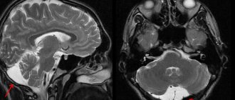
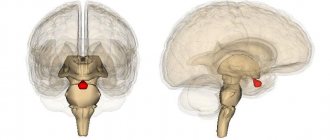
A retrocerebellar cyst is located in the brain behind the cerebellum, in the posterior cranial fossa. During magnetic resonance imaging, it is possible to clarify the exact location and size of the formation.
The report will contain all the information, but the MRI specialist cannot make a diagnosis - this must be done by your attending physician. If the diagnosis of retrocerebellar cyst is confirmed, the doctor will determine treatment tactics. For benign tumors, it is often possible to do without surgical interventions - they are required only in cases of large tumors or rapid growth. To avoid serious consequences, consult a doctor at the first alarming symptoms. Be healthy!
Suitability category for coccyx cyst
A cyst in the coccyx area is a defect in intrauterine development. In the presence of this pathology, a cavity forms in the subcutaneous case. In the absence of the influence of provoking factors, a cyst in the coccyx area does not affect the patient’s quality of life.
The likelihood of tumor inflammation increases under the influence of the following factors:
- injuries;
- presence of deep scratches in the coccyx area;
- prolonged hypothermia;
- exposure to infectious diseases;
- failure to comply with basic hygiene requirements;
- increased sweating;
- weakened immunity;
- sedentary lifestyle.
If a patient has a coccyx cyst, discomfort may occur, accompanied by itching. When the disease worsens, the following symptoms are observed:
- the appearance of a sensation of a foreign body in the coccyx area;
- increased body temperature;
- redness in the area of the gluteal fold, which is accompanied by swelling.
Like a retrocerebellar cyst of the brain, a tumor of the coccyx is a fairly serious pathology. To obtain category “B” the following conditions must be met:
- Three surgical interventions were previously performed to remove the cyst;
- At the time of examination, the disease is in the acute stage: there is a pronounced inflammatory process.