Hospitalization and treatment under the compulsory medical insurance quota. More details after viewing the pictures.
Brain oligodendroglioma is a pathological formation that originates from glial cells - oligodendrocytes. Their function is to form special lipid membranes to protect nerve cells. Tumors of this type are characterized by slow development, so the first clinical manifestations take a long time to form, the manifestations are scanty, and sick people do not immediately notice the severity of the disease. Treatment of oligodendroglioma is always complex: the main choice is surgery to remove dangerous nodes. The life prognosis is relatively favorable, much depends on the degree of malignancy and the results of radical removal of neoplasia.
Characteristics of the disease
Oligoastrocytoma is a neuroglial tumor that develops from oligodendrocytes. The neoplasm has clear boundaries with a pale pink tint.
The tumor affects people of any age group, with men between 20 and 40 years of age most often affected. The disease is not common. There are only 3% of patients with tumors in the brain. In women this is a rare phenomenon, and in children it is observed in isolated cases.
The formation is formed from non-standard cellular structures of oligodendrocytes. From them, normal protective nerve cell sheaths are formed. Such growths are heterogeneous and may have cysts that deposit calcareous components.
As the tumor grows, it grows into the cerebral cortex. It is capable of reaching large sizes and having expansive development. It is possible that a cyst may form inside the tumor. The internal development of cysts takes a long period of time. Such a tumor is difficult to treat with medication, so surgery is required to remove it. In case of untimely treatment, glioblastomas are formed.
Oligodendroglioma is localized in the frontal or temporal lobe. However, development in any area of the brain is theoretically possible. Rare formations are seen in the cerebellum, brain stem, and optic nerve. Slowly becoming larger, it gradually grows into gray matter, possibly reaching significant sizes.
The central nervous system consists of 40% neuroglia. This is the nervous tissue that surrounds neurons, participates in their intracellular metabolism, as well as in the creation and transmission of nerve impulses.
Oligodendroglia are a type of neuroglia found only in the spinal cord and brain. Oligodendrocytes protect the axons that carry nerve signals to organs. A special cell wraps part of the nerve ending in the myelin sheath.
The oligodendrocyte consists of a large number of processes and covers up to 50 axons. Under the influence of external or internal factors, a malignant formation is formed in oligodendroglial tissue.
Clinical manifestations
The symptoms of oligodendroglioma, oligoastrocytoma and anaplastic oligodendroglioma are quite similar, with the difference that with a low grade of malignancy, symptoms develop slowly. The clinical picture is caused by an increase in intracranial pressure and damage to local areas of the brain responsible for certain functions. In the first case, the signs are of a general nature, the second are called specific manifestations.
Common signs of a brain tumor include:
- headache and dizziness;
- nausea and vomiting that is not associated with poisoning and does not bring relief;
- problems with orientation in space;
- convulsions;
- epileptic seizures;
- mood disorders, psycho-emotional behavior that is not typical for humans, for example, depression, aggression and others.
Specific signs will depend on the area of the brain affected. In this case, there may be problems with hearing, speech, writing, coordination of movements, problems with memory or thinking, and so on.
Frequent areas of localization and risk group
Oligoastrocytoma is a rare cancer that occurs in 3% of all brain tumors.
Since it consists of specialized cells of the nervous system, it is quite logical that it is formed in the human central nervous system - in the white matter of the brain in the area of the brainstem or cerebellum, less often - on the optic nerve. The preferred area of localization of the formation is the frontal or temporal lobes.
Slowly increasing in size, the tumor begins to gradually grow into the gray matter, sometimes reaching gigantic proportions.
Since it consists of specialized cells of the nervous system, it is quite logical that it is formed in the human central nervous system - in the white matter of the brain in the area of the brainstem or cerebellum, less often - on the optic nerve. The preferred area of localization of the formation is the frontal or temporal lobes.
Neuroglial tumors occur at any age, predominantly in the male half of the population. Occurs in 3% of men. It develops slowly and is located in the white matter of the cerebral hemispheres.
If the pathology is not treated, it can reach significant sizes. It is diagnosed along the walls of the ventricles, can penetrate into their cavity and grow into the cerebral cortex. Very rarely localized directly in the cerebellum, optic nerve and brain stem.
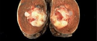
The neoplasm has a pale pink tint and a clear outline. Cysts may form inside. Develops over a long period of time. This tumor cannot be cured with medication; surgical intervention is required.
The patient first undergoes a thorough examination. Oligodendroglioma is distinguished by the presence of petrificates; most patients continue to live with the tumor for five years after diagnosis and before the start of therapy.
Symptomatika
Symptoms of oligodendroglioma result from increased fluid pressure inside the skull (intracranial hypertension). These symptoms include:
- nausea;
- vomit;
- irritability;
- headache;
- visual impairment;
- expansion of the head;
- seizures.
Oligodendroglioma may also cause weakness or paralysis on the side of the body opposite the side of the brain where the tumor is located. If the tumor is located in the frontal lobe, the patient may experience gradual changes in mood and personality. When it is located in the temporal lobe, the patient may experience difficulties with speech, hearing, coordination and memory.
Classification of tumors
In clinical neuro-oncology, neoplasms (oligodendroglioma) are classified as follows:
- Well-differentiated oligodendroglioma (grade II malignancy). The disease may have a latent course with a partially favorable course;
- Anaplastic oligodendroglioma (grade III). Brain tumors are characterized by rapid progression.
- Mixed type oligoastrocytoma (grade III), an oncological disease, tends to degenerate into an aggressive cerebral glioblastoma, which is difficult to treat and in most cases is fatal.
Treatment of oligodendrogliomas
A special feature of low-grade oligodendrogliomas is their sensitivity to chemotherapy. Usually within 3 months a reaction appears - the tumor decreases. This method of treatment can be used independently in the preoperative period or in combination with surgery, after the intervention. Tactics are selected individually by a clinical oncologist or chemotherapist, taking into account the patient’s data and all information known about the tumor.
For tumors of grade III malignancy, surgical treatment is more effective. The most radical removal leads to fewer complications and increases the patient's life expectancy compared to partial removal. The localization of some oligodendrogliomas does not allow complete removal of the tumor without neurological damage. The decisive factor here is the patient’s quality of life. Even partial removal provokes relief of symptoms.
The tumor is pinkish-reddish in color and looks similar to healthy brain matter. It is difficult to remove without damaging tissue if the boundaries of the tumor are unclear. Therefore, the operation is performed using a microscope and additional modern equipment. The equipment of the operating room and the skills of the neurosurgeon play a big role in the success of the intervention.
Additional treatment in the postoperative period, in case of contraindications to surgery or residual tumors, is radiation therapy. The use of combined treatment methods leads to an increase in average life expectancy.
Pathogenesis of pathology and causes of development
The reasons for the development of the disease are currently unknown, but it has been noted that in most cases this tumor develops in people with gene mutations in the first, nineteenth chromosome, or both at the same time.
Also, a huge risk of developing this cancer comes from etching or chronic exposure to polyvinyl chloride, which has an extremely negative effect on glial cells (auxiliary nerve cells of the nervous system). Polyvenyl chloride is a common ubiquitous material known as PVC, which is most often used as a building material, or a material from which non-food items and things that come into little direct contact with humans are made.
In addition to chemical influences, this tumor can also be provoked by radiation or high electromagnetic exposure.
Causes
Glial tumor growth is associated with a mutation of the TCF12 gene, which was experimentally proven by scientists from the British Institute of Cancer Research. This gene encodes a developmental protein, a phosphoprotein.
Factors pushing towards the development of pathology:
- Working with chemicals;
- Heredity;
- Medicines;
- Irradiation;
- Nutrition.
Li-Fraumeni syndrome and neurofibromatosis cause a genetic predisposition to the disease. Hereditary gene mutation in Li-Fraumeni syndrome contributes to the early development of intracerebral tumors in relatives in the first and second generations. The autosomal dominant type of inheritance for neurofibromatosis types 1 and 2 transmits the disease to each generation with a probability of 50%.
Hormonal contraceptive medications increase the likelihood of developing brain gliomas.
Ionized irradiation of the head as part of radiation therapy for other diseases increases the risk of developing severe brain tumors by 22 times.
Canned food, smoked meats, and protein products contain carcinogens N-nitroso compounds. Substances enter the air during the production of dyes, lubricating oils, and pesticides.
Short description
Oligodendroglioma (ODG) is a highly differentiated (WHO-2), diffusely infiltrative intracerebral tumor of predominantly adult age, consisting of cells of oligodendroglial origin. Among oligodendrogliomas, there is also a malignant variant of the tumor - anaplastic ODH (WHO-3) and the so-called “mixed tumors” - oligoastrocytoma and anaplastic oligoastrocytoma.
Code according to the international classification of diseases ICD-10:
- C71 Malignant neoplasm of the brain
- D33 Benign neoplasm of the brain and other parts of the central nervous system
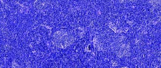
Histopathology. Characterized by moderate cellular density and the presence of cells with typical round nuclei and light cytoplasm (the so-called “honeycomb”, “fried egg”). Anaplastic ODH (WHO-3) differs from ODH (WHO-2) by the presence of significant mitotic activity, expressed by proliferation of the vascular endothelium. In addition, there may be necrosis in anaplastic ODH. Epidemiology. Oligodendroglial tumors account for 4–5% of all primary brain tumors, corresponding to an incidence of 0.3 cases per 100,000 population per year. The male/female ratio among patients is 3/2. The average age of operated patients is 43 years.
Localization. ODCs are usually localized in the cerebral hemispheres cortically - subcortically (in 50-60% of cases the frontal lobes are affected).
Characteristics of the disease
At the first neurological disorders that cause tumors, the patient first of all turns to a neurologist, who, based on a neurological examination and reflex tests, may suspect oncology. Next, a referral for examination is received:
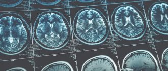
- Computed tomography, which is an advanced x-ray, which shows all brain formations and layer-by-layer images of the affected part.
- Magnetic resonance imaging, which allows extremely clear visualization of all internal structures of the brain and tumors.
- EEG – electroencephalogram – study of brain activity and its disorders.
- Audiography is a study of the degree of hearing impairment.
- Examination by an ophthalmologist.
- Analysis of cerebrospinal fluid (CSF).
- A biopsy of the tumor to determine the specific type of cells it consists of and the degree of its malignancy.
- General tests.
What is oligodendroglioma
The disease is a severe pathology, since the pathological process affects the membranes of the brain.
Oligodendroglioma is a brain cancer. Diagnosis is carried out using CT, MRI, audiography, electroencephalogram.
Treatment takes place in several stages: tumor removal, cytological examination of biomaterial, chemotherapy and radiotherapy.
The tumor begins to form from fat cells that are part of the membrane of nerve cells. In medicine, such formations are classified as primary glial.
Depending on the degree of malignancy, they are divided into several types. Each of them has its own characteristic flow characteristics.
In some cases, oligodendroglioma is characterized by a slow course, but can grow rapidly.
After treatment, patients live for a certain number of years, but after a while death occurs. Thus, oligodendroglioma is considered a dangerous disease, which, even with treatment after a certain time has elapsed after the onset of symptoms, is fatal.
Chemotherapy is effective after surgical removal of the tumor.
This prolongs patient survival. In this case, the following indications must be present: age over 40 years, tumor size greater than 6 cm, neurological deficit.
Oncologist, mammologist, chemotherapy
Butsyk Natalya Gennadievna
32 years of experience
Prognosis for patients
All patients who are faced with this disease often wonder how long do they live with oligodendroglioma?
The time without recurrence of the disease with timely assistance and quality treatment is 5 years. As for survival, in 30% of cases after treatment it can be up to 10 years.
If oligodendroglioma is diagnosed at the developmental stage and prompt surgical treatment is performed, the patient will live longer.
Since the factors for the development of the disease have not been precisely discovered to this day, and it manifests itself as a result of genetic changes, it is almost impossible to prevent its occurrence.
All patients with this type of oncology first of all wonder how long they can live with oligodendroglioma.
The answer depends on many factors: the type of tumor, its size, area of localization, specific type and degree of malignancy.
With a classic tumor, the prognosis is quite favorable. Firstly, they live with her for about five years, this period can pass from the moment of her first diagnosis to the decision to start treatment, and secondly, the likelihood of a complete recovery depends on the age of the patient, the younger it is, the higher the percentage of a favorable outcome :
- Patients over 55 years of age recover in approximately 50% of cases.
- Patients from forty-five to 55 years old - in about seventy percent of cases.
- The probability of recovery among people aged 20 to 44 years is 80%.
Complications
If oligodendroglioma is left untreated, various complications arise. First of all, as the tumor develops, there is a deterioration in memory, attention, decreased vision, and hearing. This is due to the fact that the tumor begins to put pressure on the brain structures.
Also among the complications are various personality changes, patients suffer from mental disorders. In severe cases, the tumor spreads, affects neighboring structures, and metastatic lesions develop. Ultimately death occurs.
Diagnostic methods
In order to establish a diagnosis of oligodendroglioma or oligoastrocytoma, an assessment of the neurological condition is carried out, anamnesis is taken: testing of auditory function, sense of smell, eye reflexes, the condition of the muscular and vestibular apparatus is assessed, and special attention is paid to assessing abstract thinking and memory.
If oligodendroglioma (ODG) is suspected, the following instrumental tests are mandatory:
- Visualization of brain structures using MRI;
- X-ray CT to obtain layer-by-layer images of the brain;
- Checking the bioelectrical activity of the brain - EEG;
- Diagnosis of hearing sensitivity – audiometry.
Laboratory diagnostics of ODH:
- cerebrospinal fluid analysis;
- stereotactic biopsy;
- biochemical blood test (liver and kidney functionality).
As mentioned earlier, the patient undergoes surgery. It is imperative to remove the tumor and try to preserve the volume of healthy brain tissue as much as possible. Then complex therapy is carried out to maintain the patient’s normal condition and achieve long-term remission.
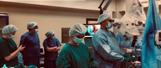
Surgical intervention is carried out using modern systems. MRI results help to accurately determine the location of the tumor, and CT results provide an opportunity to map out the path to achieving the tumor. As a result, the operation becomes safe and effective.
The patient may also be prescribed radiation therapy, which can suppress the growth of the tumor. During the procedure, ionized radiation is used, which does not have a negative effect on the body, but helps to obtain a positive result.
Oligodendroglioma is one of those types of tumors that can also be treated with a certain type of chemotherapy. It allows you to increase the effectiveness of treatment. It is recommended for use in cases of relapse of oligodendroglioma with signs of anaplasia. Modern methods of chemotherapy are highly effective and have low negative consequences.
Treatment should be carried out comprehensively. The patient may be prescribed medications that help not only during therapy, but also as a preventive measure:
- antitumor agents: Hydrozine sulfate or Proxyfein;
- antimetabolites: Hydroxycarbamides, Methotrexate;
- alkylating agents: Dacarbazine, Carmustine, Lomustine, Fotemustine;
- antitumor antibiotics: Daunorubicin.
After the tumor has been removed, it is necessary to be observed by a physiotherapist. He helps to create an individual recovery plan, which consists of psychological and motor adaptation. The patient is also required to undergo an annual preventive examination, which helps to detect relapse of the disease.
Brain oligodendroglioma has a different prognosis. It all depends on how early the diagnosis was made and appropriate therapy was carried out. The diagnostic process consists of:
- Initial examination. In its course, it is revealed in what state the reflexes, the back surface of the eyeball, and consciousness are more nervous.
- Laboratory blood tests. These studies make it possible to detect the presence of pathological processes in the body and check the functional abilities of the liver and kidneys.
- Computed tomography. This scanning method is based on X-ray radiation. The damaged area is scanned and a three-dimensional image is obtained. To improve image quality, a contrast agent can be used. The duration of the procedure is about half an hour. It is absolutely painless and very informative.
- Magnetic resonance imaging. The technique is based on the action of magnetic fields. This study can also be performed using contrast. It provides the most detailed information about the state of the brain.
- Biopsies. During the procedure, a small portion of pathological tissue is removed and sent for histological analysis. This is necessary to determine the malignancy of the tumor.
The treatment method is selected after a detailed study of the examination results.
If the first disorders are detected at the neurological level that lead to the development of tumors, the patient should consult a neurologist. During the examination, the patient will undergo a neurological examination, which includes the following tests:
- Hearing.
- Facial muscles.
- Eye reflexes.
- Equilibrium.
- Smell.
- Head movement.
- Memory test, abstract thinking.
Recommendations
Taking into account risk factors, you should:
- Avoid contact with formaldehyde, arsenic, mercury, rubber, derivatives in the production of plastics; if you cannot avoid it, then do not neglect protective equipment (doctors, firefighters, farmers).
- Avoid smoked meat products, boiled ham, and fried bacon; they contain nitrates/nitrites and secondary amines, which contribute to carcinogenesis in the body.
- Eat foods containing antioxidants that protect body cells from mutations:
vitamin C (currants, gooseberries, sauerkraut, cranberries, kiwi, dried rose hips, etc.);
- retinol, vitamin A (carrots, sea buckthorn, persimmon, chicken liver, spinach, etc.)
- tocopherol, vitamin E (wheat germ oil, cottonseed oil, almonds, etc.)
Knowing about hereditary predisposition, you should adhere to the following rules:
- Refuse to work in hazardous industries involving toxic substances; if forced contact occurs, wear protective clothing, gloves, and a mask.
- Remove smoked meats, boiled sausages, and ham from the diet.
- Include in your diet foods, vegetables and fruits with antioxidants, vitamins A, C, E: gooseberries, apples, kiwi, citrus fruits, cranberries, chicken liver, persimmon, sea buckthorn.
- Remember the dangers of smoking and avoid bad habits.
Full recovery depends on the stage at which treatment began. To detect the disease at an early stage, it is necessary to do a blood test for tumor markers every six months.
Patient prognosis and survival
The period without relapse for this tumor, if timely and high-quality treatment was provided, ranges from five years. As for survival, after therapy, it can be about ten years in 30% of cases. If oligodendroglioma was diagnosed at the initial stage and surgery was performed immediately, then the prognosis will be quite favorable.
Since the causes of the pathology have not yet been precisely identified, and it is known that it occurs as a result of genetic disorders, it is almost impossible to prevent its occurrence.
The period without relapse for this tumor, if timely and high-quality treatment was provided, ranges from five years. As for survival, after therapy, it can be about ten years in 30% of cases.
If oligodendroglioma was diagnosed at an early stage and surgery was performed immediately, the prognosis will be quite favorable.
Diagnostics
The Medsi Neurosurgery Center has high-tech equipment for diagnosing oligodendroglioma.
If the tumor is diagnosed as early as possible, the prognosis will be quite favorable.
Calcifications in the tumor can be a relatively common and characteristic sign; they are visible on MRI and CT.
Some cases require additional neuroimaging methods. They also show the functional characteristics of the tumor. Such methods include angiography, spectroscopy, tractography, and three-dimensional volumetric volumetry.
They are actively used by experienced specialists at the Medsi Neurosurgery Center.
If necessary, neurophysiological monitoring, neuronavigation, and fluorescence microscopy are used simultaneously with the operation. These methods contribute to safer interventions.