Neurophysiology is a science that studies, through electrophysiological techniques, the features of the organization, functioning and interaction of the central nervous system and the brain.
This area of medicine is closely related to neurology, psychology, physiology, biology and anatomy, however, unlike these disciplines, it is primarily engaged in theoretical research.
The subjects of study of neurophysiology are visual, auditory, tactile and olfactory perception of a person, his emotional and somatic reactions, mechanisms for receiving and processing information, etc.
The origin of neurophysiology dates back to the century before last. For a long time, scientific activity consisted of conducting and describing experiments on animals. In the course of such studies, scientists, for example, revealed the similarity of many functions of the central nervous system of animals and humans.
By the end of the 19th century, a large amount of information about neurology and physiology had been accumulated; a kind of impetus was required to give an understanding of how to use this knowledge. This impulse was the discovery of the neuron, the functional and structural unit of the nervous system.
The 20th century was an era of great medical discoveries. Russian researchers and doctors made an invaluable contribution to the development of the science of neurophysiology: I.M. Sechenov, author of the work “Reflexes of the Brain”, I.P. Pavlov, V.M. Bekhterev, N.E. Vvedensky, A.F. Samoilov.
Neurophysiological research methods invented in subsequent decades made it possible to bring the diagnosis of diseases of the brain and nervous system to a new level.
1
Consultation with a neurologist
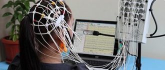
2 Electroencephalogram (Echo-EG)
3 Electroencephalogram (Echo-EG)
What does a neurophysiologist do?
A neurophysiologist is a specialist who, being both a physician and an analyst, collects and interprets neurophysiological examination data in order to make an accurate diagnosis for the patient and recommend the optimal treatment option.
Using various instrumental methods, a neurophysiologist determines the degree and nature of damage to the patient’s nervous system, analyzing functions such as vision, hearing, touch, smell, volume and coordination of movements, electrical activity of the brain and muscle cells.
Neurophysiological studies allow for accurate diagnosis, which is very important for symptoms characteristic of various pathologies. Thus, a headache may indicate both increased intracranial pressure and the presence of vascular changes or a tumor process in the brain.
The importance of neurophysiological studies for the diagnosis of neurological and other diseases cannot be overestimated.
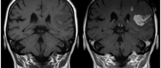
Magnetic resonance imaging of the head with contrast agent
In some cases, magnetic resonance imaging with contrast is used. The contrast agent allows you to more clearly distinguish between healthy tissues and those affected by pathology. The substance is administered intravenously before the examination using a syringe or dropper in measured doses. Gadolinium, which is part of the contrast agent, causes allergies in extremely rare cases. It is necessary to check for allergies before starting a diagnosis. An anesthesiologist can do this.
Contrast is necessarily used if an oncological tumor is to be diagnosed, or if damaged tissue is examined as a result of an infectious or inflammatory process. Examinations using contrast agents take longer.
Reasons for contacting a neurophysiologist
Nowadays, almost every person has neurological diseases.
Reasons to sign up for a consultation with a neurophysiologist may include:
- consequences of traumatic brain injury;
- disorders of vision, hearing, smell, touch;
- disturbances of memory, attention, concentration;
- impaired coordination of movements;
- muscle weakness, cramps;
- vegetative-vascular dystonia;
- headaches, dizziness;
- insomnia and other sleep disorders;
- depression, neuroses, phobias, fears, panic attacks, etc.
1 Electroneuromyography (ENMG)
2 Electroneuromyography (ENMG)
3 Electroneuromyography (ENMG)
Side effects from brain MRI
Magnetic resonance imaging examination has virtually no side effects. However, the procedure has some unpleasant features:
- The contrast agent used before diagnosis can rarely cause an allergic reaction. When contrast is administered, the patient may experience slight dizziness and nausea. However, these sensations quickly pass.
- Loud noise during operation of the tomograph can negatively affect the mood and general condition of the patient. Some people complain that they get a headache after the examination. To avoid this side effect, you need to wear earplugs before starting the scanner.
- The ability of a contrast agent to penetrate breast milk is an unpleasant “surprise” for nursing mothers. You should not breastfeed your baby for 2 days after diagnosis. It is necessary to express breast milk.
- Headaches can begin if the patient is overexcited and too sensitive by nature. It is enough to take a painkiller and rest.
Despite the fact that magnetic resonance diagnostics is an accessible medical procedure, when it comes to choosing a clinical diagnostic center, the patient faces some difficulties. For example, a clinic that is suitable for the cost is located on the other side of the city.
Our website will help you choose a medical center that meets the patient’s needs. It is enough to filter the necessary categories in the search (“Cost”, “Location”, “Rating” and others) and select a clinic. Our website specialists also provide free consultation by phone. You can make an appointment on a convenient day and receive a discount on your first appointment.
Methods of neurophysiological research
Hardware studies allow us to identify the smallest signs of pathological changes, determine the nature of the disease and the reasons for its development.
MedicCity presents all modern methods for studying the neurophysiology of the brain:
- electroencephalography (EEG);
- rheoencephalography (REG);
- electroneuromyography (ENMG);
- magnetic resonance imaging (MRI).
Echoencephalography (Echo-EG) is also used in neurophysiology.
EEG
Allows you to evaluate the activity of the cerebral cortex during wakefulness or sleep. Used for diagnosing stroke, vascular diseases, epilepsy, brain tumors, traumatic brain injuries, movement disorders, sleep disorders, etc. The only test that can be applied to an unconscious person.
REG
A method that provides information about the state of cerebral vessels (tone, degree of elasticity, activity, etc.) and cerebral blood flow. It is used in the diagnosis of vascular dystonia, migraines, arterial hypertension, disorders of the vestibular apparatus, and for traumatic brain injuries.
ENMG
Allows you to examine the functional viability of muscles and peripheral nerves. Useful in diagnosing polyneuritis, radiculopathy, intervertebral disc herniation, diabetes mellitus, etc.
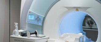
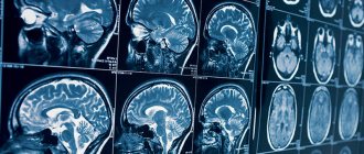
MRI
An extremely informative and practically safe research method. It is used in diagnosing the condition of the spine, joints, brain, blood vessels, and soft tissues.
Echo-EG
Ultrasonic harmless method. Provides information about pathological changes in the structure of the brain. Used in the diagnosis of tumors, injuries, developmental anomalies, etc.
Our clinic presents all the leading methods of neurophysiological research, conducted by an experienced neurophysiologist. You must register for studies in advance!
Treatment
Treatment depends on the stage of the process. This may be a conservative approach or surgery. In the first case, the focus is on normalizing the patient's lifestyle. Nutrition is adjusted, a special diet is prescribed, and a weight loss program is developed, if necessary. As drug therapy, drugs are used that normalize blood pressure, lower cholesterol levels, normalize blood counts, and replenish the supply of vitamins and microelements.
If the process has gone far enough and medications do not achieve the desired result, surgery is used. This can be open or modern endovascular intervention. A gentle method is angioplasty, during which the lumen of the vessel expands; if necessary, a stent can be installed to maintain the lumen of the vessel in the desired position. The operation is aimed at removing plaques that are blocking normal blood flow. In some cases, a severely damaged vessel may be replaced or a new blood flow path created.
Get diagnosed with atherosclerosis at Clinic No. 1
- Neurologist appointment
- Doppler ultrasound
- Coronary angiography
For one-time payment for services - 20% discount
Call
Types of neurophysiological studies
The MedicCity Clinic can offer you the following types of brain neurophysiology studies:
- EEG (rheoencephalography);
- REG (electroencephalography);
- ENMG (electroneuromyography);
- Echo-EG (echoencephalography)
EEG
Using electroencephalography, it is possible to test the functions of the central nervous system, as well as analyze functional and organic lesions of the brain.
EEG can be used to diagnose epilepsy, tumors, inflammatory and vascular diseases of the brain, and various injuries. Indications for EEG studies can be different, including seizures, convulsions, walking and talking during sleep. This study in neurophysiology is recommended for poisoning with poisons with neurotoxic effects and for traumatic brain injuries.
Electroencephalography is the only study that can be performed even if the patient is unconscious.
1 Electroencephalography (EEG)
2 Electroencephalography (EEG)
3 Electroencephalography (EEG)
REG
The main task of rheoencephalography is to identify the causes of vascular pathology of the brain. REG helps to study cerebral blood flow - by recording fluctuations in the electrical resistance of brain tissue when a weak high-frequency current is passed through them.
REG is an absolutely safe and painless study. An experienced neurophysiologist can recommend it for both low and high blood pressure, as well as for cerebral atherosclerosis, headaches and migraines.
ENMG
Electroneuromyography is designed to investigate the extent and function of lesions of the peripheral neuromotor apparatus resulting from neurosomatic and neurological diseases.
Echo-EG
Echoencephalography is an ultrasound method most often used by doctors for the primary diagnosis of damage to brain tissue.
Biofeedback-EEG therapy
In addition to neurophysiological studies, the MedicCity clinic uses biofeedback-EEG therapy, a biofeedback method that is based on information about the individual properties of brain rhythms and the distribution of biopotentials in different areas of the cerebral cortex.
With the help of biofeedback-EEG therapy, a neurophysiologist teaches the patient to control his psycho-emotional state, fight depression, panic attacks and stress.
Contacting an experienced neurophysiologist guarantees you a professional approach to diagnosing the disease, interpreting examination results and prescribing adequate therapy, strictly individual in each case.
"MedicCity" is a clinic with a powerful diagnostic and treatment base and a truly professional team. Do not hesitate to seek medical help, our doctors will delicately and skillfully help you solve all your medical problems! Don’t put off difficult questions for later, health is most important!
Degree of disease development
The disease develops gradually. Main stages:
- Lipid spots.
The easiest stage at which the artery walls begin to become saturated with lipid compounds. Localization is focal, in some areas. The changes look like yellowish spots, stripes spreading along the inner surface of the vessels. There are no symptoms. Plaques form faster in patients with excess weight, bad habits, diabetes, and high blood pressure. - Fibrous plaques.
This is the second stage of the disease, in which the previously formed lipid areas become inflamed. As a result, the immune system releases inflammatory mediators in the affected areas. The fats that have accumulated in the walls are destroyed, die, and sclerosis forms. The connective tissue grows, fibrous plaques form, protrusion occurs on the walls of blood vessels, stenosis develops, and blood flow is disrupted. - Complicated plaque.
This is the last stage in the development of the disease, during which complications appear and symptoms begin to appear. The protrusion hardens, calcifies, significantly narrows the lumen, and blood cannot circulate normally.
Raynaud's syndrome
With this syndrome, a spasm of the hands occurs in the cold or under stress; it is reversible, as is a change in skin color. During an attack, one or more fingers may turn pale, blue, etc. One finger may have one manifestation, another may have another. Sometimes Raynaud's syndrome affects the tongue or nose. Although the symptoms sometimes frighten people, treatment is simple - protection from the cold, quitting smoking, working with a psychologist, taking medications. This disease is treated by an angiologist who prescribes medications and, if necessary, refers to a psychologist.
Women are more susceptible to Raynaud's syndrome, and young women are more likely to suffer from the disease. This disease is caused by excessive α2-adrenergic activity, due to which vasospasm occurs, the mechanism of which has not yet been established by medicine.
The primary form occurs most often - in approximately 80% of cases. It develops without signs of another disease, and in case of secondary disease, patients are diagnosed with various concomitant conditions and diseases, mainly related to connective tissue.
Symptoms, in addition to changes in skin color, are a feeling of cold, goose bumps, burning sensation, a clear boundary of color change appears on the fingers, transitions can be one-, two- and three-phase, often the changes are symmetrical.
Phlebothrombosis
Phlebothrombosis is the primary formation of a blood clot in the deep vessels of the legs; the vein is blocked by a blood clot, but no inflammation is observed. Usually the disease focuses on the deep veins of the leg, its back surface. Phlebothrombosis is dangerous because it increases the risk of developing pulmonary embolism and postthrombophlebitis syndrome. This disease is typical for older people, but now it is rapidly getting younger. Phlebothrombosis can be caused by blood stagnation, damage to the vessel wall, and increased blood clotting. The main triggers are oncology and chemotherapy, pregnancy, cardiovascular disorders, the postpartum period, obesity, hormone therapy or oral contraceptives.
Symptoms are pain in the legs, which becomes stronger with movement, slight swelling, and the pain is bursting in nature. If several vein trunks are affected, the skin may turn blue and swelling may increase. After physical activity, the condition worsens, the person tries to move the affected leg less. When the femoral vein is damaged, swelling quickly increases, the venous pattern becomes clearly visible, that is, the veins become full, the skin turns blue, body temperature rises, and the lymph nodes enlarge. With the listed symptoms, the patient urgently needs the help of an angiologist who treats this disease in the initial stages or gives a referral to an angiologist surgeon.
Phlebothrombosis
Hemangiomas
Hemangioma is a benign vascular tumor that occurs mainly in children, more often in girls. Its type is lymphangioma, that is, a benign tumor of the lymphatic bed. Tumors appear immediately after birth, they are mainly found on the head and neck, sometimes they are found on internal organs.
Both tumors are noticeable visually - it is a thickening that is higher than the level of the skin. When pressed, the formation decreases and then restores its size. Hemangiomas grow quickly, and it is impossible to predict their behavior.
Lymphangioma grows more slowly and is located in places where there are many lymph nodes.
The reasons are hormonal characteristics of childhood, especially with prematurity, viruses that a woman suffered during pregnancy, and side effects of drugs during pregnancy.
If you find a tumor in your child, you need to contact an angiologist who treats this disease or, if necessary, refers you to a vascular surgeon.
Symptoms that should prompt you to contact an angiologist
You should contact an angiologist if you have:
- swelling, heaviness, fatigue in the legs for no apparent reason;
- spider veins and networks;
- the venous pattern has intensified, it hurts to touch, it is hot to the touch;
- veins become inflamed;
- varicose veins;
- trophic ulcers appear;
- the skin of the limbs changes color;
- the general condition worsens – the temperature rises, weakness appears;
- swelling does not go away even after rest and a night’s sleep;
- Legs hurt - with exertion, at rest;
- night cramps in the legs.
Phlebitis
Phlebitis is an inflammation of the vein wall with its gradual destruction; it can be acute or chronic. The main danger of the disease is the high risk of blood clot formation and complete blockage of the vessel. Most often, phlebitis is caused by past infections and inflammation. However, the inflammatory process can also begin if a substance that irritates the vessel wall enters the bloodstream. Phlebitis can affect deep and superficial veins.
Depending on which particular vessel wall is inflamed, phlebitis is divided into three types:
- endophlebitis – inner wall;
- periphlebitis – outer wall;
- Panphlebitis is a common form, all membranes are affected.
An angiologist treats any form of this disease; you need to go to a doctor if there are any signs of unhealthy veins, and he will accurately determine the disease.