05/10/2021 | Category Symptoms of cervical osteochondrosis
Many who had an ultrasound of the neck saw in the conclusion the phrase “impaired venous outflow.” Let us immediately note that if this is discovered, this is already good, it means that the doctor looked carefully, not in a hurry, and did not miss anything. Sometimes there is no such record, although in fact there is a problem. The lack of recording may be due to various reasons, including due to outdated equipment; we will not dwell on this, but further we will describe the factors and symptoms by which it can be understood that the venous outflow is impaired, even if this is not indicated by the ultrasound results.
What is a venous outflow disorder?
First of all, let's understand the anatomy. Imagine a spine. Veins run next to it, through which blood should flow freely from the head down. If the vertebrae are displaced, then the veins can be pinched, deformed, and because of this their capacity is reduced. Blood accumulates in the posterior cranial fossa.
With normal intracranial pressure, there is no outflow of blood through the compressed vessels. In order for the outflow to be restored, the blood must flow with greater pressure, that is, the pressure must increase. We all know that increasing blood pressure is not beneficial for the body, to put it mildly, and here it is a necessary measure to restore blood circulation, otherwise more serious problems will arise.
In the long term, stagnation of blood and impaired circulation in the brain are fraught with many bad consequences, but there is no need to be afraid. The human body is excellent at recovering on its own, plus it informs in advance about possible problems with unpleasant and even painful sensations. Therefore, you need to listen to your body and simply help it be healthy, which is not difficult to do.
After an increase in pressure, a sharp outflow occurs through the posterior or jugular veins. Changes in the condition of the jugular veins are easier to see on an ultrasound than changes in the vessels near the spine, on the back of the neck.
Helpful advice
When you go for an ultrasound, be sure to ask to look at the jugular veins. If they write to you that they are dilated, this is an accurate sign of venous stagnation and circulatory disorders in the brain.
Reference. It is more difficult to see a similar sign of a disorder in the spine. Therefore, it may be that the problem is already there, but it is not yet visible when examining the back of the neck.
The jugular veins dilate and lose elasticity when large volumes of blood are dumped through them. As a result, blood circulation worsens even more, and the body signals this with pain.
Anatomical features
To understand the disease, it is important to understand the anatomy. Knowing the features of the structure of all parts of the head, both hemispheres of the brain and blood vessels, it will become easier to even identify this disease.
The venous wall of the brain does not have a valve apparatus, which is why the blood disperses freely through all adjacent vessels on different sides of the head. Almost all of them are located at a distance from each other and from the arteries. Venous sinuses are intermediate points for blood with cerebrospinal fluid from the ventricles. Vessels emerge from them. They come in two types: superficial and deep. The first are located inside the pia mater in the interlobar grooves; they are responsible for the flow of blood from the cerebral cortex to the cerebellum. The latter are formed due to the white matter with subcortical nodes.
Diploic and emissary veins are also distinguished, which conduct blood flow to the sinuses and are responsible for the connection between the superficial and deep vessels. There are also extracranial veins that provide blood supply to certain areas of the brain. In this case, all the vessels are intertwined into one, called a tank. Through it, blood flows to the central part of the brain, many sinuses and the basal plexus.
For the stable functioning of blood circulation, intervenous and intersinus anastomoses are of great importance. At the exit from the brain near the neck, three plexuses are formed: the foramen ovale of the skull, the canals of the hypoglossal nerve and the carotid artery.
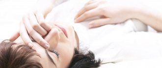
Symptoms of impaired venous outflow
Headache
The most common symptom is pain due to increased intracranial pressure. The blood does not leave the head on time, and new portions of it continue to flow - the pressure rises, the body signals this with a headache.
A case from the practice of Dr. Shishonin
The man was diagnosed with migraine and prescribed medication. The patient took the pills with discipline, but there was no noticeable improvement. Migraine attacks did not become less frequent and were often as severe as before starting to take the drugs. The attack was very difficult to relieve with analgesics; you had to take the maximum allowable doses, and even they did not always work. The pain went away on its own after some time.
Dr. Shishonin recommended going for an ultrasound under the compulsory medical insurance policy and asking to look at the jugular veins. An ultrasound showed that they were dilated. The cause of the migraine became clear - increased intracranial pressure.
After conservative non-drug treatment, improvement occurred within two months.
Extreme migraine
A very painful symptom occurs due to loss of elasticity of the jugular veins. Up to a certain point they can withstand the increased load, and then they can no longer cope. Swelling appears in the brain stem. They are not yet dangerous to human life and health, but they cause severe pain in the ternary nerve.
This is an extreme degree of migraine, it is almost impossible to tolerate and difficult to relieve with analgesics. A person becomes incapacitated, no matter how strong his will is. In addition, nausea occurs, the patient is physically unable to drink liquids or eat food. A migraine can last a day and goes away on its own when the pressure rises to such an extent that blood discharge does occur. If such conditions are repeated frequently, it becomes potentially dangerous.
Ptosis of the eyelid
A case from the practice of Dr. Shishonin
The person suddenly developed ptosis and the mobility of the eyelid was impaired. The hospital carried out a full examination - a microstroke was not confirmed, and no tumor was found.
An ultrasound scan examined the veins in the neck and revealed gross venous congestion. Conservative treatment without pills gave results after six months. If a person had paid attention to the headaches that he often had earlier, the problem could have been solved faster.
Labored breathing
This symptom itself is ambiguous; there can be many reasons. But if it is accompanied by regular pain in the back of the head or temples, then the cause is most likely venous stagnation.
Lack of air due to a violation of venous outflow can occur either unexpectedly and without cause (not during physical activity, not in a stuffy room), or, as they say, out of the blue - during a walk, in a well-ventilated room. If such situations recur, and under different conditions, you need to check and treat the neck first.
What else you need to know about symptoms
There are many signs of circulatory problems. Any discomfort in the head, neck, chest, or upper back may indicate this problem. You need to undergo an examination, and in any case it will be useful to do simple exercises. Unlike pills, carefully performed gymnastics and gentle self-massage have no side effects, that is, in any case the condition will not worsen.
The main principle of the doctor is Primum non nocere (“First of all, do no harm”). Therefore, Dr. Shishonin recommends making maximum use of the natural recovery abilities inherent in our body.
Hypoplasia of the ACA
The cause of sudden blockage of a cerebral vessel is often an abnormal decrease in its lumen. The reason for this is not cholesterol plaques, but hypoplasia of the cerebral artery - a pathological narrowing of the vessel. The disease is observed in 80% of older people. In addition to their congenital defect, age-related changes in blood vessels are added.
PMA hypoplasia – what is it and how does it manifest? ACA hypoplasia manifests itself in insufficient development of the right cerebral artery. The vessel has an abnormal shape. With this pathology, the structure of the blood vessels supplying intracranial structures may be disrupted. ACA hypoplasia is a congenital pathology that occurs in utero. With hypoplasia of the ACA, the nutrition of the cerebellum, brainstem and occipital lobes is disrupted. As a result of the pathology, the risk of aneurysm formation or stroke increases. Hypoplasia of the ACA is a dangerous condition, to which neurologists at the Yusupov Hospital and neurosurgeons at partner clinics pay special attention.
How can you help yourself without pills?
There are two simple ways. The first can be used by each person independently without preparation. The second one is a little more difficult, but regular viewers and readers of Dr. Shishonin will master it very quickly.
It is better to use both methods: working on the points on the sides of the front of the neck, and working under the back of the head. This way, you will not only get rid of headaches and other symptoms of impaired venous outflow, but you will also feel more energetic, your mood and memory will improve. The main thing is to regularly devote time to yourself and help your body recover. The capabilities of the human body are almost limitless, but you need to develop them by doing gymnastics and self-massage.
Self-massage in the collarbone area
Working out certain points improves blood flow through the jugular veins. This will help immediately reduce or completely eliminate headaches caused by blood stagnation. If you do this self-massage regularly, the veins will become more elastic, resilient and begin to work like pumps, draining blood from the head in time. Accordingly, headaches will stop without taking medications. This is important because any drugs have side effects, including those delayed over time. It is imperative to use the body’s natural ability to heal itself.
Self-massage technique step by step:
- Place two fingers in the middle of the collarbone - just above the jugular fossa.
- Lightly press on the point - the pressure should not be strong, but should feel pleasant.
- Start working the point with circular rotations clockwise or counterclockwise - whichever is more comfortable for you.
- Do 20-30 rotations.
- Move to the other side of the neck and repeat steps 1-4.
Important! Please note again that you cannot put pressure on the point. Throughout the self-massage there should be pleasant sensations, and after its completion - a feeling of vigor or pleasant relaxation (depending on the time of day). This is a marker that you are doing everything right.
How often to perform
Everything is individual here. For some, it will help the first time and the headache will subside for a long time, for others, several sessions in a row are needed, and then maintain the condition of the veins with preventive self-massage.
If you have a diagnosed migraine, you need to start working on the point as soon as the first signs of pain appear. As soon as there is the slightest hint of imminent pain, we immediately do it. This will certainly reduce the intensity of pain and will most likely stop its further development. That is, a terrible migraine attack, for which even pills don’t help, simply won’t happen, your head will hurt a little, but it will go away after working on the clavicular point.
To prevent migraines, if they happen several times a month or a year, it is useful to do self-massage every week. This will help maintain the elasticity of the veins and blood vessels, so the blood will circulate correctly, without stagnation. The risk of not only headaches, but also increased intracranial pressure will be reduced.
Treatment methods
Depending on the speed of the disease and the patient’s well-being, the following treatment methods may be prescribed:
- Medicines;
- physiotherapy;
- massage of the collar area and back;
- physical therapy (physical therapy);
- acupuncture;
- special diet.
Treatment is carried out using one of the listed methods, or in combination.
Drug therapy
Drug therapy is based on the patient taking venotonic drugs. They help improve blood circulation. The doctor prescribes drugs such as: Detralex, Tanakan, Vinpocetine, Asniton, Phezam. Diuretics may also be prescribed to relieve swelling in the face and neck.
ATTENTION! The drugs are selected individually and strictly as prescribed by the doctor.
Physiotherapy
In addition to taking medications, physical therapy can help you feel better. A general practitioner or neurologist may prescribe:
- Electrophoresis;
- Valerian masks;
- Magnetotherapy;
- Laser therapy;
- Electrical stimulation.
The effectiveness of physiotherapy has been tested for decades, and compared to medicinal treatments, it is a more “gentle” method.
- What to do and how to recognize impaired venous outflow with cervical osteochondrosis?
Medication and physical therapy can be prescribed in combination.
Massage
Massage of the collar zone is prescribed to a patient with venous stagnation quite often. Massage is prescribed only in the absence of acute inflammation , otherwise this procedure can become extremely painful and harm the patient’s health.
Massage helps improve blood circulation, relieve muscle tension, and eliminate pain. It is advisable that the procedure be performed by an experienced massage therapist or chiropractor. The massage course lasts no more than 10 days.
Self-massage is also possible, 2-3 times a day: light stroking in the direction from the base of the neck to the head. Circular movements are also made from bottom to top to improve blood flow.
Exercise therapy: effective exercises
Therapeutic exercise is prescribed to a patient if there is a violation of the outflow in the brain, if there are no other contraindications and acute pain symptoms are relieved with the help of medications. Exercise therapy is aimed at strengthening the neck muscles and is initially performed in a sitting position to avoid dizziness and fainting.
Effective exercises to improve blood flow include:
- Smooth circular rotations of the head;
- tilts back and forth, left and right;
- turns the head and neck left and right.
REFERENCE. A course of exercise therapy can be taken at a specialized medical center, where a physical therapy doctor will select the necessary set of exercises, because to improve well-being it is necessary to strengthen the entire body as a whole.
Treatment of vertebrogenic causes
Vertebrogenic headache (cervicocranialgia) is associated with the presence of SCH. The muscles of the neck and back are subject to tension and therefore pain occurs.
The causes of vertebrogenic headaches can be severe physical exertion, injuries to the back muscles, and as a result, protrusions, prolapses or hernias in the intervertebral discs.
- Venous discirculation of the brain
The main symptom of vertebrogenic headaches is a noticeable limitation of movements of the head or neck, as well as pain that occurs when palpating areas of the neck.
Treatment is achieved with medications, and after acute symptoms are relieved using the above methods of restorative treatment.
Self-medication for cervicocranialgia is unacceptable!
The course of therapy is prescribed by a neurologist or vertebrologist.
IMPORTANT INFORMATION! Massage for cervicocranialgia can lead to a deterioration in well-being and is prescribed only at the rehabilitation stage.
Working out the back of the neck
As we have already found out, the problem arises due to compression of blood vessels, which is a consequence of the incorrect position of the vertebrae. Gymnastics will help solve and even more so prevent such a situation. For urgent help, you can gently influence the vessels. Let's take a closer look at how to do all this.
Gymnastics for the neck
Gymnastics should be performed regularly. Spend just 3-5 minutes a day and you will almost 100% protect yourself from headaches. A bonus is the prevention of high blood pressure – both intracranial and arterial.
There are many sets of exercises for the neck; you can see in detail, with step-by-step instructions and explanations, in Dr. Shishonin’s publications on cervical osteochondrosis. You've probably already read these articles and watched the videos, so let's briefly remind you. Tilts and rotations of the head, movements of the neck should not be performed abruptly, but carefully, so that there are pleasant sensations similar to those you experience when stretching after sleep.
Triggering a reflex response
The back of the neck can be treated with a piece of ice. The temperature effect causes a reflex reaction of the vessels: they react by restoring their shape. Dr. Shishonin spoke in detail about these techniques in specialized articles on cervical osteochondrosis.
Here we just remind you that you need to work with ice carefully. Don’t be afraid, you physically won’t be able to harm yourself, but don’t overdo it, otherwise the effect will be worse than from a delicate influence.
Cerebral vasospasm
Elderly people, middle-aged and brain-aged people are often bothered by headaches, noise and dizziness, increased fatigue, memory impairment, and decreased performance. Often patients do not take such complaints very seriously. Meanwhile, these may be signs of vasospasm in the left cerebral arteries, MCA (middle cerebral artery) and ACA (anterior cerebral artery). Cerebral vasospasm (narrowing of the lumen of the arteries at the base of the brain after subarachnoid hemorrhage due to rupture of a saccular aneurysm) can secondary cause cerebral ischemia.
After an aneurysm ruptures, the patient experiences a temporary period of improvement or stabilization until symptomatic vasospasm occurs. Neurological symptoms of cerebral spasm from the fourth to the fourteenth day after the first rupture of the aneurysm. The resulting neurological symptoms correspond to cerebral ischemia in specific arterial territories. The severity of cerebral vasospasm determines the likelihood of developing cerebral ischemia and infarction.
Signs of vasospasm in the left arteries of the brain, MCA and ACA often occur in those patients in whom early magnetic resonance or computed tomography of the brain showed layers of coagulated blood 1 mm thick or more in the sulci of the brain or spherical blood clots larger than 5 mm3 in the basal cisterns.
Doctors at the Yusupov Hospital determine the localization and severity of vasospasm in the ACA and MCA using magnetic resonance or computed tomography. To ensure an accurate prognosis, a CT scan of the brain is performed between 24 and 96 hours after subarachnoid hemorrhage.
Clinically pronounced cerebral vasospasm is manifested by symptoms that relate to one or another pool of blood supply to the brain of a certain artery. When the trunk or main branches of the middle cerebral artery (MCA) are involved, the patient develops the following symptoms:
- Contralateral hemiparesis - weakness of the muscles of half the body on the side opposite to the intracerebral hemorrhage;
- Dysphasia is a speech disorder due to spasm of the arteries of the dominant hemisphere of the brain;
- Anosognosia, apraktoagnosia - a recognition disorder due to spasm of the arteries of the non-dominant hemisphere of the brain.
Signs of vasospasm in the left cerebral arteries, MCA and ACA may not be pronounced due to the fact that collateral blood flow is formed in the brain through fusion of areas of adjacent cerebral blood supply.
Ischemia due to vasospasm of the ACA is manifested by abulia. The patient is awake, lies with his eyes closed or open, and responds to instructions with a delay. He cannot actively conduct a conversation, but answers questions with short phrases that he pronounces in a whisper, chews food for a long time and often holds it between his gums and cheek. All focal neurological symptoms resulting from cerebral vasospasm in patients may occur suddenly, reaching maximum severity within a few minutes, or develop over several days.
If the entire brain area in the MCA basin (middle cerebral artery) is subject to ischemia or infarction, its edema develops, which can lead to increased intracranial pressure. Early magnetic resonance or computed tomography of the brain can predict an unfavorable outcome if a large blood clot is detected in the Sylvian cistern or in the lumen of the Sylvian fissure and a second significant clot in the basal frontal fissure, located between the cerebral hemispheres. The simultaneous presence of clotted blood in these areas is combined with severe symptomatic spasm of the MCA and ACA. In such a situation, superficial collaterals in the cerebral cortex from the ACA are not able to compensate for ischemia in the MCA territory.
If spasm of the cerebral arteries occurs against the background of subarachnoid hemorrhage, drug prevention and treatment are ineffective.
Because patients with cerebral vasospasm have increased blood volume and swelling of the brain parenchyma, even the small increase in intracranial volume that occurs with vasodilator exposure can aggravate neurological disorders. If a patient has severe symptomatic cerebral vasospasm, neurologists do not prescribe vasodilators.
All efforts of doctors are aimed at increasing cerebral perfusion pressure by increasing mean arterial pressure. This is achieved by increasing plasma volume and prescribing vasopressor drugs (phenylephrine, dopamine). Since treatment aimed at increasing perfusion pressure leads to an improvement in the picture of the neurological status in some patients, but high blood pressure is associated with the risk of recurrent hemorrhage, when using this method of treatment, neurologists at the Yusupov Hospital determine cerebral perfusion pressure and cardiac output, and conduct a direct study of the central venous pressure. In severe cases, the patient's intracranial pressure and pulmonary artery wedge pressure are measured.
Administration of the osmotic diuretic mannitol, while maintaining adequate intravascular volume and mean arterial pressure, increases the patient's serum osmolarity. In severe cases, barbiturate coma is used to reduce intracranial pressure.