Symptoms and signs
This pathology is often called both a syndrome and a symptom. Both names are applicable, because there may be more than one symptom. In medical institutions, when diagnosing children, it is designated as a symptom. The setting sun symptom in newborns is often also called Graefe's syndrome.
This is the official term. It is used by doctors when observing characteristic symptoms in a child or an adult patient. Currently, Graefe syndrome is called hydrocephalic, it refers to a neurological pathology. In Graefe syndrome, nerve cells degenerate.
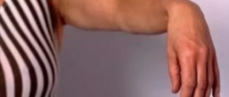
Typically, such babies have motionless eyes and often tilt their heads back. The main sign of pathology is a white strip of sclera located between the iris and the upper eyelid. It is easy to notice when the child looks down. Often this is the only sign.
If no further symptoms are observed, you can expect that the disease will soon disappear on its own. If, in addition to the white stripe above the pupil, other signs are observed, it is necessary to show the baby to the pediatrician as soon as possible. There can be many signs that accompany the main symptom.
Namely:
- wandering gaze;
- decreased tone of the arms and legs;
- reduced visual acuity;
- slightly trembling body;
- frequent regurgitation;
- quiet crying for no apparent reason;
- cyanosis of the skin between the upper lip and nose, as well as on the arms and legs;
- pronounced descending strabismus;
- open sutures of the skull;
- convulsions that appear in attacks;
- throwing the head back;
- frequent, shallow breathing;
- increased head diameter;
- too prominent fontanelle;
- marble skin color.
The setting sun symptom in newborns is characterized by 2 main manifestations:
- If it is too pronounced and is always observed, it can be assumed that the cause of the pathology is damage to the brain and nervous system.
- If the expression is less pronounced and the signs appear rarely, this will not be considered a disorder.
The symptom of the setting sun in newborns is symptoms of the disease.
The influence of the brain on eye motor disorders can be caused by organic changes or damage to the 3rd and 4th pairs of cranial nerves. They are responsible for the downward movement of the pupils: for lowering the gaze.
The development of setting sun syndrome occurs under the influence of many factors that have a negative impact on the nervous system that has not yet had time to strengthen, thereby provoking the appearance of pathology.
As the child’s body grows and develops, the central nervous system gradually adapts to the conditions, as a result of which the disease disappears.
It is necessary to remember: setting sun syndrome can develop not only in children. Its symptoms often appear in some adults.
In this case, the list of main features will be as follows:
- frequent headaches that occur at night;
- dizziness that occurs in attacks throughout the day;
- slow thought process;
- difficulty looking down;
- weakened memory and impaired concentration;
- hypertonicity of the muscles of the arms and legs;
- general lethargy of the body and frequent desire to sleep;
- involuntary attempts to walk on toes;
- manifestation of signs of dyspepsia, which include nausea and vomiting;
- development of diplopia: a condition in which there is double vision.
The development of pathology is based on the defective functioning of the nervous system. Cerebrospinal fluid can collect in the brain in large quantities. Its total volume is approximately 150 ml.
The body produces up to 180 ml per day. The rate of production depends on the perfusion pressure in the brain. The rate at which fluid is absorbed is influenced by venous and intracranial pressure. The increased volume of cerebrospinal fluid is the result of a violation of normal outflow.
There are 2 types of setting sun syndrome:
- The 1st appears when the position of the body changes. It disappears on its own a few weeks after the baby is born;
- Type 2 – when symptoms appear, regardless of the position of the patient’s body. In this case, medical attention is required.
Syndrome or symptom?
Very often, many people confuse the concept of symptom and setting sun syndrome. A syndrome is a whole complex of symptoms that indicate certain disturbances in the functioning of the body.
Most newborns have a wandering gaze, and this is due to the fact that their nervous system is not yet fully developed. The baby needs a little time to adapt to the world around him. As a rule, before the 21st day of birth, the wandering gaze disappears. If the pediatrician made a note in the medical history about the presence of setting sun syndrome in the baby, then there is no need to worry, this is done only in order not to miss the pathology.
Also, do not confuse the syndrome with Gefre's syndrome. Indeed, these two phenomena have quite similar symptoms, but have different factors of genesis. Today, the pathology, described back in the 19th century by the ophthalmologist Gefre, is called hydrocephalic syndrome and is classified as a neurological pathology in which degeneration of nerve cells in the brain is observed in newborns. Typically, such children lack mobility in their gaze; they throw their heads back.
In light of this, when we talk about the “setting sun,” this does not mean at all that the child has a pathology. 99% of children experience a wandering gaze at the beginning of life. Such a symptom may be associated with a genetic feature, for example, if one of the relatives had such an anomaly. In such cases, no treatment is required.
If the child was born prematurely, the syndrome can be observed for a longer period of time, up to approximately 28 days from birth. In very rare cases, the defect appears at a later age. The syndrome may appear against the background of certain infectious diseases, due to head trauma or metabolic disorders.
Causes
The symptom of a setting sun in a newborn is an individual feature of the structure of the eyes or insufficient maturity of the nervous system. After some time, the pathology disappears on its own, so young parents should not worry too much about the child’s condition. However, visits to the doctor should not be neglected.
One of the causes of setting sun syndrome is hypoxia, which can result from birth injuries.
The following factors can influence the development of the setting sun symptom:
- pathologies in the mother that appeared late in pregnancy;
- worsened chronic diseases during pregnancy;
- ischemia;
- infectious diseases.
In addition, setting sun syndrome can also occur as a result of the following diseases suffered by the baby:
- meningitis;
- disrupted functioning of the endocrine system;
- brain cyst;
- injuries received during birth;
- spinal cord injuries;
- disruptions in the hormonal system;
- neoplasms in the brain;
- metabolic disorders;
- fetal hypoxia.
Often, setting sun syndrome begins to develop as a result of premature or, conversely, late birth. Heredity plays a significant role. Many diseases that the mother suffers from increase the risk of this syndrome in the baby.
Often, signs of setting sun syndrome appear in a baby who was born before the 36th or after the 42nd week. If signs of setting sun syndrome appear in adults, it means that it is acquired.
Most often, pathology is caused by the following factors:
- metabolic disorder;
- head injuries;
- infectious diseases.
The risk group includes people with a weakened nervous system. It may function poorly or be underdeveloped.
Additional symptoms requiring immediate medical attention
Suspicion that a child has Gefre's syndrome may arise when the baby makes involuntary movements of the eyes, which he can move while in both a horizontal and vertical position.
If the baby has problems with the vegetative-vascular or cardiovascular system, then blue discoloration of the limbs and the area between the nose and lips may be observed. The skin may acquire a marbled color if the child has problems with blood supply.
In cases where a pathology is actually diagnosed in a newborn, cerebrospinal fluid can provoke the development of severe diseases, such as coma, paralysis and epilepsy. If cerebrospinal fluid penetrates into the venous system, the child may lose hearing, vision, and there will be obvious deviations in the development of the psyche and physiology.
Treatment methods
Before starting therapy, parents should make sure that the observed signs actually indicate setting sun syndrome. It is necessary to look at the corresponding photographs and understand what the symptoms look like. After looking at the photo, you need to pay attention to your child’s eyes.
When the first signs appear, you should consult a doctor. To accurately identify the signs of sundowning syndrome, it is necessary to conduct a diagnosis.
Diagnostic measures should begin with a visual examination. Since the pathology has many striking distinctive features, a specialist will be able to accurately identify it. To do this, the patient must undergo hardware and laboratory diagnostics.
In this case, the following methods are used:
- Blood and urine examination.
- Lumbar puncture.
- Determination of visual acuity and fundus examination.
If the disease is detected, specialists will use instrumental diagnostics:
| Method | Description |
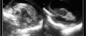
| Ultrasound test | Allows you to study the brain without radiation. It is performed on infants to study the structure of the brain and anatomical formations. The technique is safe and painless, so it is ideal for repeated use. |
| X-ray of the skull | Required to assess the condition of the bones. This procedure provides little information, but in some cases it cannot be avoided. With its help, you can choose the right treatment tactics and monitor the effectiveness of the measures taken. |
| Dopplerography | This method provides a lot of information and allows you to study the vessels of the brain, assessing the rate of blood flow to it. This procedure is safe. |
| CT and MRI | They make it possible to identify defects and fractures that have appeared in bone tissue, but do not make it possible to identify vascular diseases. |
| Rheoencephalography | Vascular examination. |
| Electroencephalography | A technique aimed at identifying tumors and epilepsies. Through the electrodes used, it is possible to obtain the necessary information about the electrical activity of the brain. |
After an accurate diagnosis is made, the child is transferred under the close supervision of a doctor. If sunburn symptoms are present, your pediatrician may prescribe conservative treatment. At home, you can massage your child and do gymnastics with him.
A mandatory requirement is to maintain the correct daily routine. Situations in which the child’s intracranial pressure may increase should not be allowed. A child suffering from this pathology will benefit from water procedures: swimming and diving.
Sometimes treatment with drugs belonging to the group of nootropics may be required. They have a positive effect on brain circulation. Treatment with diuretics is also possible.
The symptom of a setting sun in a newborn with the underlying cause of intracranial injury will require immediate medical intervention. First, a neurosonography procedure will be performed, which will allow us to examine the structure of the brain. After this, a decision will be made on the necessary treatment.
Signs of brain damage also include nystagmus, a disorder of vascular nature. With such a symptom, the pediatrician suspects vascular lesions in the head or cervical spine.
You can overcome the disease if you approach its treatment comprehensively. As a rule, pathology is treated in special neurological centers.
What is it and how is it dangerous for the child’s health?
Graefe syndrome is a very serious disease of neurological origin, which is caused by paralysis of the eye muscles. In the presence of such a pathology, the baby experiences a lag of the upper eyelid from the iris itself.
The reason for this phenomenon lies in the low level of development of the newborn’s nervous system. In addition, there may be convulsions, severe squint, a weakened muscular system, which can be judged by the child’s lack of active movements of the limbs, and other signs.
In the case under discussion, it is necessary to include long-term and thoughtful treatment in order to prevent the situation from worsening and to avoid a threat to the health and life of the baby.
As for Graefe's symptom in infants, it consists of a once-noted manifestation of the syndrome. The main sign of this pathology is the lag of the child’s upper eyelid from the eyeball when it is working.
This phenomenon can be noticed when the baby lowers his eyes down, but the eyelid still remains slightly raised and does not completely close the eye.
Medicines
Drug treatment is based on the use of the following drugs:
- diuretics. These are substances that effectively remove excess water from the body. A common remedy is Furosemide;
- nootropics. They normalize the activity of the nervous system and neurometabolic processes occurring in the brain;
- vitamins. As a rule, these are complexes aimed at supporting the body. The most famous of them are Kombilipen and Milgamma;
- agents that increase vascular tone . They normalize blood flow to the brain. The most famous examples are Actovegin and Curantil;
- antibiotics and antiviral agents . Prescribed only when absolutely necessary;
- sedatives . These include Tazepam, Diazepam.
The doctor should choose the medicine based on the results of diagnosis and subsequent monitoring of the patient.
If a patient experiences a severe attack of the syndrome, emergency treatment is necessary. This may be a dehydration procedure in which Lasix is injected into the muscles. Instead, other means that have a similar effect can be used.
Preventive actions
You can reduce the risk of your baby developing serious pathologies even at the stage of pregnancy planning. To do this, parents should fully prepare the body, treat existing diseases, take vitamins, and give up bad habits.
During pregnancy, the expectant mother must be attentive to her health, follow all doctor’s instructions, correctly distribute work and rest schedules, eat right and remain calm.
After the birth of a child, you should be attentive to his health, do not ignore warning signs, visit specialists monthly for examination and follow their recommendations.
The adult population is also susceptible to the disease, so if you suspect a pathology, you should seek medical help. To reduce the risk, it is recommended to give up bad habits, correctly distribute work and rest, eat well, strengthen the immune system, avoid traumatic brain injuries and promptly treat infectious diseases.
Graefe's syndrome (Graefe's disease, hydrocephalic syndrome) is a neurological disease characterized by bilateral paralysis of the extraocular muscles against the background of degenerative processes in nerve cells. Graefe's symptom (setting sun symptom) is one of the manifestations of the syndrome. This name is due to the special position of the eyes, in which the child seems to be looking down, and part of the iris is located behind the lower eyelid. This pathology is most often detected in newborns, but sometimes the disease affects adults.
Treatment using folk remedies
In newborns, traditional medicine helps normalize the condition of the setting sun symptom. However, these prescriptions should not be considered as primary therapy. They should be used simultaneously with drugs of official medicine in order to increase efficiency.
The following folk remedies help to successfully overcome the signs of setting sun syndrome:
- boiled plantain leaves (you can use both dry and fresh);
- mulberry decoction;
- mint infusion with the addition of a small amount of eucalyptus essential oil and hawthorn berries;
- lemon;
- garlic;
- infusion of string and nettle.
All of these remedies normalize intracranial pressure and effectively combat the symptoms of pathology.
Let's understand the terminology
Hernia in children: how to determine, photo of hernia in newborn boys
Graefe syndrome - in modern medical practice, the diagnosis is understood as a severe neurological disease associated with bilateral paralysis of the eye muscles, which occurs as a result of degenerative processes of a number of nerve cells. The syndrome requires many months of consistent treatment, otherwise severe consequences for human health and life may develop.
Graefe's disease is identical to the concept of syndrome.
Graefe's symptom is a single manifestation of the syndrome of the same name. The symptom is that the upper eyelid lags behind the eyeball in its functioning. This is noticeable when the eyeball is lowered down - the eyelid remains raised, as a result of which the eye does not close completely.
The phenomenon is also called the “setting sun symptom.” Unlike the syndrome, the symptom is not life-threatening and often manifests itself because the development of the infant’s unequal system is defective. It is likely that the symptom will gradually disappear spontaneously as you grow older.
Possible consequences and prognosis
In most cases, even without treatment, the symptoms of setting sun syndrome in a baby disappear on their own. The child's eyeball may return to normal within a month. Sometimes it may take a little longer.
If no negative factors are observed during the development of the newborn, then the nervous system will soon fully recover and all signs will disappear.
However, there are times when symptoms do not go away and other problems may appear:
- vomit;
- frequent crying for no reason;
- tense fontanelle at rest;
- weakening of muscles;
- throwing the head back;
- strabismus.
In all of the above cases, the doctor prescribes additional tests.
If symptoms become more severe over time, surgery may be considered. In particular, neuroendoscopic surgery can be performed; another surgical option is bypass surgery.
How to Test a Patient for a Symptom
If in a pronounced case the symptom is easily observed in a calm state, then in a doubtful case, “sunset” is especially indicative when rotating the head with the eyes following an attractive object.
The gaze cannot fixate on an object slowly moving downwards, and the eyes periodically “float” down and partly inward, exposing a strip of sclera of inadequate width.
The combination of a positive test with nystagmus and convergent strobism should be the reason for a more in-depth neurological study.
Neuroendoscopic surgery
The essence of this operation is to perforate the bottom of the third ventricle. The risks of any complications in this case are minimal. However, the expected effect is not easy to achieve, so after the operation is completed, a shunt will need to be installed after some time.
Bypass surgery
Shunting is the installation of a special system that prevents the retention of cerebrospinal fluid in the brain. It gradually passes into the atrium or into the abdominal cavity.
Currently, bypass surgery is the most effective method of getting rid of the symptom of the setting sun and its secondary manifestations. However, this procedure is too risky. After the shunt is installed, the child is entitled to disability, because a foreign body will now be present in his body.
The main danger of shunting is that the installed shunt can stop performing its main function at any time. Cerebrospinal fluid will again begin to accumulate in the brain and surgical intervention will be immediately required.
Complications
If the necessary measures are not taken in a timely manner, setting sun syndrome, accompanied by additional symptoms, can lead to various serious complications.
The most common illnesses that appear are:
- loss of vision;
- hearing loss;
- slow development of muscles and nervous system;
- epilepsy;
- coma;
- severe forms of paralysis;
- urinary incontinence.
In some cases, death is possible. For children, the prognosis for the course of pathology in most cases is positive. After some time, the blood pressure in the child’s body will return to normal. If this disease is observed in an adult, then it is necessary to take the patient more seriously.
In this case, the risk of serious complications increases significantly. Treatment should be started as soon as possible. The sooner the right steps are taken, the greater the likelihood that serious health problems will be successfully prevented.
With timely diagnosis and proper treatment, setting sun syndrome in a newborn will have a favorable outcome. The main condition is compliance with the pediatrician’s recommendations.
What is Sunset Syndrome in Infants?
Sometimes in infants, and especially premature ones, a thin white stripe appears above the iris.
It is noticeable when the child lowers his eyes down, looks up, or is surprised by something. Lagging movement of the eyelids from the apple in a baby does not cause discomfort and is considered only one of the symptoms of pathology. With Graefe's syndrome, it is difficult to remove cerebrospinal fluid from the brain; the fluid that accumulates in the ventricles interferes with the functioning of the organ, which makes itself felt:
- impaired reflexes;
- blue skin near the lips;
- marbled limbs;
- pronounced strabismus.
The baby often spits up, has trouble holding his head up, and suffers from seizures. When a child is stressed, his chin trembles and his eyes wander.
results
A timely diagnosis and urgent pulse therapy with metipred in combination with azathioprine made it possible to quickly relieve acute symptoms and preserve, at least partially, visual functions in these patients.
In our observations, azathioprine was used as a drug that produces fewer side effects compared to other drugs, allowing faster (in a week) to achieve a clinical effect, characterized by a decrease in the intensity and area of intraocular inflammation.
Systemic use of corticosteroids in high doses in our observation demonstrated a clinically obvious immunosuppressive effect, which prevailed over the anti-inflammatory effect of the drug.
We believe that the recurrence of panuveitis in the second patient was associated with the rapid discontinuation of prednisolone and azathioprine, which indicates an underestimation by doctors at the outpatient stage of the clinical picture and insufficient knowledge of the features of pharmacotherapy.
conclusions
1. The use of pulse therapy with metipred is the basis for the treatment of Vogt-Koyanagi-Harada syndrome.
2. Treatment of autoimmune uveitis with small doses of azathioprine in addition to prednisolone can reduce the rate of disease progression and minimize the side effects that occur when using high doses of prednisolone (arterial hypertension, toxic effects on the kidneys).
3. Important in the treatment of patients with Vogt-Koyanagi-Harada syndrome is active interaction with an immunologist and the appointment of pathogenetically oriented therapy, long-term (at least 2-3 years) dynamic observation. Prescribing antibacterial, non-steroidal anti-inflammatory, antihistamine therapy, local administration of corticosteroids to patients with autoimmune panuveitis is inappropriate.
Hydrocephalus in children: just the facts
Hydrocephalus is a disease that occurs due to excessive production and excessive accumulation of cerebrospinal fluid in the intrathecal spaces and/or ventricles of the brain (in the subarachnoid or subdural space), leading to thinning (atrophy) of the medulla and divergence of the skull bones [1–4].
ICD-10 provides the following codes for various types of hydrocephalus: G91 (G91.0 - communicating hydrocephalus, G91.1 - obstructive hydrocephalus, G91.2 - normal pressure hydrocephalus, G91.3 - post-traumatic hydrocephalus unspecified, G91.8 - other types hydrocephalus, G91.9 - hydrocephalus unspecified).
Classification of hydrocephalus
There are several options for classifying hydrocephalus. First of all, a distinction is made between congenital and acquired hydrocephalus.
Congenital hydrocephalus is present at the time of birth of the child and is the result of inflammatory diseases of the central nervous system (CNS), malformations of the brain, as well as intracranial (birth) trauma, hemorrhages in the brain.
Acquired hydrocephalus develops after birth due to etiologically and pathogenetically diverse pathologies (neuroinfections, tumor processes, vascular diseases, traumatic brain injuries, etc.).
The disease can be active or passive. In the first case, there is an increase in intracranial pressure in the presence of dilatation of the ventricles and subarachnoid spaces. With passive hydrocephalus, it is assumed that the described changes are observed in the absence of intracranial hypertension.
There are open and closed forms of hydrocephalus. Open (communicating) hydrocephalus is diagnosed in the absence of disturbances in the connection of the ventricular system of the brain with the subarachnoid space, and closed (occlusive or non-communicating) - when this communication is disrupted.
External and internal forms of hydrocephalus are determined by the localization of the accumulation of cerebrospinal fluid (CSF); External hydrocephalus is an accumulation of CSF in the intrathecal spaces of the brain, internal hydrocephalus is an accumulation of CSF in the ventricles of the brain.
Pathogenetic classification of hydrocephalus: 1) hypersecretory forms (excessive formation of CSF); 2) aresorptive forms (impaired CSF absorption processes); 3) occlusive forms (the presence of an obstacle to the movement of CSF from the source of its formation to the areas of its resorption).
Depending on the existing level of occlusion of the cerebrospinal fluid pathways, five variants of hydrocephalus are considered: 1) occlusion of one or both foramina of Monroe; 2) blockade of the cavity of the third ventricle; 3) stenosis or occlusion of the Sylvian aqueduct; 4) occlusion or non-opening of the openings of the fourth ventricle; 5) violation of the patency of the subarachnoid spaces.
Depending on the achieved level of control over intracranial hypertension, it is customary to distinguish between compensated, subcompensated or decompensated hydrocephalus.
A separate variant of the disease is normal pressure hydrocephalus (“symptomatic”). With it, an increase in intracranial pressure is initially observed (a consequence of excessive accumulation of CSF in the ventricles of the brain) - with or without ventriculomegaly, which gradually decreases, but remains moderately elevated (150–200 mmH2O). There are two types of normal pressure hydrocephalus - idiopathic (cause unknown) and secondary (consequence of subarachnoid hemorrhage, trauma, tumor and/or infection of the central nervous system, complication of neurosurgical intervention, etc.) [1–4].
S. Oi (2010) proposed a “multi-category classification of hydrocephalus”, reflecting modern ideas about this disease (Table) [5, 6].
Etiology and pathogenesis of hydrocephalus
The most important etiological factor of hydrocephalus is intra- and perinatal pathology of the central nervous system. Pregnancy pathology and oxygen starvation of cerebral tissue are considered as etiological causes of congenital hydrocephalus; intrapartum factors leading to hypoxic-ischemic and/or traumatic brain damage; gestational immaturity of brain structures most susceptible to the described damage.
The development of hydrocephalus is caused by inflammatory diseases of the brain and its membranes, as well as intrauterine and neuroinfections, congenital malformations of the central nervous system, vascular pathology, tumors of the brain and spinal cord, traumatic injuries (including intracranial birth injuries), genetic factors, etc.
Cases of hydrocephalus associated with cerebral dysgenesis, brucellosis, mumps and other infections, diffuse villous hyperplasia of the choroid plexus, vascular anomalies, intracranial hemorrhages, etc. have been described.
Post-traumatic hydrocephalus (progressive excess accumulation of CSF in the cerebrospinal fluid spaces and brain matter due to traumatic brain injury) is caused by impaired circulation and resorption of CSF [1–4, 7].
According to C. Schrander-Stumple and JP Fryns (1998), hereditary X-linked (congenital) hydrocephalus occurs in 4% of all registered cases of the disease (according to other data in 5–15% of observations), up to 40% of cases of hydrocephalus are etiogenetically caused [8 ]. Hydrocephalus occurs in Dandy–Walker syndrome, Arnold–Chiari syndrome, etc. At least 43 mutations/loci associated with hereditary forms of hydrocephalus (in humans and laboratory animals) have been described; Under experimental conditions, 9 genes associated with this cerebral pathology were found, while in humans there is only one [1–4].
The progression of hydrocephalus is accompanied by structural and morphological changes in the brain of varying severity: 1) thinning of the cortex and white matter (up to its complete elimination); 2) atrophy of the choroid plexus; 3) atrophy/subatrophy of the basal ganglia, brainstem, cerebellum; 4) severe disorders of capillary blood flow; 5) thickening and/or fusion of the meninges; 6) excessive growth (hypertrophy) of glial tissue. In severe cases, the formation of hydroanencephaly is possible, when there is only ependyma and a thin layer of the pia mater.
The most intensively functioning and highly vascularized periventricular region suffers the most from hypoxia. The consequence of atrophy of the periventricular white matter of the brain is passive expansion of the ventricular system with the formation of ventriculomegaly.
With post-traumatic hydrocephalus, pathological processes in the brain are morphologically characterized by dilation of the ventricular system, periventricular edema and obliteration of the subarachnoid fissures. Obliteration of the CSF flow paths is determined by the following pathogenetic factors: subarachnoid hemorrhage, intracranial hematomas, focal and/or diffuse brain damage, cicatricial adhesions and atrophic processes (including after extensive craniotomy and resection trepanations), meningoencephalitis and ventriculitis. The time frame for the development of post-traumatic hydrocephalus (normotensive, hypertensive or occlusive) usually varies from 1 month to 1 year [1–4, 7].
Clinical syndromology of hydrocephalus
The main symptoms of hydrocephalus (congenital and acquired) are determined by two groups of factors: 1) the causes of the disease; 2) directly hydrocephalic syndrome.
The first group mainly includes focal manifestations (usually in the form of spastic paresis of the ascending type in the lower and/or upper extremities). The severity of group 2 symptoms depends on the form, stage and degree of progression of hydrocephalus. In the congenital form of the disease, signs of hydrocephalus may be present both at birth and appear later - by the age of 3–6 months.
More often, the first sign of the disease is a disproportionately rapid increase in head circumference. To assess it in children, special tables (centile) are used.
In children of the first year of life, there may be a predominance of the “cerebral” parts of the skull over the “facial” (as a result - a forced position with the head thrown back), increased venous pattern and congestion of the saphenous veins of the head, tension of the greater and other fontanelles, divergence of the skull bones, Graefe’s symptom . These symptoms are accompanied by a lag in psychomotor development (of varying severity), and less often in physical development. Optic nipple atrophy is a severe complication of untreated or treatment-resistant progressive hydrocephalus.
Cephalgic syndrome is more typical of acquired hydrocephalus in children over 1 year of age (when the fontanelles and cranial sutures are already closed).
Clinical manifestations of post-traumatic hydrocephalus are characterized by neurological and mental disorders caused by primary brain injury. In part, they are a reflection not so much of hydrocephalus itself, but rather a consequence of a traumatic brain injury (TBI) or premorbid pathology.
A. P. Konovalov et al. (1999) distinguish three variants of post-traumatic hydrocephalus: 1) against the background of resolved or mild residual symptoms of severe TBI, with dominance in the clinic of any specific symptom complex; 2) against the background of slowly resolving severe symptoms of severe TBI with the addition of intellectual-mnestic and ataxic syndromes; 3) against the background of a vegetative state (which prevents exit from it). Hypertensive and occlusive post-traumatic hydrocephalus (excluding normotensive hydrocephalus) is characterized by headache, vomiting, and dizziness. All patients exhibit psychopathological symptoms (intellectual-mnestic disorders, euphoria or lethargy, aspontaneity, akinetic mutism, etc.); as well as gait disturbances and ataxia (with a characteristic “sticking of the feet to the floor”), dysfunction of the pelvic organs.
With normal pressure hydrocephalus, there are usually no classic symptoms characteristic of congenital or acquired hydrocephalus, but since ventriculomegaly has a negative effect on adjacent areas of cortical tissue, the disease has its own characteristics (classic triad: gait disturbances, urinary incontinence, decreased intelligence of varying degrees of severity) [ 1–4].
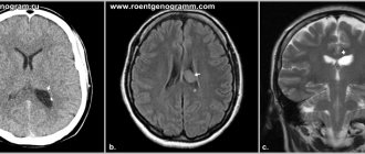
Diagnostics and examination methods
Establishing a diagnosis of hydrocephalus (in addition to physical data) is based on neuroimaging data (neurosonography - with an open fontanel, computed tomography and magnetic resonance imaging of the brain - CT and MRI), which are considered in conjunction with the symptoms of the disease described above. These neuroimaging methods have replaced the previously used skull radiography. It is resorted to only in rare cases; Survey radiography of the skull allows one to indirectly judge secondary changes in the bones of the skull (in the absence of CT and MRI).
It is especially important to monitor the adequate functioning of shunts installed during neurosurgical intervention. At the slightest suspicion of shunt failure, the patient should be referred to a neurosurgeon.
Isotope cisternography with the introduction of a radioactive isotope (during a lumbar puncture) and subsequent observation of the removal of CSF from the brain makes it possible to establish the diagnosis of normal pressure hydrocephalus.
Diaphanoscopy (transillumination of the skull) has now been practically abandoned. Lumbar puncture is a traditional research method that allows you to assess blood pressure and analyze the CSF. An increase in echo pulsation up to 70–80% during an ultrasound examination of the brain is not a diagnostic sign of hydrocephalus.
The study of auditory evoked potentials often reveals their disturbances, which indicates a special sensitivity of the brain stem to intracranial hypertension.
An ophthalmological examination of the fundus allows one to identify changes characteristic of hydrocephalus (congestion in the fundus, atrophic processes, signs of inflammation, as well as hemorrhage, changes in the tone and caliber of blood vessels, etc.), which makes it possible to assess the course of the pathological process.
If a congenital infection (intrauterine infection) is suspected, serological and virological studies are justified.
Abroad, a method of non-invasive monitoring of intracranial pressure through the large fontanel in children of the first year of life, based on the principle of applanation, is used. The measurement is made using a special device (fontanometer), and the study itself is called “phonogram” or “fontanometry” [1–4, 7].
Differential diagnosis
Hydrocephalus is differentiated from the following main pathological conditions: subdural intracranial hemorrhage, megalencephaly (primary), hydroanencephaly, meningitis, brain tumors, familial (constitutional) macrocephaly, vitamin D-deficient rickets, rickets of premature infants, etc.
Less common are other types of pathology from which it is necessary to differentiate hydrocephalus: achondroplasia, Soto syndrome (cerebral gigantism), numerous so-called “neurocutaneous” syndromes, a group of leukodystrophies (Alexander’s disease, Canavan’s disease, globoid and methochromatic forms of leukodystrophies), gangliosidosis, mucopolysaccharidosis (Tay’s disease –Sachs and Sandhoff), urine disease with the smell of maple syrup, etc. [1–4, 7].
Treatment
Therapeutic measures for progressive hydrocephalus are divided into surgical and therapeutic (drug and non-drug).
If there are signs of an ongoing inflammatory process in the central nervous system, children are prescribed appropriate therapy (antibacterial agents, specific drugs, as well as glucocorticosteroids and human intravenous immunoglobulins - according to indications).
Progressive forms of hydrocephalus (occlusive), as a more serious variant of the pathology, require timely neurosurgical intervention (shunt surgery). The purpose of surgery is to create an adequate outflow of CSF using special shunts (drainage systems made of synthetic materials). There are shunts that divert CSF from the ventricles of the brain to various loci of the body (ventriculoperitoneal, lumboperitoneal, ventriculoatrial). Thus, some shunts transport excess accumulation of cerebrospinal fluid into the peritoneal cavity, others - into the right ventricle of the heart. Ventriculoperitoneal shunting is the main method of neurosurgical treatment of hydrocephalus in the Russian Federation and abroad.
There is a method of surgical treatment called “endoscopic third ventriculostomy” (perforation of the lower part of the third ventricle of the brain to eliminate the blockage of CSF flow and increase its outflow); another name for the method is “endoscopic ventriculocisternostomy of the bottom of the third ventricle.” The purpose of the described operation is to create pathways for the outflow of CSF from the third ventricle into the cisterns of the brain, through which the resorption of cerebrospinal fluid occurs.
Other types of endoscopic treatment of hydrocephalus in various clinical situations are aqueductoplasty, ventriculocystocysternostomy, septostomy, endoscopic removal of an intraventricular tumor, as well as endoscopic installation of a shunt system.
Among the neurosurgical procedures in infants, ventricular puncture (removal of CSF from the ventricles of the brain through the fontanelle) can be used. This external drainage procedure is used extremely rarely, as it can be accompanied by a large number of complications (infection, etc.).
There are isolated reports of the use of intradural spinal endoscopy in the treatment of hydrocephalus and associated conditions in children.
In the acute occurrence of intracranial hypertension, drugs with a diuretic effect are used. These include: furosemide (intramuscular) - sometimes in combination with a solution of magnesium sulfate, glycerol (per os), mannitol (intravenous drip).
If long-term treatment of hydrocephalus is necessary, acetazolamide (Diacarb) is first prescribed, a diuretic from the group of carbonic anhydrase inhibitors (an enzyme that catalyzes the reversible reaction of carbon dioxide hydration and subsequent dissociation of carbonic acid). The action of acetazolamide is associated with the suppression of carbonic anhydrase in the ventricular plexuses of the brain and with a decrease in CSF production (hypolyquor effect). The side effects of acetazolamide (acidosis and shortness of breath) are systematically corrected by the administration of sodium bicarbonate (0.5 g 3 times a day).
Patients with hydrocephalus under the age of 3 years need to be prescribed vitamin D and calcium supplements. In addition to calcium, supplementation of potassium and magnesium preparations (Asparkam, Panangin) is absolutely necessary during acetazolamide therapy.
Symptomatic treatment for hydrocephalus is determined by individual indications and usually includes: massage, physical therapy, various types of physiotherapy, biofeedback method, as well as stimulating therapy (nootropic, metabolic, vascular drugs, etc.), etc. Anticonvulsants for children with hydrocephalus prescribed if there are appropriate indications (symptomatic epilepsy, etc.).
Neurodietological measures should be aimed at maintaining nutritional status, adequate fluid intake, supplementation of vitamins and minerals, and, if necessary, nutritional support (clinical nutrition: enteral and/or parenteral) [2–4].
Prevention
Since in a significant number of cases hydrocephalus can be diagnosed during fetal development, sonographic examination of the fetus is recommended (starting from the 17th week of gestation). In some cases, MRI examination is indicated, although P. Peruzzi et al. (2010) indicate that it does not have pronounced diagnostic advantages compared to ultrasound methods [9].
In order to prevent hydrocephalus in children, timely detection and treatment of intrauterine infections in their mothers is necessary. It is necessary to fully implement the prevention of childhood injuries and neuroinfections. To prevent myelomeningocele, preventive administration of folic acid supplements is indicated [1–4, 7].
Forecast
Hydrocephalus is a cerebral pathology associated with significant neurological deficits, potential disability and decreased quality of life. There is a risk of irreversible changes in the nervous system and a decrease in intelligence (even mental retardation).
With congenital and acquired hydrocephalus, the prognosis is determined by the early onset and adequacy of treatment (pharmacological or neurosurgical).
A serious complication of hydrocephalus can be epilepsy, induced both by hydrocephalus itself (structural and morphological changes in brain structures) and by the installation of a drainage shunt [1–4, 7].
According to M. Mataro et al. (2001), the average IQ values in patients with hydrocephalus are reduced in a number of verbal and nonverbal functions. Among patients who received adequate therapy, normal levels of intellectual function are observed in 40–65% of cases [10].
Literature
- Kliegman RM, Stanton BF, St Geme III JW et al., eds. Nelson textbook of pediatrics. 20 th ed. Philadelphia. Elsevier. 2016. 3474.
- Shamansurov Sh. Sh., Studenikin V. M. Congenital and acquired hydrocephalus. Ch. 11. In the book: Neurology of early childhood. Tashkent: O'Qituvchi, 2010. 156–164.
- Studenikin V. M., Shamansurov Sh. Sh. Congenital hydrocephalus. Ch. 9. In the book: Neonatal neurology. M.: Medforum, 2014. 120–135.
- Studenikin V.M., Shelkovsky V.I., Kuzenkova L.M. Hydrocephalus and hydrocephalic syndrome in children // Doctor.ru. 2006; 5:2–5.
- Oi S. A proposal on “Multi-categorical Hydrocephalus Classification”: McHc. Critical review in 72,576 patterns of hydrocephalus // J. Hydrocephalus. 2010; 2 (2): 1–21.
- Oi S. Hydrocephalus research update: controversies in definition and classification of hydrocephalus // Neurol. Med. Chir. (Tokyo). 2010; 50:859–869.
- Kandasamy J., Jenkinson MD, Mallucci CL Contemporary management and recent advances in pediatric hydrocephalus // BMJ. 2011; 343:d4191.
- Schrander-Stumple C., Fryns JP Congenital hydrocephalus. Nosology and guidelines for clinical approach and genetic counseling // Eur. J. Pediatr. 1998; 157(5):355–362.
- Peruzzi P., Corbitt RJ, Raffel C. Magnetic resonance imaging versus ultrasonography for the in utero evaluation of central nervous system anomalies // J. Neurosurg. Pediatr. 2010; 6 (4): 340–345.
- Mataro M., Junque C., Poca MA et al. Neuropsychological findings in congenital and acquired childhood hydrocephalus // Neuropsychol. Rev. 2001; 11 (4): 169–178.
V. M. Studenikin, Doctor of Medical Sciences, Professor, Academician of the Russian Academy of Economics
LLC NPSMC "Dream Clinic", Moscow
Contact Information
Hydrocephalus in children: just the facts / V. M. Studenikin
For citation: Attending physician No. 4/2018; Issue page numbers: 66-69
Tags: cephalalgia, brain, cerebrospinal fluid, intracranial hypertension
Causes of occurrence and mechanism of development
Modern medicine today believes that the main reason for the development of respiratory failure remains the immaturity of the lungs and the still imperfect functioning of the surfactant.
It may be that there is enough surfactant, but there is a defect in its structure (normally it consists of 90% fat, and the rest is protein), which is why it does not cope with its purpose.
The following factors may increase your risk of developing RDS:
- Deep prematurity, especially for children born before the 28th week.
- If the pregnancy is multiple. The risk exists for the second baby of twins and for the second and third of triplets.
- Delivery by caesarean section.
- Large blood loss during childbirth.
- Serious illnesses in the mother, such as diabetes.
- Intrauterine hypoxia, asphyxia during childbirth, infections (intrauterine and not only), such as streptococcal, which contributes to the development of pneumonia, sepsis, etc.
- Meconium aspiration (a condition when a child swallows amniotic fluid with meconium).