Types of paroxysmal activity
Paroxysmal states in neurology are the process of an increase in the amplitude of brain activity on the electroencephalogram. An interesting fact is that the amplitude of the waves not only increases sharply, but also does not appear chaotically. In addition to the waves themselves, the source of their origin is also recorded. Sometimes some doctors deliberately narrow paroxysmal activity to epileptic seizures, however, this is not true.
An interesting fact is that a child may have paroxysms as a variant of the norm, since paroxysmal activity of the brain will not be supported by pathological changes in the structures of the brain.
For adults, it makes sense to talk about paroxysms as a pathological process occurring in the cerebral cortex. If we talk about paroxysm, as the most general concept, we can summarize the following: paroxysm is an intensified attack, occurring at the maximum of its tension and repeating a certain number of times.
Thus, the paroxysmal state will have the following characteristics:
- in the cerebral cortex there is an area with excitation processes that prevail over inhibition processes;
- excitation of the cortex is characterized by a sudden onset and an equally unexpected decline in activity;
- When studying brain impulses, a characteristic pattern is noticeable on the electroencephalogram, in which one can trace the waves reaching their highest amplitude.
Taking into account all the characteristics, the phenomenon of paroxysmal states is classified into two large categories - epileptic and non-epileptic.
Epileptic type of activity is manifested in a sick person by typical conditions - seizures that appear from time to time. These are convulsive conditions that occur with a certain frequency, and sometimes repeat one after another.
Epilepsy can be a congenital pathology, but it can also be acquired if a person has suffered a severe traumatic brain injury, suffers from a brain tumor, intoxication, or has experienced conditions of severe ischemia. Epilepsy, in turn, is also divided into convulsive and non-convulsive; the picture of such conditions is very diverse.
What it is
Paroxysmal activity is a value that is recorded on the electroencephalogram. Paroxysmal brain activity is a change in the normal wave, and is manifested by peaks, peaked waves, pathological complexes and a slowdown in the electrical activity of the brain.
In a broad sense, paroxysmal activity is abnormal electrical activity of the brain.
The focus of paroxysmal activity occurs in many pathological human conditions:
- Neurotic (depression, social phobia, panic attacks) and severe mental (schizophrenia) disorders.
- Immaturity of the brain.
- Epilepsy and epileptiform disorders.
- Dementia.
- Severe intoxication with drugs, alcohol, metals.
- Encephalopathy.
- Chronic stress, severe physical fatigue or mental exhaustion.
- Increased intracranial pressure.
- Psychopathic personality changes.
- Autonomic disorders.
When paroxysmal activity is recorded, doctors mean by this a phenomenon in which the processes of excitation in the cortex and subcortex greatly predominate over the processes of inhibition. Signs of paroxysmal activity: sudden onset, transience, sudden termination and tendency to relapse.
On the electroencephalogram, paroxysmal brain activity appears as a series of waves, the amplitude of which quickly approaches a peak. Paroxysmal activity covers EEG rhythms: alpha, beta, delta and theta rhythms.
To study the nuances, doctors compare peaked waves with normal ones. Fundamental indicators of activity are considered: basic activity, symmetry, rhythm, amplitude. Changes in activity during brain stimulation are also recorded: light or sound stimulation, a state of hyperventilation, opening or closing the eyes.
Paroxysmal syndrome
In order to understand the essence of this diagnosis, you need to understand some terms. By paroxysm, or attack, we mean a transient dysfunction of any system or organ that occurs suddenly. This condition is divided into two main types: epileptic and non-epileptic.
But speaking generally, we are talking about a situation where a certain painful attack sharply intensifies to the highest degree. In some cases, the term “paroxysmal state” is used to describe recurrent symptoms of a specific disease. We are talking about health problems such as swamp fever, gout, etc.
In fact, paroxysms are a reflection of emerging dysfunction of the autonomic nervous system. The most common causes of such attacks are neuroses, hypothalamic disorders and organic brain damage. Crises may be accompanied by migraines and attacks of temporal lobe epilepsy, as well as severe allergies.
Despite the fact that there are several forms through which the paroxysmal state manifests itself, symptoms with similar characteristics can be found in all cases. We are talking about the following signs: stereotypicality and tendency to regular relapses, reversibility of disorders and short duration. Regardless of the background of what disease the paroxysm made itself felt, these symptoms will be present in any case.
Analysis of the rhythm of bioelectric impulses
Biorhythms of the brain are divided into several groups, each of which is named after its Latin letters. So, there are alpha rhythms, beta rhythms, theta and delta rhythms. Depending on the identified rhythm of activity, one can assume what pathology such impulses are associated with.
When deciphering an electroencephalogram, rhythms are the main focus. When reading diagnostic results, it is very important to take into account the symmetry of the appearance of activity in both hemispheres, the basal rhythm, and changes in rhythms during functional loads on the body.
Alpha rhythms normally have an oscillation frequency of 8 to 13 Hertz (Hz). The amplitude of normal oscillations is up to 100 μV. Rhythm pathologies are spoken of in the following cases:
- if the rhythm is associated with neuroses of the third type;
- with interhemispheric asymmetry of more than a third, there is reason to talk about a tumor or cystic neoplasm, a post-stroke condition with tissue scarring, or a previously suffered hemorrhage in this location;
- if the rhythm is unstable, doctors suspect a concussion.
Amplitude disturbances are also recognized as a sign of pathology. Although officially it is the maximum possible 100 μV, but with a value less than 20 and more than 90 units, doctors already suspect pathological abnormalities.
Beta rhythms are also present during normal brain activity and, under certain parameters, do not at all indicate a paroxysmal state. This rhythm is most pronounced in the frontal lobes of the brain.
The amplitude is normally small - from 3 to 5 μV. A normal reserve is an increase in activity by 50 percent, i.e. Even with an amplitude of 7 µV, beta rhythms can be conditionally considered normal, but already when this figure is exceeded they are classified as paroxysmal activity.
For example, waves of this type of diffuse nature with a length of up to 50 µV indicate a concussion. Short spindle-shaped waves will indicate the presence of encephalitis - an inflammatory disease of the meninges, and the frequency and duration of the wave’s existence illustrate the severity of the inflammatory process.
When beta-active waves with a high amplitude of about 30-40 μV and a frequency of about 16-18 Hz are detected in a child, they speak of delays in the child’s mental development.
Theta and delta waves are predominantly recorded in humans during sleep. Therefore, when examined by a doctor while awake, they are not normally recorded. If such waves appear, then this indicates degenerative processes in the brain.
A paroxysmal state usually occurs when the brain matter is compressed, so the doctor may suspect swelling of the brain or a tumor. Theta and delta waves differ in that they indicate severe and profound changes in the brain. Like all waves, theta and delta waves before the age of 21 are not considered a pathology, since in children and adolescents they are a variant of the norm.
In people older than this age, the presence of such waves indicates acquired dementia. In parallel, this is confirmed by synchronous flashes of theta waves with high amplitude. In addition, such waves indicate the presence of neurosis.
Terminology and related concepts
In adults (after 21 years), bioelectrical activity of the brain (BEA) should normally be synchronous, rhythmic and not have foci of paroxysms. In general, paroxysm is an intensification to the maximum of any pathological attack or (in a narrower sense) its recurrence. In this case, paroxysmal brain activity means that:
- when measuring the electrical activity of the cerebral cortex using EEG, it is discovered that in one of the areas the processes of excitation prevail over the processes of inhibition;
- the process of excitation is characterized by a sudden onset, transience and sudden ending.
In addition, when checking the state of the brain on an EEG, patients develop a specific pattern in the form of sharp waves that very quickly reach their peak. Pathologies can be observed in different rhythms: alpha, beta, theta and delta rhythms. In this case, additional characteristics can suggest or diagnose the disease. When deciphering and interpreting the EEG, clinical symptoms and general indicators must be taken into account:
- basal rhythm,
- the degree of symmetry in the manifestation of electrical activity of neurons of the right and left hemispheres,
- changing schedules during functional tests (photostimulation, alternating closing and opening of eyes, hyperventilation).
Alpha rhythm
The norm for alpha frequency in healthy adults is 8-13 Hz, amplitude fluctuations are up to 100 μV. Alpha rhythm pathologies include:
- Paroxysmal rhythm, which, as well as weak expression or weak activation reactions in children, may indicate a third type of neuroses.
- Interhemispheric asymmetry exceeding 30% may indicate a tumor, cyst, manifestations of a stroke, or a scar at the site of a previous hemorrhage.
- Disruption of sine waves.
- Unstable frequency - allows you to suspect a concussion after a head injury.
- A permanent shift of the alpha rhythm to the frontal parts of the brain.
- Extreme amplitude values (less than 20 µV and more than 90 µV).
- Rhythm index with a value less than 50%.
Beta rhythm
During normal brain function, it is most pronounced in the frontal lobes. It has a symmetrical amplitude of 3-5 µV. Pathologies are recorded when:
- paroxysmal discharges,
- interhemispheric asymmetry in amplitude above 50%,
- increasing the amplitude to 7 μV,
- low-frequency rhythm along the convexital surface,
- sinusoidal graph.
In this list, a concussion is indicated by diffuse (non-localized) beta waves with amplitudes of up to 50 μV. Encephalitis is indicated by short spindles, the frequency, duration and amplitude of which are directly proportional to the severity of inflammation. For psychomotor delay in the development of a child - high amplitude (30-40 μV) and frequency of 16-18 Hz.
Theta and delta rhythms
These rhythms are normally recorded in sleeping people, but when they occur in people who are awake, they speak of dystrophic processes developing in the brain tissue and associated with high pressure and compression. At the same time, the paroxysmal nature of theta and delta waves indicates deep brain damage. Until the age of 21, paroxysmal discharges are not considered a pathology. But if a disorder of this nature is recorded in adults in the central parts, then acquired dementia can be diagnosed. This can also be evidenced by flashes of bilaterally synchronous high-amplitude theta waves. In addition, paroxysms of these waves also correlate with the third type of neuroses.
Summarizing all paroxysmal manifestations, two types of paroxysmal conditions are distinguished: epileptic and non-epileptic.
Epileptic type of paroxysmal activity
A pathological condition characterized by convulsions and seizures, sometimes repeated one after another, is epilepsy. It can be congenital or acquired as a result of traumatic brain injury, tumors, acute circulatory disorders, or intoxication. Another classification of epilepsy is based on the localization factor of the paroxysmal focus that provokes seizures. Epileptic seizures, in turn, are also divided into convulsive and non-convulsive with a wide typological spectrum.
Grand mal seizure
This type of seizure is most common in epilepsy. Several phases are observed during its course:
- aura,
- tonic, clonic phases (atypical forms),
- confusion (twilight disorder or stupor).
1. An aura is a short-term (counted in seconds) clouding of consciousness, during which the surrounding events are not perceived by the patient and are erased from memory, but hallucinations, affective, psychosensory, depersonalization facts are remembered.
Some researchers (for example, W. Penfield) believe that the aura is an epileptic paroxysm, and the subsequent grand mal seizure is a consequence of the generalization of excitation in the brain. The clinical manifestations of the aura are used to judge the localization of foci and the spread of excitation. Among several classifications of the aura, the most common division is into:
- viscerosensory - begins with nausea and an unpleasant sensation in the epigastric zone, continues with an upward displacement, and ends with a “blow” to the head and loss of consciousness;
- visceromotor - manifests itself in a variety of ways: sometimes - dilation and contraction of the pupil not associated with changes in lighting, sometimes - alternating skin redness and heat with paleness and chills, sometimes - “goose bumps”, sometimes - diarrhea, pain and rumbling in the abdomen;
- sensory – with various manifestations of auditory, visual, olfactory and other characters, dizziness;
- impulsive – manifested by a variety of motor acts (walking, running, violent singing and shouting), aggression towards others, episodes of exhibitionism, kleptomania and pyromania (an attraction to arson);
- mental - where the hallucinatory type manifests itself in visual hallucinations of scenes of holidays, manifestations, disasters, fires in bright red or blue tones, in olfactory and verbal hallucinations, and the ideational type of mental aura - in the form of a thinking disorder (reviews of survivors describe it as “blockage of thoughts ", "mental blocker").
The last, mental, type of aura also includes deja vu (deja vu - the feeling of what has already been seen) and jamais vu (jamais vu - the opposite feeling of never seen, although objectively familiar).
It is important that these disorders fall under the definition of “aura” only if they become precursors to the generalization of a seizure. The transition from an aura to a grand mal seizure occurs without an intermediate stage. If the stage of a convulsive seizure does not occur, then these disorders are classified as independent nonconvulsive paroxysms.
2. Rudimentary (atypical) forms of a grand mal seizure are possible in the form of tonic or clonic phases. Such forms are typical when manifested in early childhood. Sometimes their manifestation is expressed in non-convulsive relaxation of the body muscles, sometimes with a predominance of spasms in the left or right part of the body.
3.
Epileptic state (status) . A dangerous condition that, if prolonged, can lead to the death of the patient due to increasing hypoxia or cerebral edema. Before this, status epilepticus may be accompanied by somatovegetative symptoms:
- temperature rise,
- increased heart rate,
- a sharp decrease in blood pressure,
- sweating, etc.
In this status, seizures of 30 minutes or more follow each other, and this sometimes lasts up to several days, so that the patients do not regain consciousness, being in a stunned, comatose and stuporous state. At the same time, the concentration of urea in the blood serum increases, and protein appears in the urine. Each subsequent paroxysm occurs even before the disturbances after the previous attack have time to subside. Unlike a single seizure, in the case of status epilepticus the body is not able to stop it. For every 100 thousand people, status epilepticus occurs in 20.
Minor seizures
The clinical manifestation of minor seizures is even wider than that of major seizures, which introduces significant confusion in their definition. This is also facilitated by the fact that representatives of different schools of psychiatry put different clinical content into the basic concept. As a result, some consider only those that have a convulsive component to be minor seizures, while others derive a typology that includes:
- typical – absence seizures and pycnoleptic – petit mal seizures,
- impulsive (myoclonic), and retropulsive,
- akinetic (which includes pecks, nods, atonic-akinetic and salam seizures).
- Absence seizures are conditions associated with a short-term sudden loss of consciousness. This may look like an unexpected interruption in conversation in the middle of a sentence, or an action "in the middle" of the process, the gaze begins to wander or stops, and then the process continues from the point of interruption. Sometimes, at the time of an attack, the tone of the muscles of the neck, face, shoulders, arms changes, and sometimes mild bilateral muscle twitching and autonomic disturbances occur. As a rule, such seizures end by age 10 and are replaced by grand mal seizures.
- Impulsive (myoclonic) seizures. They manifest themselves as unexpected shuddering with jerky movements of the hands, bringing them together and spreading them apart, in which a person cannot hold objects. In the case of a longer seizure, consciousness turns off for a few seconds, but quickly returns and, if a person falls, he quickly gets to his feet on his own. The basis of such movements, which can be repeated in “volleys” of 10-20 for several hours, is the “anti-gravity reflex” - exaggerated straightening.
differs in specific movements directed forward (propulsion). The resulting movement of the torso or head is explained by a sharp weakening of postural muscle tone. It occurs more often at night in boys under 4 years of age. Later, large convulsive seizures appear along with them. At the same time, nodding and pecking - sharp tilts of the head forward and down - are more typical for children under 5 months. Another type - Salaam seizures - got their name by analogy with the position of the arms, body and head, which are characteristic of a person bowing in a Muslim greeting.
Akinetic (propulsive) type
One person never experiences seizures of different clinical natures or a transition from one type to another.
Focal seizures
This epileptic form has three types:
- Adversive convulsive . It is distinguished by a specific rotation of the body around its axis: the eyes turn, followed by the head, and behind it the whole body, after which the person falls. The epileptic focus in this case is located in the anterior temporal or frontal region. However, if the paroxysmal focus is in the left hemisphere, the fall occurs more slowly.
- Partial (Jacksonian) . It is distinguished from the classic manifestation by the fact that the tonic and clonic phases affect only certain muscle groups. For example, a spasm moves from the hand to the forearm and further to the shoulder, from the foot to the lower leg and thigh, from the muscles near the mouth to the muscles of the side of the face where the spasm began. If generalization of such a seizure occurs, it ends in loss of consciousness.
- Tonic postural cramps . When paroxysmal activity is localized in the brainstem, powerful convulsions immediately begin, ending in breath-holding and loss of consciousness.
Nonconvulsive forms of paroxysms
Paroxysms associated with clouding of consciousness, twilight states, dream delirium with a fantastic plot, as well as forms without a disorder of consciousness (narcoleptic, psychomotor, affective paroxysms) are also quite widespread and varied.
– short-term twilight states of paroxysmal nature. A person performs automatic actions, completely detaching himself from the world around him. These can be actions associated with chewing, swallowing, licking (oral automatisms), rotating in place (rotatory automatisms), attempts to shake off “dust”, methodical undressing, running in an uncertain direction (so-called “fugues”). Sometimes aggressive, antisocial behavior is observed with simultaneous complete detachment from the environment.
Outpatient automatisms- Dream (special) states. They appear as dream-like delirium. With them there is no complete amnesia - the person remembers his visions, but does not remember the surrounding environment.
Provoking factors
So, understanding that the basis of such a problem as a paroxysmal state is in fact always cerebral disorders, it is worth paying attention to those diseases that can lead to a sudden deterioration in physical condition, without the appearance of noticeable symptoms before.
It is this fact that allows us to assert that with all the abundance of various pathologies that serve as the background for a crisis, it is almost always possible to trace a single etiological picture.
You need to understand that doctors pay enough attention to this problem, so a study was carried out on the condition of a significant number of patients in order to identify common etiological factors that lead to the occurrence of paroxysms. The examinations were focused primarily on working with diseases such as vegetative-vascular dystonia, migraine, epilepsy, neuralgia and neuroses, etc.
What diseases lead to a crisis?
- Some hereditary diseases actively provoke the appearance of central paroxysms. Of these, the most common are systemic degenerations of the central nervous system (Wilson-Konovalov disease, Tourette's disease), metabolic diseases caused by heredity (phenylketonuria, Gaucher disease, leukodystrophies, glycogenosis, galactosemia), a group of epileptic diseases and others.
- Organic diseases of the nervous system. In the first row are traumatic brain injuries, post-traumatic cerebrovascular diseases, and causalgia. Neoplasms in the brain and spinal cord, vascular pathologies of brain networks, strokes, ischemic diseases, neuralgia of the trigeminal, glossopharyngeal and upper laryngeal nerves.
- The manifestation of paroxysms is characteristic of a number of diseases of the psychovegetative syndrome: vegetative-vascular pathologies, Charlen's syndrome, Slader's syndrome, neuroses, migraines, depressive states, hysteria, affective disorders.
- Paroxysmal conditions in certain diseases of internal organs - congenital heart defects, myocardial infarction, renal failure, uremia, acute hepatitis, hepatic coma, pneumonia, bronchial asthma, malignant lung diseases, blood diseases.
- Diseases of the endocrine system and metabolic disorders - pheochromocytoma, Cushing's disease, menopausal syndrome, hypoxia, hypercapnia;
- Paroxysms are characteristic of a whole range of infectious encephalitis, neurosyphilis, complications after vaccination, parasitic infestations (cysticercosis, echinococcosis).
- They often provoke a paroxysmal state of intoxication due to alcohol and drug poisoning, long-term use of certain medications, and technical poisoning.
Migraine-like paroxysms
Headaches are one of the most common signs of cerebral pathologies. Several main etiological causes contributing to the occurrence of headache have been identified: vascular disorders, muscle tension, liquorodynamic causes, neuralgic etiology, mixed and central.
Each etiological factor is characterized by a separate mechanism for the occurrence of pain, but the basis is always a dysfunction of the nerve cells of the brain. In particular, migraine is characterized by vascular disorders, when high or low blood pressure in the network of cerebral capillaries provides regular insufficient trophism of neurons, or pressure from dilated blood vessels occurs on brain tissue.
Paroxysms during migraine belong to the non-epileptic series and are expressed in the form of regular attacks of pain in the area of one side of the head. The pain is excruciating and very long-lasting, sometimes lasting for several days. A feature of migraine-like paroxysms is their sufficient resistance to treatment - it can be extremely difficult to stop the pain.
An extraordinary feature of migraine is the fact that the paroxysmal state in this pathology can be both a clinical sign and also join the complex of symptoms of other cerebral pathologies. This situation makes it much more difficult to make a correct diagnosis - it is extremely difficult to discern third-party diseases behind migraine attacks.
Diseases with characteristic manifestations of paroxysm
As mentioned above, in the vast majority of cases, a sharp exacerbation of symptoms occurs due to dysfunction of the brain. In addition, manifestations that are directly related to cerebral disorders are often recorded, and this is one of the key features of this condition.
In addition, you need to understand that there are both primary and secondary paroxysmal genesis. Primary is caused exclusively by congenital factors of manifestation, such as disorders in the brain and genetic disposition, which is formed during the development of the embryo. Secondary paroxysm is a consequence of the influence of internal and external factors. It manifests itself already during life.
The peculiarities of such a problem do not end there. Such paroxysmal states are recorded in neurology, which accompany the disease throughout the entire period of its course. Also, a sharp increase in symptoms may be one-time in nature and result from a state of shock in the central nervous system. One striking example is acute blood loss or a sudden increase in temperature.
Panic disorder (episodic paroxysmal anxiety)
To treat paroxysmal conditions, consultation with a neurologist is necessary. Before prescribing treatment, the neurologist must know exactly the type of attacks and their cause. To diagnose the condition, the doctor clarifies the patient’s medical history: when the first episodes of attacks began, under what circumstances, what their nature is, and whether there are any concomitant diseases. Next, you need to undergo instrumental studies, which may include EEG, EEG video monitoring, MRI of the brain and others.
After performing an in-depth examination and clarifying the diagnosis, the neurologist selects treatment strictly individually for each patient. Therapy for paroxysmal conditions consists of medications in certain doses. Often the dosage and the drugs themselves are selected gradually until the required therapeutic effect is achieved.
Typically, treatment of paroxysmal conditions takes a long period of time. The patient should be constantly monitored by a neurologist for timely adjustment of therapy if necessary. The doctor monitors the patient’s condition, assesses the tolerability of the drugs and the severity of adverse reactions (if any).
The Yusupov Hospital has a staff of professional neurologists who have extensive experience in treating paroxysmal conditions. Doctors are proficient in modern effective methods of treating neurological pathologies, which allows them to achieve great results. The Yusupov Hospital performs diagnostics of any complexity.
The clinic is located near the center of Moscow and receives patients around the clock. You can make an appointment and get advice from specialists by calling the Yusupov Hospital.
Panic disorder is a mental disorder in which the patient experiences spontaneous panic attacks. Panic disorder is also called episodic paroxysmal anxiety disorder. Panic attacks can occur from several times a day to once or twice a year, while the person is constantly expecting them. Severe anxiety attacks are unpredictable because their occurrence does not depend on the situation or circumstances.
This condition can significantly impair a person's quality of life. The feeling of panic can be repeated several times a day and last for an hour. Paroxysmal anxiety can occur suddenly and cannot be controlled. As a result, a person will feel discomfort while in society.
The manifestations of paroxysmal sleep disorders are very diverse. These may include:
- nightmares;
- talking and screaming in sleep;
- sleepwalking;
- motor activity;
- night cramps;
- shuddering when falling asleep.
Paroxysmal sleep disorders do not allow the patient to regain strength or rest properly. After waking up, a person may feel headaches, fatigue and weakness. Sleep disorders are common in patients with epilepsy. People with this diagnosis often have realistic, vivid nightmares in which they run somewhere or fall from a height.
During nightmares, your heart rate may increase and you may perspire. Such dreams are usually remembered and can be repeated over time. In some cases, during sleep disorders, breathing disturbance occurs; a person may hold his breath for a long period of time, and there may be erratic movements of the arms and legs.
Paroxysmal and permanent autonomic disorders in the sleep-wake cycle. Kotova O.V.
Oksana Mikhailovna Drapkina , professor, doctor of medical sciences:
- Let's move on to neurology. Kotova Olga Vladimirovna: “Paroxysmal and permanent autonomic disorders in the sleep-wake cycle.”
Olga Vladimirovna Kotova , associate professor, candidate of medical sciences:
- Dear colleagues. I want to introduce myself first. I represent the Sechenov First Medical University, I work in the laboratory of pathology of the autonomic nervous system, previously it was called the department of pathology of the nervous system. This department was founded and led for a long time by Academician Wayne, who also worked a lot on sleep. The connection “sleep - autonomic disorders” was our topic, our laboratory, and remains this topic.
At the beginning of the lecture I wanted to present you with such a clinical example. A 62-year-old woman with type 2 diabetes mellitus, suffering for more than ten years, whose sugar levels fluctuate and then normalize, arterial hypertension, coronary artery disease (I think this is a fairly trivial example at an appointment with a therapist), who, among her complaints related to feeling unwell, says she is not sleeping well. And when they start to find out how long she has been sleeping poorly, it turns out that she has been sleeping poorly for the last six months. When we begin to find out the reasons why she sleeps poorly, it turns out that at one time she privatized an apartment with her husband, daughter and grandson, and assigned her shares to her daughter and grandson. Now she had a fight with her daughter, and her daughter told her six months ago: “I will evict you” - since then she has stopped sleeping normally. It is unlikely that this condition is related to her diabetes or her physical poor health. I would like to immediately draw attention: we find out the reasons, we find out that trigger mechanism, that factor that makes our patient say that he sleeps poorly. Because daytime wakefulness, our state during the day, reflects our state at night.
There are studies in which approximately eight thousand respondents took part, although this was conducted in 1989, but is still very relevant today. All respondents have some kind of mental disorder. 40% of these patients have insomnia, and 46% have extreme sleepiness. And there is even a combination that the patient does not sleep at night - he sleeps during the day, accordingly, he disrupts night sleep even more, and in general the entire sleep-wake cycle is disrupted. And only 16% of patients have no complaints about sleep. Therefore, we can conclude that people with persistent insomnia are at risk of developing depression or anxiety disorders. That is, these are the disorders that the therapist most often sees at his appointment, without calling them such disorders, but at least one can suspect it. According to other data, two thirds of patients with mood disorders have difficulty sleeping.
If we talk about anxiety, the most common condition, anxiety is normal, physiological, and this is our defense mechanism. We see some kind of danger, in particular a dog - we must collect ourselves and run away from this dog. To do this, our blood pressure increases, our muscles begin to work, and we run away. But if this dog is not there, if this danger is not there, then anxiety develops into pathological anxiety. What it is? This is a groundless, vague excitement, a premonition of danger, an impending catastrophe, a fearful expectation of something, but the patient cannot say what. This is not associated with a real threat and may be perceived as pointless concern. This is all amplified by an inadequate situation, due to internal reasons, but, naturally, we can always find some external provoking circumstance. And this is always combined with motor restlessness and autonomic reactions. Everything that I described, the patient may not tell us, and we must actively find out.
What is autonomic dystonia syndrome? The syndrome that is very often diagnosed in both very young and very elderly people. This is primarily a functional disorder of the nervous system, which is characterized by a deterioration in the general condition and well-being of a person. And this always leads to a decrease in the quality of life, and is manifested by inorganic dysfunction of various organs and systems. We see this most clearly in young patients. When, for example, a young woman comes and begins to complain about interruptions in her heart function and lack of air. We are trying to give her some kind of therapeutic diagnosis, and we do not find any organic changes.
Pathological anxiety is, unfortunately, the lot of general practitioners. And general practitioners in 40% of cases can only see anxiety disorders and another 30% are anxiety and depression. It is very important to identify this. Anxiety disorders are very often combined with sleep disorders. If we talk about the USA, then this is 10-40% of the population who complain about sleep.
A few words about vegetative dystonia. Doctors, always remember that this is not a uniform condition. Academician Wayne always urged you not to call everything vegetative dystonia. These are three large blocks: angiotrophoalgic syndrome, progressive autonomic failure and psychovegetative syndrome - the syndrome that is most often discussed. And when a therapist diagnoses vegetative dystonia syndrome, most often we mean psycho-vegetative syndrome.
How does vegetative behavior manifest itself in this syndrome? A wide variety of symptoms, and they can be associated with a wide variety of organs and systems. If we talk about the cardiovascular system, these could be cardiology, blood pressure fluctuations, acrocyanosis, rhythm disturbances. If we talk about the respiratory system, then hyperventilation syndrome is most often represented in this system. If we talk about the gastrointestinal system, then these are dyspepsia, abdominalgia, dyskinetic disorders. If there is thermoregulation, then this is low-grade fever, hyperhidrosis, chills. If we are talking about the vestibular system, then a person may complain of dizziness, instability, or some kind of vestibular disorders. Genitourinary system – frequent urination, cystalgia, and decreased libido, up to impotence in men. If we talk about movement disorders, our patients usually don’t talk about it, but we either see it ourselves, we see fussiness, tremors. In other cases, our patients say that they cannot relax, they are tormented by muscle tension and pain. And very often such patients come to see a neurologist and complain that their back hurts and their head hurts. Upon examination, we begin to detect hypertonicity in the muscles of the corresponding localization. And not only the localization that the patient presents to us. He says, I have a headache or neck pain - we begin to touch the muscles of the forearms, thighs, lower legs and there we also find very pronounced muscle tension.
What mental and behavioral symptoms of anxiety can we identify? These are irritability, impatience, a feeling of being on edge, being on the verge of a breakdown, tension, and stiffness. Our patient may experience rapid fatigue, anxiety over trifles, inability to concentrate and memory deterioration, inability to relax, and naturally, difficulty falling asleep and disturbed nighttime sleep.
How is anxiety combined with sleep disorders? In 70% of cases, sleep disorders occur simultaneously with the development of anxiety. In 15% - following the onset of an anxiety disorder. More often, presomnia disorders are detected, that is, disorders before the initiation of sleep in the form of difficulty in starting to fall asleep. The process of falling asleep can take up to two hours or more. The formation of pathological “bedtime rituals”, “fear of bed”, “fear of non-occurrence of sleep” may occur, which further aggravates the non-occurrence of this very sleep. And, unfortunately, our patients very often do not perceive a two-hour lack of sleep as an abnormal condition, because they most often have been living in this state for many months and have forgotten how it can be different, how you can fall asleep in the 10-15 minutes that are allotted for the normal process of falling asleep.
In another case, anxiety contributes to the development of insomnia. That is, a person is anxious, anxious thoughts haunt him, and he cannot fall asleep. On the other hand, people who suffer from insomnia tend to have higher levels of anxiety, initially high levels of anxiety, than people without sleep disorders.
Of course, a very common combination of two affective states is anxiety and depression. What do they lead to and what do we actually see with the combination of anxiety and depression? The course of somatic diseases worsens, and diagnosis of morbidity becomes difficult. Somatic diseases can be provoked, that is, diseases can be initiated. Contact with the doctor is deteriorating. The patient's compliance and adherence to prescribed therapy is significantly reduced. And this, in general, increases the frequency of visits to internists.
If we talk about a panic attack, this is a peak of anxiety that a person can feel. Most often, when visiting a therapist, our patient says: “My blood pressure is rising.” In any case, you should always ask: “Why did you decide that your blood pressure is rising? Why do you associate your poor condition with increased blood pressure?” The patient usually cannot verbalize his feelings himself, and we have to ask. Here are the 13 features of the DSM-IV, which is the American classification of psychiatric disorders that we should ask about. If we have four or more symptoms associated with increased blood pressure, we can say that it was a panic attack. Moreover, if we are talking about fairly young people who have never complained about pressure before and have never controlled it.
Subtypes of panic attacks. Based on many epidemiological studies and twin studies, two subtypes of panic attacks have been identified. The first are panic attacks, which are characterized by general somatic symptoms. The second are panic attacks, where respiratory disorders predominate in the picture of the attack.
It must be said that with this type of respiratory panic attack, our patients very often come to a therapist, then to a pulmonologist. They undergo an examination of the respiratory system, find some very nonspecific disorders, and, unfortunately, such patients, quite young, are often diagnosed with bronchial asthma, which results in the prescription of hormonal drugs, on which we have no effect, because these drugs are not capable of stop a panic attack. And in the future, we see, in addition to the fact that we did not treat panic attacks, also addiction to hormonal drugs, that is, additional pharmacological problems.
What are the characteristics of respiratory panic attacks? They are more likely to have a family history of panic disorder. They have a lower level of comorbidity with depression, that is, depression is detected very often in them. Usually these are longer-term illnesses, because, I emphasize again, the correct diagnosis is not immediately made. This is a more severe course of the disease, unfortunately. This is a large number of spontaneous panic attacks, that is, unprovoked panic attacks, and a better response to antidepressants if there is a respiratory subtype of panic attacks.
If we talk about hyperventilation syndrome and panic attacks. Individuals with idiopathic hyperventilation have significantly higher anxiety and depression scores than healthy controls. And, in fact, this is the cause of panic attacks. That is, idiopathic hyperventilation occurs, there is a higher level of anxiety - as a result, sooner or later a panic attack occurs.
Origin of common symptoms in acute hyperventilation and panic disorder. Patients with panic disorder suffer from episodes of chronic hyperventilation. They have hypocapnic alkalosis. This induces acute hyperventilation, and as a result we develop a panic attack.
With panic attacks and hyperventilation syndrome, the same symptoms occur, in particular: shortness of breath, palpitations, tremor, paresthesia, dizziness. Hyperventilation syndrome occurs in 40% of patients with panic disorder. And provocative hyperventilation tests, specifically 30 breaths per minute for four minutes, provoke the same panic attacks as in patients with panic disorder, that is, in patients with hyperventilation.
If we talk about panic disorder or panic attacks and sleep disorders. Our patients very often have insomnia, sleep paralysis, hypnagogic hallucinations, somnambulism and nightmares.
If we talk only about nighttime panic attacks, then they occur in up to 70% of cases, if the patient has panic attacks at all. Then this is generally a very difficult differential diagnostic task, because if something happens at night, it always frightens the patient extremely, and he wants to figure out what is happening to him.
If we talk about sleep again, then patients with nocturnal panic attacks have indicators of total power on polysomnography that are significantly higher during NREM sleep. Higher total power of low-frequency oscillations during REM sleep, and decreased sleep efficiency and a reduction in stage 4 sleep compared to healthy individuals.
But other studies have not revealed significant differences in sleep architecture between patients with daytime panic attacks, and there are, in general, nonspecific sleep disorders that can be identified not only in panic disorder.
A few words about the differential diagnosis of epileptic seizures and panic attacks. It would seem, for a therapist, what is epilepsy and why should it worry? I can tell you that panic attacks occur in 5% of the population, and epilepsy has a lifetime incidence rate of 3% of the population. That is, quite a lot of patients have both epilepsy and panic attacks. And the saddest thing is that very often these two conditions are combined. In 60% of cases, epilepsy is represented by simple or complex focal seizures, that is, not classic seizures when a person falls, loses consciousness, has convulsions, foams at the mouth, and so on. And, unfortunately, very often doctors say: “You don’t fall, you don’t lose consciousness, what kind of epilepsy do you have?” But there are attacks when the picture of a seizure is dominated by fear and autonomic disorders, and there is either no loss of consciousness at all, or it is extremely short-lived. Witnesses either do not pay attention to this or do not even know what to pay attention to. Therefore, we must question our patients extremely carefully about whether they are losing consciousness or not.
The next slide provides differential diagnostic criteria for panic attacks and epileptic seizures according to Thompson. The first is the short duration of the attacks. Anything that is short, lasting one or two minutes, should always alert us to epileptic paroxysms. The presence of motor automatisms, which are more often noticed by witnesses - these can be swallowing movements, chewing, fiddling with clothes, and a vacant gaze. The onset of the disease is more often after 45 years. There may be a history of febrile seizures and an insufficient response to a trial of treatment for panic attacks.
There are several cases described in the literature, I came across such sources, when patients allegedly with panic disorder were treated for decades with antidepressants and tranquilizers - they did not get any effect. And only with very long-term monitoring of electroencephalograms was it possible to detect the presence of epiactivity in the brain. Then they called and provoked these paroxysms, and saw changes specific to epilepsy on the electroencephalogram, and, in fact, made the correct diagnosis and began to treat such patients correctly.
What is epilepsy? This is also a vegetative crisis. How does hemodynamics change in epilepsy? First, fluctuations in the level of cerebral blood flow velocity in the middle cerebral artery increase. Patients with epilepsy are significantly more likely to have strokes. The variability of blood pressure and heart rate increases in patients with epilepsy compared to healthy people during an orthostatic test. The existence of an epileptic generator, unfortunately, makes it possible to form a disintegration syndrome, which is clinically manifested by cardiovascular disorders. We have seen patients who first begin to have some kind of cardiac attack, a heart attack, and it all ends with a generalized tonic-clonic classic attack.
Returning to affective disorders, if we talk about anxiety disorders, I would like to mention generalized anxiety disorder. This is a disorder that was identified not so long ago and attention began to be paid to it not so long ago. It is characterized by excessive and uncontrollable worry about one's health, loved ones, personal worries, financial problems and one's own future.
Epidemiology of GAD. Approximately 3% to 5% of the population may become affected during their lifetime. The age of onset is usually before 25 years. I’ll even say more, more often it’s generally children’s age. Women are more susceptible to the disease than men. If we talk about children, children generally are not given this diagnosis at all. They are diagnosed with vegetative dystonia syndrome, and such patients are also not managed correctly. And, already growing up, they come to adult neurologists with their childhood problems. The risk of chronicity of the process is very high. The course is complicated by comorbid disorders - up to 90% of cases also include panic disorder, agoraphobia, social phobias, major depressive disorder, and dysthymia. And about two-thirds of patients are at risk of relapse within a year.
According to other data, in the USA the incidence is 30%. The 12-month prevalence rate is 18%. The cost of treating anxiety disorders (again in the US) amounted to $42 billion in 1990.
What are these costs associated with? Firstly, these are direct costs: hospitalization, calling an ambulance, prescription drugs, and a huge number of consultations with doctors who cannot give the patient their diagnosis: gastroenterological, cardiological, and so on. Indirect costs include absenteeism from work, school, decreased productivity, unemployment, substance abuse, drug addiction, and even suicide.
You are presented with the DSM-IV criteria. This condition must have lasted six months or more, and three or more of the following symptoms presented on this slide must have occurred. Including sleep disturbance, please pay attention to this.
Here are the criteria according to ICD-10, encrypted with the letter F, and accordingly, this is a psychiatric diagnosis. But usually our patients say that they will not go to a psychiatrist because they do not feel they have a mental disorder, and internists, neurologists, and therapists are forced to treat such patients.
Speaking of sleep, 70% of patients with generalized anxiety disorder complain of sleep disturbances, so this is a reason to include insomnia in the list of main symptoms of an anxiety disorder, and patients have both difficulty falling asleep and frequent awakenings during the night.
With polysomnography, we identify nonspecific disorders. And polysomnography itself is extremely rarely done for generalized anxiety disorder. This is a very expensive procedure, which is why it is actually not done almost anywhere. It’s not so much about expensive equipment, but rather a very labor-intensive process, because the room is occupied for the whole day, and the doctor is in charge. The doctor must describe all this, decipher everything correctly. Medical staff must connect a huge number of sensors. But if we talk about the clinic, a positive correlation has been established between the level of anxiety and the number of awakenings. And if we don’t talk about just sleep disorders, but talk about sleep apnea syndrome, then, in general, the gold standard is polysomnography, because there are absolutely clear polysomnographic criteria.
If we talk about pharmacotherapy for anxiety disorders, antidepressants come first: these are SSRIs (selective serotonin reuptake inhibitors), serotonin and norepinephrine reuptake inhibitors; tricyclic, tetracyclic antidepressants; antipsychotics, anticonvulsants, and so on, up to antihistamines. But we must always remember that many psychotropic drugs, which our therapists, unfortunately, do not really like to prescribe, and which are used to treat anxiety, can cause sleep disorders or aggravate existing sleep disorders.
According to Western literature, cognitive behavioral therapy is recognized as more effective methods, and it comes first, and then only sleeping pills, in very short courses, if insomnia occurs.
I would like to present to you one longitudinal study (from 2003 to 2007, 10 thousand patients). Patients with generalized anxiety disorder were prescribed SSRIs in almost 50% of cases, serotonin and norepinephrine inhibitors in 6%, and benzodiazepines in 34%. Benzodiazepines were the initial treatment in one third of patients. The average duration was about a month. But one in five patients starting treatment with benzodiazepines remained on treatment for more than 90 days. And I think that this is not a discovery at all for your practice either. Our patients become accustomed to benzodiazepines and consider them a panacea.
This study examined healthcare costs in the year before diagnosis of generalized anxiety disorder and in the year after diagnosis. Please note that preliminary expenses amounted to five thousand dollars, and subsequent expenses amounted to seven thousand dollars. The increase in this money was directly related to pharmacotherapy in only 18% of cases, and everything else is an exacerbation of existing diseases, or the discovery of some new diseases, or the search for these new internal diseases. Because the patient usually doesn’t stop: “I have pain, let’s find why I have pain. And it doesn’t matter to me how many examinations you give me.”
If we talk about anxiety disorders and insomnia (also data from foreign literature), the most commonly used drugs are Trazodone and Mirtazapine (antidepressants) and benzodiazepines. Actually, benzodiazepines were the standard treatment for insomnia for about 20 years, and were a common method of treatment, including anxiety disorders.
I think that you all know about all the disadvantages of benzodiazepines - I want to remind you again. High risk of dependence and sedation, increased risk of industrial and road traffic injuries; neonatal and infant mortality when used in late pregnancy; A significant proportion of patients receiving benzodiazepines are at risk of increased anxiety after discontinuation of such therapy. If we are talking about elderly patients and long-term use, then there is a risk of developing addiction, cognitive deficits, and an increase in the frequency of falls in older people. That is, these are already such global problems.
What's the alternative? If there is insomnia in anxiety disorders, let's use sleeping pills very briefly, no more than five days, and preferably three days, and explain to patients that we need to review sleep hygiene, that is, how we treat this sleep. If we talk about sleeping pills, I would like to tell you about Donormil - it is doxylamine succinate, a histamine receptor blocker. It has a hypnotic, sedative and M-anticholinergic effect. It is very important that it has a sedative effect - that is, having anxiety, the patient can calm down a little more when taking the drug. The drug is available in tablets of 15 milligrams. And what is very important, it not only increases the duration and improves the quality of sleep, but also does not disrupt the physiological phases of normal sleep and circadian rhythms. The severity of its sedative effect is comparable to barbiturates, that is, quite high. Has relative contraindications in relation to sleep apnea. And as we have already said, we do not detect these apneas, that is, we do not register them, so we can only make assumptions and must use more or less safe sleeping pills. No addiction or dependence. The use of the drug is not accompanied by deterioration of cognitive function, and it can be used throughout the entire period of pregnancy. In particular, doxylamine is very actively used in Western literature for the treatment of toxicosis in the first half of pregnancy.
Principles to be followed in case of sleep disorders. Firstly, this is the prescription of medications during the day with their maximum concentration immediately before bedtime and the regulation of the functional states of the brain. If we prescribe a tranquilizer at night, we reduce the severity of emotional-vegetative reactions, especially in the REM sleep phase, when various cardiovascular accidents most often occur. And if we are talking about wakefulness, then we must maintain a long-term state of relaxed wakefulness, and not anxious wakefulness, which, in fact, helps to improve sleep.
Let me finish here. Thank you for your attention.
Results of examination of children's condition
When a child is bothered by frequent headaches, the pediatrician refers him to a consultation with a pediatric neurologist. Finding out the cause of pain is not easy, but now there are harmless, reliable and informative methods for examining even the smallest children.
Electroencephalography and echoencephaloscopy are prescribed by specialists for deviations in speech, mental, and motor development. These research methods make it possible to identify many other disorders in the functioning of the central nervous system.
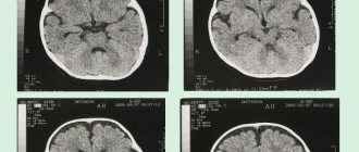
EEG allows you to study brain activity and give an accurate assessment of the child’s development. READ ALSO: what does an MRI of a child’s brain show?
In order to understand what non-epileptic paroxysmal states look like in children, it makes sense to pay attention to several current examples.
First of all, these are short-term breath holdings. This problem can be caused by severe fear, frustration, pain, or any surprise. During this condition, the child may scream, while the scream itself is delayed while exhaling, which is often followed by loss of consciousness.
Attacks of this kind are most often recorded in the age period from 6 months to 3 years. The good news is that their presence does not increase the risk of cognitive decline or epilepsy.
Paroxysmal state in a child - what is it? It is worth paying attention to one more example that clearly demonstrates a similar problem. We are talking about loss of consciousness. Fainting in this case is the result of acute circulatory failure in the brain area. In fact, this is nothing more than a manifestation of vascular lability.
Fainting occurs mainly in adolescents; among children who are at an early age, such conditions are rare. As for the causes of this problem, they include a sharp transition from a horizontal to a vertical position, as well as a state of strong emotional arousal.
Fainting begins with a feeling of darkening in the eyes and dizziness. In this case, both loss of consciousness and loss of muscle tone occur at the same time. There is always a possibility that short-term clonic convulsions may occur during depression of the child’s consciousness. As a rule, children do not remain unconscious due to fainting for more than 1 minute.
Reflex epilepsy is another problem that can be caused by a paroxysmal state in a child. It is unnecessary to say that this is a rather dangerous condition. Stressful situations and flashes of light can provoke such manifestations. But complex activities and auditory stimuli are unlikely to cause reflex epilepsy.
Paroxysmal state in a child - what is it? It is worth paying attention to one more example that clearly demonstrates a similar problem. We are talking about loss of consciousness. Fainting in this case is the result of acute circulatory failure in the brain area. In fact, this is nothing more than a manifestation of vascular lability.
Fainting occurs mainly in adolescents; among children who are at an early age, such conditions are rare. As for the causes of this problem, they include a sharp transition from a horizontal to a vertical position, as well as a state of strong emotional arousal.
Reflex epilepsy is another problem that can be caused by a paroxysmal state in a child. It is unnecessary to say that this is a rather dangerous condition. Stressful situations and flashes of light can provoke such manifestations. But complex activities and auditory stimuli are unlikely to cause reflex epilepsy.
Prevention
Prevention lies in early diagnosis. Often, success directly depends on the patient and his attitude towards his health. Basic recommendations that should be followed during prevention:
- moderate regular physical activity;
- proper nutrition;
- rejection of bad habits;
- avoidance of situations that lead to stress and cause anxiety;
- compliance with the instructions of the attending physician.
The main thing is to diagnose tachycardia in a timely manner in order to move on to prevention in time. You can get acquainted with the programs of our center by following the link.
Different forms of seizures
When considering the syndrome of paroxysmal states, it is worth paying attention to those diseases that most often accompany such crises.
- headache;
– myoclonic syndromes and other hyperkinetic states;
– autonomic disorders;
– muscular dystonic syndromes and dystonia.
In most cases, these problems are recorded in patients who have not reached the age of majority. But recently, more and more often, the paroxysmal state makes itself felt for the first time already in adulthood. It is also possible for the symptoms of the above diseases to dynamically progress, which become more severe against the background of chronic and acute cerebrovascular accidents or age-related cerebral disorders.
It is also important to take into account the fact that in some cases, non-epileptic paroxysmal conditions may be a consequence of the effects of certain medications prescribed to neutralize circulatory failure, as well as diseases such as parkinsonism and some mental disorders caused by old age.
Epilepsy is not the only form of manifestation of disorders of the central nervous system. There are other paroxysmal conditions in neurology that can be classified as epileptic.
One striking example is sensory (sensitive) Jacksonian seizures. Their manifestation occurs when a person is conscious. Symptoms include tingling and numbness in the face, limbs and half of the torso. In some cases, sensory seizures can turn into motor ones, which will significantly complicate the patient’s condition.
Attention should also be paid to Jacksonian epilepsy. In this case, both sensory and motor seizures are possible. The latter are especially problematic because they involve muscle spasms in the part of the face and limbs that are located on the side opposite the epileptic focus. In this case, disturbances in consciousness, as a rule, are not observed. In some cases, motor seizures can become generalized.
Complex absence seizures can be atonic, myoclonic, or akinetic. The first make themselves known through a sudden fall, the cause of which is a sharp decrease in the postural tone of the legs. As for the myoclonic form, it is characterized by rhythmic short-term muscle twitching, accompanied by a loss of consciousness. Akinetic absence seizure is a seizure with immobility, which can also result in falls.
Small absence seizures are also possible, in which a person also plunges into an unconscious state. There are no feelings of discomfort after its completion. The patient often cannot remember the moment of the attack.
Kozhevnikov epilepsy is characterized by limited short seizures that are clonic in nature. They most often capture the muscles of the arms, but the tongue, face and even legs can be affected by this process. Loss of consciousness during such convulsions is a rare occurrence.
Such paroxysmal activity can be divided into several types - muscle dystonia, autonomic disorders, headaches, myoclonic syndromes. They usually first appear at a young age and progress in old age.
This is usually affected by cerebrovascular accidents seen in older people. Therefore, to prevent such conditions, patients are prescribed drugs in advance to activate cerebral blood flow. This is done with extreme caution, since the wrong medicine can provoke a seizure.
- headache;
– myoclonic syndromes and other hyperkinetic states;
– autonomic disorders;
– muscular dystonic syndromes and dystonia.
Epilepsy is not the only form of manifestation of disorders of the central nervous system. There are other paroxysmal conditions in neurology that can be classified as epileptic.
Kozhevnikov epilepsy is characterized by limited short seizures that are clonic in nature. They most often capture the muscles of the arms, but the tongue, face and even legs can be affected by this process. Loss of consciousness during such convulsions is a rare occurrence.
Treatment of paroxysmal conditions is the domain of highly qualified specialists. Therefore, if signs of a single seizure become noticeable, especially when it is the first, the patient must be urgently hospitalized in a neurosurgical or neurological department. There they will be able to examine him and determine the current treatment plan.
It is important to ensure that the patient does not suffer any injuries before being taken to the hospital. It is also worth putting a spoon wrapped in a bandage into the mouth or using a mouth dilator.
In most cases, the treatment process for patients with status epilepticus begins in the ambulance. If doctors are not yet around, and the person is still having a seizure, then the first thing to do is to rule out the possibility of aspiration of vomit or mechanical asphyxia due to tongue prolapse.
As for non-epileptic forms, the causes of paroxysmal states can be completely different. It all depends on the key disease, the symptoms of which are aggravated. Therefore, the best thing that can be done is to take the person to the hospital as quickly as possible, where he can be examined and an accurate diagnosis made.
Forms of treatment and first aid
Treatment is not aimed at paroxysmal activity, but at its causes and subsequent manifestations:
- In case of a head injury, the damaging factor is eliminated, blood circulation is restored, and symptoms are determined for further treatment.
- Therapy for paroxysms associated with pressure is aimed at treating the cardiovascular system.
- Epileptic nature, especially with the manifestation of a grand mal seizure, suggests a visit to the neurological or neurosurgical department. Witnesses of a seizure should use a mouth dilator or use a spoon wrapped in a bandage to avoid injury, prevent asphyxia due to a sunken tongue or vomit, and call an ambulance. Treatment of patients with similar epileptic manifestations begins in the emergency room, where antiepileptic drugs (anticonvulsants) are used. These same remedies are effective for getting rid of panic attacks and fainting.
- Autonomic paroxysms are treated with drugs that affect the GABAergic systems (Clonazepam, Alprozolam). Many note the effectiveness of Finlepsin and Cavinton in the treatment of paroxysmal conditions of a non-epileptic nature.
Epilepsy and paroxysmal conditions
This is a rather difficult diagnosis in terms of the level of its negative impact on a person. But first, it’s worth remembering what epilepsy is. We are talking about a chronic pathological disease of the brain, which is characterized by seizures that have a different clinical structure and are constantly recurring. This condition is also characterized by psychopathic paroxysmal and non-convulsive manifestations.
It is possible to develop two forms of epilepsy: genuine and symptomatic. The latter is a consequence of traumatic brain injury, intoxication, brain tumors, acute circulatory disorders in the head, etc.
It is worth understanding that the special relationship between the epileptic focus and different parts of the nervous system determines the occurrence of repeated seizures of various clinical structures. Some features of the pathological process can lead to this result.
In addition, other paroxysmal conditions may occur
Treatment of polymorphic disorder in hospital and at home
Acute polymorphic psychotic disorder is a type of psychosis in which the patient experiences symptoms of many mental states at once, alternating one after another at a fairly fast pace, so that it is not possible to single out any one leading one.
Most often, the diagnosis of acute polymorphic psychotic disorder is first given to a person who first comes to the attention of specialists. The disorder is quite severe, entails a persistent inadequate reaction of the patient to all stimuli, and it becomes impossible to work and attend school.
The peculiarities of polymorphic mental disorder are that it is completely curable and responds well to treatment with timely access to appropriate specialists. This condition is also characterized by a pronounced, acute onset and severe course.
The symptoms of the disorder are varied; states from euphoria to depression can quickly replace each other, and they may be accompanied by hallucinations, delusions and other signs of acute psychosis. The disorder may be preceded by a strong emotional outburst, most often of a negative nature - the loss of a loved one, car accidents, natural disasters, violence.
There are two main types of acute polymorphic disorder:
Acute polymorphic disorder without symptoms of schizophrenia
In this state, patients exhibit characteristic symptoms of psychosis, but the intensity and types of delusions, hallucinations and behavioral disturbances do not correspond to schizophrenia. There are rapid changes in mood, varied delusions, hallucinations (auditory, tactile, olfactory or visual), the condition may resemble depression, apathy, mania, paranoia or schizophrenia and is characterized by a rapid increase and change in various symptoms (polymorphism). However, in this case there are no signs of schizophrenia with symptoms specific to it; the patient retains the ability to self-identify and may even have some criticism of his condition.
Multiple disorder with symptoms of schizophrenia
The symptoms described above are accompanied by the classic symptoms of schizophrenia, when patients are under the influence of hallucinations and delusions of oppression, persecution, exposure to fields, hypnosis, ideas of abduction, stalking, theft, or reading the minds of strangers. Common symptoms are cases when the patient hears voices commenting on his actions; it seems to him that his thoughts have become accessible to others. In this case, the forms of delirium are constantly changing, thinking is fragmented, the patient may experience pseudohallucinations, anxiety, excitability, and irritability prevail in the emotional sphere.
Polymorphic disorder is considered a dangerous condition due to the presence of schizophrenic symptoms, as it can become a manifestation of a more severe illness. Therefore, adequate and timely treatment is required, which can be provided to you at the ROSA clinic in Moscow.
Here you will be helped to remove the diagnosis of acute polymorphic psychosis quickly, confidentially and with maximum efficiency, which is guaranteed by the extensive experience of the medical staff in the treatment of acute disorders of any complexity.