Gliosis is a process that starts in brain tissue as a response to neuronal damage, which allows it to be regarded as a protective, compensatory function of the body. When the cells of the nervous tissue die for any reason (ischemic and atrophic changes, abscesses, infectious lesions, traumatic brain injuries), new tissue is formed in place of the free areas formed from glial cells.
Glial cells (astrocytes, oligodendrocytes, microgliocytes) closely interact with neurons, take on some of the latter’s functions, and protect neurons from damage. Thanks to glial cells, metabolic processes in brain tissue continue after a person has suffered severe pathologies of the central nervous system. Brain gliosis is not an independent disease. This is a consequence of pathological changes that have occurred in the nervous tissue.
General information
Gliosis of the brain is a pathological process manifested by the replacement of neurons with glial elements.
Gliosis is not an independent disease; rather, it is a protective reaction to the death of neurons, that is, a secondary process. At the same time, the process of destruction of neurons stimulates the filling of the resulting voids with glial cells, which leads to a change in the quantitative ratio of elements of the nervous tissue. Brain tissue consists of a collection of neurons, ependymal membrane and glial cells. Normally, glial tissue is responsible for trophic/secretory functions and metabolism in brain cells, and also performs the function of protecting neurons. It accounts for about 40% of the total brain mass. The proliferation of glial cells forms foci of gliosis in the white matter of the brain, which, depending on the etiology of the primary disease, can be focal/diffuse in nature. Foci of gliosis can be located in any area (structures) of the brain. And the more glia are formed, the less efficiently the central nervous system functions. With a large proliferation of glia, a cystic-gliotic atrophied area of brain tissue can form. The more glia are formed in the white matter, the worse the functioning of the entire nervous system. A cystic-glial-atrophic area is formed.
There are no statistical data on the incidence of various types of gliosis. It can only be noted that supratentorial foci of gliosis are most common.
Supratentorial lesions - what are they? Supratentorial are foci of gliosis of vascular origin. Various pathological processes in the vessels of the brain ( thrombosis , fibrosis , necrosis ) that cause circulatory disorders contribute to the formation of small hyper/hypertensive areas, which are accompanied by such disorders as poor coordination, dizziness , and changes in handwriting.
Single foci of gliosis
Focal gliosis is characterized by single, clearly defined islands in the white matter, the size and location of which is determined by the causes of neuronal death. They occur in people of any age and, perhaps, as a manifestation of natural age-related degenerative processes in brain tissue, ischemia of cerebral structures, local inflammatory process, chronic hypertension demyelination process , and as a consequence of injuries during childbirth in infants. Typically, single (limited foci) of gliosis of the brain are an accidental finding on CT/MRI, since in most cases they are not clinically manifested and do not cause discomfort, with the exception of their localization in the frontal lobe, where the centers associated with feelings and sensations are located, which can manifested by the appearance of hallucinations. A single gliosis of the brain is not prone to proliferation.
Multiple foci of gliosis
The appearance of multiple foci of gliosis is caused by injuries and acute/chronic circulatory disorders of the brain, that is, of vascular origin. As the pathology/disease that causes neuronal death develops, both the number and size of the altered areas increase. They occur predominantly against the background of severe atherosclerosis , stroke , cerebral infarction , with compression of cerebral vessels and as a result of age-related changes.
Multiple foci of gliosis for the most part enhance the clinical symptoms of the underlying disease and cause dysfunction of the central nervous system.
Treatment
Cerebral gliosis itself is not a disease, but a complication that was caused by chronic or acquired diseases of the nervous system. Therefore, there is no specific medicine or procedure to eliminate such tumors.
Treatment is aimed at the specific disease that caused the development of gliosis. It should be noted that medications are prescribed directly by a doctor.
During drug treatment, it is necessary to take special agents that can maintain and improve the condition of blood vessels. Also, with this disease, the brain may experience a lack of oxygen, so patients are often prescribed antioxidants that neutralize oxidative processes, and nootropics that help improve brain activity.
Surgery
Surgical intervention is used when large single foci of gliosis appear and in case of their negative impact on an organ or system that cannot be ignored, for example, during seizures. But most often, surgery is resorted to if it is impossible to control the patient’s well-being with the help of medications.
Complementary and alternative treatments at home
In addition to traditional methods of treatment, a patient suffering from this disease must eat in accordance with a special diet and take preventive measures to maintain normal functioning of the body and prevent the development of pathologies against the background of gliosis.
Nutrition and supplements
With gliosis of the brain, it is necessary to normalize your daily nutrition. The most important condition here is the exclusion of fatty foods and dishes from the diet, because fatty compounds disrupt the functioning of neurons and lead to their death.
Alcohol and herbal infusions
As mentioned earlier, a patient with this disease may have problems with cerebral circulation.
In addition to medications that normalize this process, you can also take tinctures from various herbs, which will be a source of useful substances for the functioning of blood vessels. For example, you can buy ready-made herbal tinctures at the pharmacy, mix them and take them as an additional remedy, if this is possible for medical reasons.
For this alcohol infusion you need:
- tinctures of valerian roots, motherwort, evasive peony, hawthorn - in 100 ml containers;
- eucalyptus - 50 ml;
- mint - 25 ml;
- Corvalol - 30 ml;
- whole cloves - 10 pieces.
Before use, this mixture should sit for about two weeks in a cool place, avoiding exposure to sunlight. Take 3 times a day, 30 drops diluted in a glass of water, half an hour before meals. The total duration of the course is from 1 to 3 months.
To prevent the development of atherosclerosis against the background of weakened blood vessels and gliosis, patients need to take a herbal infusion. It may include components such as:
- immortelle, oregano, mint, flax seed - one part each;
- hawthorn and birch leaves - two parts each;
To prepare the tincture, you need to mix the mixture and add one tablespoon per 200 ml of water. Afterwards, you need to boil the broth and leave it for about 2 hours. Before taking, you need to strain and distribute the herb into 3 doses per day. The course of treatment lasts 1 month.
Herbs and herbs (treatment with folk remedies)
Also, to improve blood circulation, you can take herbs such as:
- Dandelion root helps lower cholesterol and strengthens blood vessels. Its decoction should be taken 50 g before meals.
- Dill seeds restore blood circulation and help with abnormal blood pressure.
- St. John's wort has the ability to relieve vascular spasms and restore tissue.
- lemon balm nourishes the cerebral cortex and the walls of blood vessels, helps calm the nerves and restore nerve cells;
- Celandine helps to recover from a post-stroke condition. Its decoction should be drunk 2 times a day.
- Sweet clover is saturated with many vitamins that nourish the heart muscle and its main vessels, and cleanse the lymph. It should be taken with caution and in small doses; the herb must be infused in the proportion of a teaspoon per glass of boiling water for two hours. After drinking 3 times a day before meals, one third of a glass, for about 30 days.
- Anise lofant helps with various diseases, cleanses blood vessels and ducts, it is especially useful for those who have had a heart attack or have problems with changes in blood pressure. Its collection, from 50 to 200 g, must be poured with 0.5 liters of cognac or vodka and left in a dark place for 20 days, not forgetting to shake every day. The infusion should be taken for about 30 days, 2 times a day, 30 minutes before meals, a teaspoon in 30 ml of water.
- Sophora japonica not only restores the balance of blood vessels, but also removes free radicals from the body. Its infusion can also be made with vodka or cognac. You need to take 100 g of fruit, pour in 0.5 liters of alcohol and leave for 3 weeks, after straining, take 3 times a day, about 35 drops, an hour after eating.
- mistletoe helps with convulsions, paralysis and sclerosis. One tablespoon of herb should be poured into a glass of chilled boiled water and left overnight. Drink 1/3 glass 3 times a day before meals, for about a month.
- Dioscorea Caucasica helps with problems with blood vessels and heart rhythm, vision and headaches. The root of this herb must be crushed and poured boiling water at the rate of a teaspoon to a glass of boiling water, and then kept in a steam bath for about 20 minutes. Take a tablespoon 3 times a day, after meals, for about 4 months with breaks of a week.
If alcoholic beverages are contraindicated, herbal infusions can also be made with water, pouring the decoctions overnight with 1 glass of boiling water and taking 50 ml 3 times a day half an hour before meals.
Exercises
Intense physical activity with gliosis is undesirable, because it can provoke additional complications or attacks of chronic diseases.
Any therapeutic exercises must be agreed with the attending physician, who can determine its necessity. But to maintain good health and the proper flow of processes in the body, it is recommended to take daily walks of 30-60 minutes in remote places from roads and highways.
It is also recommended to undergo a massage course to normalize muscle tone and tissue metabolism.
Classification
The classification is based on the nature of the proliferation of glial cells and their localization.
Based on morphological characteristics, the following are distinguished:
- The isomorphic form is characterized by an ordered proliferation of neuroglia.
- Anisomorphic form - has a pronounced cellular structure and is characterized by chaotic growth; most often localized subcortically/paraventricularly. Periventricular gliosis - formed in the ventricles of the brain. Often periventricular gliosis is accompanied by the development of cysts. Fibrous form—the predominance of the fibrous type structure is noted.
According to the localization of the pathological process, the following are distinguished:
- Periventricular gliosis - glial cells are formed in the ventricles of the brain. Periventricular gliosis is often accompanied by the development of cysts.
- Perivascular (vascular) gliosis is the growth of glia along atherosclerotic vessels. It is often diagnosed on MRI as microangiopathy with the presence of foci of gliosis.
- Subependymal gliosis is predominantly localized on the inner lining of the ventricles of the brain.
- Marginal gliosis - located on the surface of the brain.
- Marginal gliosis - foci of degeneration are localized in the intrathecal region.
Based on quantitative criteria, single and multiple foci of gliosis are distinguished. Histologically, hypodense (without a definite structure, cannot be stained) and hyperintense (clearly structured, well stained) areas of gliosis are distinguished.
Based on the nature of the process/prevalence, the following are distinguished:
- A focal type of flow, involving a limited area of the brain (usually in the temporal/parietal regions).
- Diffuse type of course - characterized by multiple lesions of various sizes/localizations. Most often of vascular origin and may have the appearance of a cystic-gliotic formation.
Symptoms
Symptoms may be absent if the cyst is small or located in an area remote from functionally important parts of the brain. An increase in the size of the lesion may be accompanied by a mass effect - compression of the surrounding brain tissue with the appearance of corresponding symptoms. Symptoms depend on the location of the pathological focus:
- Temporal lobe. Increased pain in the head area after physical or mental stress, vestibular disorders (dizziness, inability to maintain balance).
- Frontal lobe. Memory deterioration, inability to concentrate, speech dysfunction, psycho-emotional disorder.
- Parietal lobe. Impaired fine motor skills, inability to perform precise, measured, smooth movements.
- Occipital lobe. Visual impairment (loss of visual fields, appearance of foreign objects in the field of view).
Changes in the structure of white matter located in the left or right frontal lobe are often detected in elderly patients, which is associated with destructive processes (atrophy, ischemia, necrosis) affecting the brain. General symptoms of gliosis transformations:
- Sleep disturbance.
- Sudden changes in blood pressure values.
- Visual, auditory dysfunction.
- Deterioration of cognitive abilities.
- Motor coordination disorder.
- General weakness, increased fatigue.
- Impaired urination and defecation function.
In newborns, pathological processes associated with the replacement of normal brain tissue by cystic-glial formations are accompanied by signs:
- Apathy, lethargy, low motor activity.
- Absence or weakening of response to external stimuli.
- Hydrocephalic syndrome (decreased muscle tone, weak natural reflexes - grasping, swallowing, tremors of the limbs, convulsive syndrome). Visual disturbances may be observed - strabismus, fountain-like repeated regurgitation.
With supratentorial localization, pathological processes associated with the proliferation of neuroglia are manifested by visual disorders. Damage to the white matter in the temporal lobe is accompanied by convulsive syndrome, epileptic seizures, and dizziness. The location of lesions in the anterior part of the frontal lobe negatively affects cognitive abilities.
The patient's memory and mental activity deteriorate, and it becomes difficult for him to concentrate. Localization of the lesion in the parietal lobe can manifest itself as apraxia (impaired fine motor skills while maintaining the ability to perform basic movements) and sensitivity disorders.
Causes
The pathological process of replacing neurons with neuroglial cells may be based on a variety of reasons:
- Age-related changes caused by the natural process of neuron death.
- Diseases of hereditary origin - lysosomal storage disease ( Tay-Sachs disease ), inherited in an autosomal recessive manner, characterized by massive death of neurons in children, which is the cause of their death); genetically determined pathology - tuberous sclerosis .
- Birth injuries at birth.
- Traumatic brain injuries.
- Multiple sclerosis (destruction of myelin and the formation of foci of demyelination in various brain structures).
- Cerebral infarction (ischemic stroke).
- Hemorrhagic stroke is a hemorrhage into the structures of the brain, which forms foci of gliosis of vascular origin.
- Brain swelling.
- Epilepsy.
- Arterial hypertension/encephalopathy caused by a persistent increase in blood pressure.
- Neuroinfections ( encephalitis , meningitis ). oxygen starvation of brain tissue ( hypoxia ).
- Hypoglycemia (low blood sugar).
- Surgical interventions on brain structures.
- Atherosclerosis of cerebral arteries.
- Abuse of products containing animal fat.
Preventive measures
The main preventive measure is to avoid situations that can lead to the development of cystic-glial transformation. Therefore, after cerebral accidents (TBI, ischemic or hemorrhagic stroke), constant monitoring by a neurologist and periodic MRI studies of the brain are necessary.
This recommendation is most relevant for children and the elderly, since the former have an incompletely formed nervous system that is more susceptible to traumatic factors, and the latter have a significantly reduced reparative ability.
Symptoms
Clinical symptoms vary widely and are determined mainly by the symptoms of the underlying disease that caused gliosis. As a rule, single small foci of gliosis do not produce specific neurosymptoms and are discovered by chance during an MRI examination of the brain. At the same time, multiple (extensive) foci of gliosis almost always manifest neurosymptoms in the form of:
- Unreasonable long-term intense headaches that are not controlled by taking antispasmodics.
- Blood pressure instability (sudden changes in a short period of time).
- Periodically occurring dizziness .
- Decreased performance/increased fatigue.
- Changes in visual/auditory perception.
- Disorders of coordination, attention and memory.
- Motor disorders (with large lesions, up to convulsive conditions).
Clinical symptoms are largely determined by the localization of the focus of gliosis:
- Localization of foci of gliosis in the temporal lobes is characterized by frequent, intense, long-term headaches. With the vascular genesis of the process, the pain syndrome is combined with sudden changes in blood pressure.
- Supratentorial gliosis manifests itself mainly with visual disturbances – hallucinations; distortion of the outlines, sizes, shapes of objects; loss of visual fields, difficulty recognizing an object by appearance.
- Gliotic foci in the white matter of the brain can cause dizziness , an episyndrome characterized by increased convulsive activity with the possible development of epileptic seizures. Such symptoms develop as a complication of traumatic brain injury/surgery.
The localization of gliosis foci in the frontal lobes is often due to age-related changes. It is generally accepted that if diseases that can cause the death of neurons and the proliferation of glial cells have not been identified, then such a process should be considered a primary pathology that develops as the body ages and manifests itself as impaired concentration, attention, memory, fine motor disorders, and slowed reactions.
Clinical case
Many cases have been described where, after a minor injury, a huge cyst was discovered several months later. So a 48-year-old man hit his head on the asphalt. I did not lose consciousness, there was no nausea or vomiting. He considered the injury minor and did not see a doctor.
After 2 years, he began to notice periodic headaches, dizziness, and lack of smell. I went to the doctor and an MRI scan of the brain was ordered.
Conclusion: in the right frontal lobe there is a zone of cystic-glial changes, probably of a post-traumatic nature, measuring up to 2.4×1.9×1.6 cm. Thus, a minor TBI led to dysfunction of the sense of smell.
Cases of birth trauma are common. The clinical picture is usually erased immediately after birth and appears only by 1-2 years. Parents note developmental delays, constant drowsiness of the child, focal (localized) symptoms are possible. Often, cysts in the brain cause the development of epilepsy.
So, parents with a 5-year-old child turned to a neurologist with complaints of epilepsy attacks. It was found that the disease was caused by a cyst. After carefully collecting anamnesis, the doctor learned that the labor was protracted, the child was born with a triple torsion of the umbilical cord, and was not breathing.
High percentage of development of cystic-gliotic transformation in “silent” strokes.
Tests and diagnostics
Diagnostics is based on imaging methods (MRI/CT) of the brain. These methods allow you to determine:
- location and size of pathological foci;
- the presence of marginal (perifocal) infiltration;
- structure (homogeneity) of the white matter of the brain;
- perivascular spaces of various areas;
- areas of the basal ganglia;
- the presence of other pathological formations;
- condition of the ventricles of the brain/pituitary gland, functionality/condition of the blood vessels of the brain.
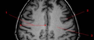
The image below shows multiple foci of gliosis.
MRI makes it possible to establish not only the presence of areas of gliotic changes, but also to identify the cause. MRI images provide a clear picture of the presence of areas in the brain that do not receive sufficient nutrition due to vascular obstruction by atherosclerotic plaques; allow you to identify inflammatory processes, areas of hematoma, and identify neoplasms. This, to a certain extent, makes it possible to eliminate the provoking factor and develop tactics to minimize the risks of further spread of the process.
Gliosis of the brain - what is it?
To understand what gliosis changes in the brain are, you need to understand how the central nervous system (CNS) works.
Neurons are the main cells of the nervous system, a kind of “minicomputers” that receive and process information coming from the environment and issue commands to all internal organs. The functioning of the body as a whole depends on the coordinated work of neurons.
For reference. But besides neurons, the central nervous system contains glial cells. These are auxiliary structures of nervous tissue. They perform supporting, trophic (nourish, regulate metabolic processes of neurons) functions, and participate in the development of nerve cells.
Glial cells are located next to neurocytes and can replace them after death: if neurons die, glial cells grow and fill their place.
Like scars on the skin, glial changes in the brain can be figuratively considered scars on the “body” of nervous tissue.
Such cellular replacement, especially if multiple foci are formed, leads to disruption and changes in the central nervous system.
Diet
There is no special diet, but it is recommended to adhere to the diet recommended for the underlying disease that initiates the process of formation of glial lesions ( Diet for stroke , Diet for hypertension , atherosclerosis , diabetes mellitus , Diet for obesity , etc.). In general, it is recommended to adhere to a low-calorie diet with sufficient vitamin content (vegetables, fruits) and a minimum content of animal fats in the diet.
Prevention
No drugs or methods of specific prevention of neuronal death have been developed. To maintain the activity of neuro-processes and brain vessels, we can only recommend:
- Lead a healthy lifestyle.
- Monitor your body weight.
- Maintain age-appropriate physical activity.
- Constantly monitor blood pressure, cholesterol , and blood sugar levels.
- Minimize bad habits.
- Practice a healthy balanced diet by minimizing foods containing animal fats.
- Train brain functions: solve puzzles/crosswords, learn poetry, foreign languages.
Forecast
The prognosis of life with cerebral gliosis is largely determined by the underlying disease and can vary significantly depending on the volume of gliosis foci and the intensity of neuronal replacement. As a rule, the prognosis for life with single foci outside important brain structures is relatively favorable, while extensive foci of gliosis in areas of the brain responsible for important functions of the central nervous system can be unfavorable, for example, in the temporal lobe, initiating frequent epileptic seizures.
Patient complaints
General symptoms are usually mild. Typically, patients complain of constant headaches, a feeling of dizziness, weakness, and worsening mood. Often, a headache signals the development of hydrocephalus (hydrosis of the brain) - a condition when the outflow of cerebrospinal fluid from the intracranial space is disrupted.
In the brain, each region performs a specific function, so the type of neurological disorder can suggest the location of the cystic gliosis lesion.
List of sources
- Vereshchagin N.V., Gulevskaya T.S. Pathology of the brain in atherosclerosis and arterial hypertension. – M.: Medicine, 1997. – 228 p.
- Vasiliev V.N., Kindyashova V.V., Kozhevnikova V.V., Tikhomirova O.V., Lomova I.P., Serebryakova S.V. Clinical and diagnostic significance of microfocal brain lesions of vascular origin among management specialists // Medical, biological and socio-psychological problems of safety in emergency situations. 2014. No. 3. pp. 27-32.
- Gladky P. A., Sergeeva I. G., Tulupov A. A. Infectious lesions of the brain. Tutorial. Novosibirsk 2015. – 24 p.
- Putilina M.V. Chronic cerebral ischemia//Attending physician. 2005. No. 5.
- Yanishevsky S.N. Brain damage in patients with type 2 diabetes // International Journal of Endocrinology. No. 8(64). 2014 pp. 89-97.
The best medications for therapy
For all the measures described above to be effective, you need to support the sick body with special medications. Doctors have mixed opinions about which medications are best to use to treat cerebral gliosis. But they agree on one thing - treatment should be medicinal, not folk. The five most popular prescribed drugs include the following:
| Drug name | Active substance | Manufacturer |
| Cavinton | Vinpocetine | Hungary |
| Cinnarizine | Cinnarizinum | Bulgaria and Russia |
| Actovegin | Deproteinized calf blood haemoderivative | Austria |
| Glitsed | Glycine | Ukraine |
| Vinpocetine | Vinpocetine | Russia, India, Belarus |
To the described vasoactive drugs, you need to add antiplatelet agents to improve blood flow, amino acids, and nootropics. Vitamins E or B provide good support for treatment. But the most important thing is to choose the right main medicine. To do this, you should familiarize yourself with contraindications and side effects, and also consult your doctor. As stated earlier, his decision should be based only on complete brain studies.
Cavinton
Cavinton increases the consumption of glucose and oxygen by the tissues of the frontal lobes. That is why it is used in the treatment of the consequences of ischemic stroke, vascular dementia, atherosclerosis and other diseases. Sometimes the drug is used in ophthalmology: it treats chronic diseases of the blood vessels of the eye and its retina. The medicine should only be used after consultation with a doctor, otherwise the risk of side effects is too high. Know that:
- distributed in the form of tablets or concentrates for infusion;
- the dosage is determined strictly on an individual basis;
- the medicine costs from 228 to 850 Russian rubles.
With the correct dosage, the drug rarely produces side effects. But sleep disturbances, dizziness, tachycardia, nausea, and heartburn may occur. Less common are increased sweating, rapidly passing allergic skin reactions and general weakness. By the way, it may be a manifestation of an underlying disease.
The medicine does not have a negative effect on patients with kidney or liver diseases.
Cavinton is strictly prohibited for patients with severe coronary heart disease, serious arrhythmias, and the acute phase of a stroke. It is dangerous to prescribe it to pregnant women or nursing mothers. But this is acceptable if the risk to the mother’s health is more serious than the likely side effects for the child. Pregnant women may experience placental bleeding from taking it, and there is a risk of spontaneous abortion.
Cinnarizine
A selective blocker of slow calcium channels called Cinnarizine is used not only in the treatment of gliosis and its consequences. It is used in the treatment of post-stroke conditions, traumatic brain injuries, migraines, vestibular disorders, kinetosis, and encephalopathy. The medicine quickly relieves dizziness, tinnitus, nausea and vomiting. Cinnarizine has the following characteristics:
- Available in the form of tablets of 50 milligrams;
- tablets are taken orally after meals, the exact dose depends on the severity of the patient’s condition;
- It is difficult to answer exactly how much Cinnarizine costs, but usually its price ranges between 37 and 101 rubles.
Cinnarizine has few contraindications. It is not given only to pregnant, lactating women, and people with allergies. The medicine should be used with caution if a person is diagnosed with Parkinson's disease. But side effects are possible. Among the most common are drowsiness, lack of hunger, fatigue, dyspepsia and dry mouth. In exceptional cases, lichen planus and serious weight gain are recorded.
- Treatment of cerebral vessels: folk remedies, traditional medicine
Actovegin
This medicine is prescribed for cognitive impairment, which includes dementia and the consequences of strokes. It is occasionally used for diabetic polyneuropathy or circulatory disorders. Thanks to the active substance, the medicine increases the rate of blood flow in the capillaries. After systemic use, it improves the morphological and physiological state of the central nervous system. Remember, that:
- Available in the form of digestible tablets or injection solutions;
- first, the body should receive 10-20 milliliters of Actovegin, and then five milliliters per day;
- packaging of the drug will cost 568-1928 Russian rubles.
Actovegin rarely leads to side effects. But in particularly sensitive patients, drug fever, symptoms of shock, urticaria, and sudden redness in the injection area are recorded. Sometimes doctors observe myalgia, but the exact frequency is unknown. Actovegin is prohibited in case of pulmonary edema, decompensated heart failure, oliguria, anuria or long-term fluid retention in the body.
Glitsed
Glitsed is prescribed for cerebral circulatory disorders, traumatic brain injuries, and neurocirculatory dystonia. It helps in the fight against psychological overload, improves memory, relieves insomnia and facilitates vigorous physical activity. As part of the treatment of cerebral gliosis, the drug has only an auxiliary effect. A brief description of Glitsed is as follows:
- sold in the form of flat white or cream tablets;
- as a rule, one tablet is prescribed three times a day;
- the cost starts from 136 rubles, but you can find cheaper offers on the Internet.
The medicine rarely has side effects, especially if used in the treatment of cystic glial changes in the brain. But allergic reactions on the skin are possible; indigestion, headache, and nausea occur less frequently. In exceptional cases, increased irritation and poor concentration are observed. The overdose was either not recorded or not observed.
Vinpocetine
According to the instructions, Vinpocetine is prescribed for insufficient cerebral circulation, for damage to the blood vessels and retina. Sometimes they treat vegetative-vascular dystonia against the background of climatic syndrome. For patients with gliosis of the brain, Vinpocetine is prescribed to improve metabolism in the affected organ and normalize blood flow. It is important to remember the following characteristics of the medicine:
- the drug is available in the form of tablets for oral administration or solution for injection;
- The dosage regimen is individual, since the dosage varies depending on the severity of the patient’s condition;
- the average price is 50-164 rubles.
Vinpocetine is contraindicated in case of coronary heart disease, stroke or severe signs of arrhythmia. It is dangerous to give it during pregnancy or lactation. Even if the dosage and rules of administration are followed, side effects are possible. Typically, adults experience insomnia, dizziness, increased heart rate, surges in blood pressure, profuse sweating, or allergic reactions.