Mechanism of development of central nervous system lesions
Residual damage to the central nervous system is always caused by unfavorable factors that preceded it.
In most cases, the basis for the pathogenesis of such symptoms is cerebral ischemia. In children, it develops during the period of intrauterine development. Due to insufficient blood supply to the placenta, the fetus receives little oxygen. As a result, the full development of nervous tissue is disrupted, and fetopathy occurs. Significant ischemia leads to intrauterine growth retardation and the birth of a child before the gestational age. Symptoms of cerebral hypoxia can appear already in the first days and months of life. Residual organic damage to the central nervous system in adults often develops due to traumatic and infectious causes. Sometimes the pathogenesis of nervous disorders is associated with metabolic (hormonal) disorders.
Complications, prevention
In the absence of proper treatment, the patient will inevitably develop complications. This can only be avoided by working with an experienced doctor and taking preventive measures that are effective both in improving the patient’s condition and in preventing the onset of the disease.
Many people face the unpleasant consequences of the disease. Only a serious approach to treatment and the help of an experienced doctor help reduce the risk of complications. And with severe residual encephalopathy, it is extremely difficult to avoid the consequences of the disease, and the treatment itself in such cases can last for years.
What complications may the patient experience:
- Episyndrome and epileptic seizures;
- Brain dysfunction;
- Cerebral paralysis;
- Vegetovascular dystonia;
- Hydrocephalus;
- Autism (Asperger's syndrome);
- Cerebrasthenic syndrome;
- Retardation in intellectual development;
- Brain gliosis;
- Psychospeech deviations.
Young children or teenagers may suffer the most from complications. For adults, the consequences are less dangerous. In special cases, the patient may even fall into a coma.
Prevention
You can protect yourself from the onset of the disease or its complications with simple preventive measures. Because they are applicable for healthy people and those who have already developed residual organic encephalopathy; some recommendations will be relevant only for the first category. Regular adherence to the rules and advice will eliminate all risks associated with this disease.
Preventive measures include recommendations:
- Visit your doctor at the first sign of health problems.
- Follow all directions and treat illnesses as soon as possible.
- Spend time outdoors regularly.
- Be more physically active and play sports
- Make the right diet with plenty of vitamins and minerals.
- Do special gymnastics that activates blood circulation in the brain.
- Avoid dangerous places and head injuries.
- Monitor the health of the fetus during pregnancy.
- To refuse from bad habits.
- Normalize the quality of sleep, eliminate stress from life.
This is enough to protect the body from many diseases. Every person should include such prevention in their life.
Pathology in ICD-10
Like all pathologies, disorders of neuropsychic development have a specific code in the international classification of diseases. It is worth understanding the broadness of the concept of “residual organic damage to the central nervous system.” The code (ICD-10) for this pathology is G96.9. This code means the diagnosis “unspecified damage to the central nervous system.” In more specific cases, the ICD-10 code changes to a specific nosology.
The International Classification of Diseases, Tenth Revision (ICD-10) treats residual encephalopathy controversially. Pathology is considered under the code G93.4. He equates it to encephalopathy of unspecified pathogenesis. But the presence of additional factors forces us to assign other codes to the disease.
- G93.8 – destruction of CNS cells caused by exposure to radiation;
- T90.5, T90.8 – cell death due to injury.
Different types of brain lesions
The residual type of encephalopathy according to the ICD has code G93.4. If changes in the brain are caused by a head injury, then the doctor may indicate the code T90.5 or T90.8. The disease is considered dangerous because can lead to serious complications. It is divided into degrees that correspond to the severity of the disease: mild, moderate and severe.
The main classification involves division into 2 types: perinatal and acquired. They determine the genesis of the disease and occur with special symptoms, which is important to consider when making a diagnosis.
Perinatal type
Congenital residual encephalopathy manifests itself in childhood and increases the risk that the child will become disabled. It occurs when the child’s brain is damaged during pregnancy or during childbirth, as well as in the first days after birth.
An increased likelihood of this disease occurring is present in those whose mother experienced severe symptoms of toxicosis or experienced premature birth. The main reasons for the development of the disease are as follows:
- Birth injury;
- Fetal hypoxia;
- Genetic brain mutations;
- Fetal asphyxia;
- Intrauterine infection;
- Rhesus conflict.
If an illness associated with pregnancy occurs, special attention should be paid to the affected baby in order to reduce the risk of complications.
Acquired type
The residual genesis of the acquired type is secondary. As a rule, the disease is detected in adults from 18 to 50 years old. At risk are people who have suffered spinal or head injuries, brain damage or infections.
Main reasons:
- Congenital cerebral pathologies;
- Brain injuries;
- Stroke;
- Toxic effects on the brain;
- Inflammation of nerve tissue in the brain due to infections or endocrine diseases;
- Vascular damage due to atherosclerosis;
- Abuse of psychotropic drugs or antipsychotics;
- Hypertension combined with manifestations of hypertensive crisis;
- Pathologies of the liver or kidneys.
In adults, the disease occurs with less consequences, but in some cases dangerous complications can still occur.
Since there are a lot of brain disorders, it was advisable to come up with a classification that would cover all diseases:
- organic brain damage;
- defeat of an infectious nature;
- cerebrovascular diseases;
- tumors (both benign and malignant);
- various congenital pathologies;
- brain diseases that are associated with trauma or abnormal structure;
- ionizing radiation;
- intoxication with various substances;
- negative impact of electromagnetic fields;
- mixed defeat.
EEG interpretation
Decoding an electroencephalogram is the process of interpreting it, taking into account the clinical symptoms that the patient has. When analyzing the EEG, neurophysiologists at the Yusupov Hospital take into account:
- basal rhythm;
- the level of symmetry in the electrical activity of brain neurons in the right and left hemispheres;
- commissure activity;
- EEG changes during functional tests (hyperventilation, photostimulation, opening and closing eyes).
Neurologists-neurophysiologists make the final diagnosis only taking into account certain clinical signs of the disease that worry the patient.
Changes in the alpha rhythm on the EEG are the following signs:
- constant recording of the alpha rhythm in the frontal lobes of the brain;
- violation of sinusoidal waves;
- interhemispheric asymmetry above 30%;
- unstable frequency;
- paroxysmal or arc-shaped rhythm;
- rhythm index less than 50%;
- amplitude is less than 20 µV or more than 90 µV.
Severe interhemispheric asymmetry may be evidence of a tumor, brain cyst, heart attack, stroke, or scar at the site of an old hemorrhage. High frequency and instability of the alpha rhythm can appear after traumatic brain injury. A disorganized type of EEG (impaired organization of the alpha rhythm or its complete absence) indicates acquired dementia.
In children, delayed psychomotor development is indicated by:
- alpha rhythm disorganization;
- moving the focus of activity from the occipital and parietal regions;
- increased synchrony and amplitude;
- excessive response to hyperventilation;
- weak short activation reaction.
A decrease in the amplitude of the alpha rhythm on the EEG, a weak activation reaction, and a shift in the focus of activity from the back of the head and crown are signs of psychiatric pathology. Excitable psychopathy is manifested by a slowdown in the frequency of the alpha rhythm against the background of normal synchrony. Inhibitory psychopathy is characterized by EEG desynchronization, low frequency and alpha rhythm index. Increased synchronization of the alpha rhythm in all parts of the brain, a short activation reaction are a sign of neuroses.
In patients, neurophysiologists determine the following pathological types of beta rhythm:
- paroxysmal discharges;
- low frequency, distributed along the convexital surface of the brain (adjacent to the frontal, temporal, parietal and occipital bones of the skull);
- amplitude more than 7 µV;
- violation of symmetry between the hemispheres in amplitude;
- sinusoidal type of beta rhythm.
Beta rhythm disturbances on the EEG indicate brain pathology. The presence of diffuse beta waves with an amplitude no higher than 50-60 μV indicates a concussion. Short spindles in the beta rhythm indicate encephalitis. Beta waves with a frequency of 16–18 Hz and high amplitude in the central and anterior parts of the brain are signs of delayed psychomotor development of a child.
Normally, the theta rhythm and delta rhythm can only be recorded on the EEG of a sleeping person. In a state of wakefulness, such slow waves appear in the presence of degenerative processes in the brain tissue, which are combined with compression, high blood pressure and lethargy. Paroxysmal theta and delta waves in a patient while awake are recorded when the deep parts of the brain are damaged.
High amplitude delta waves are evidence of a tumor. The predominance of theta and delta waves on the EEG with maximum activity in the occipital region, flashes of bilaterally synchronous waves, the number of which increases with hyperventilation, are a sign of delayed psychomotor development of the child.
Residual organic damage to the central nervous system: treatment of pathology
Therapy for focal brain damage is aimed at eliminating the cause of the changes and restoring the functions of the organ.
For example, if the pathology is caused by a disease characterized by increased blood pressure, then the patient is prescribed to take medications that lower blood pressure. These may be diuretics, calcium channel blockers, or beta blockers.
Restoration of brain activity and elimination of pathological phenomena is carried out with the help of drugs that increase metabolism in nerve tissues: nootropics. Also used are agents that improve blood circulation, rheological properties of blood, and reduce the need for oxygen.
Symptomatic treatment is aimed at reducing the manifestations of pathology: taking anticonvulsants, antiepileptic drugs, antidepressants, and tranquilizers for anxiety.
The most common manifestations of impaired blood supply to the brain are focal changes in the brain substance of a discirculatory nature, which are characterized by impaired blood circulation in certain areas of the brain substance, and not in the entire organ.
As a rule, these changes are a chronic process that develops over quite a long time, and in the first stages of this disease, most people cannot distinguish it from other diseases of the nervous system.
Doctors distinguish three stages in the development of focal changes of a discirculatory nature:
- At the first stage, in certain areas of the brain, due to vascular diseases, a slight disruption of blood circulation occurs, as a result of which the person feels tired, lethargic, and apathetic; The patient experiences sleep disturbances, periodic dizziness and headaches.
- The second stage is characterized by deepening of vascular damage in the area of the brain, which is the focus of the disease. Symptoms such as decreased memory and intellectual abilities, disturbances in the emotional sphere, severe headaches, tinnitus, and coordination disorders indicate the transition of the disease to this stage.
- The third stage of focal changes in the brain substance of a discirculatory nature, when a significant part of the cells in the focus of the disease due to circulatory disorders has died, is characterized by irreversible changes in the functioning of the brain. As a rule, in patients at this stage of the disease, muscle tone is significantly reduced, there is practically no coordination of movement, signs of dementia (dementia) appear, and sensory organs may also fail.
In addition to changes of a discirculatory nature, a disease with similar symptoms is single focal changes in the brain substance of a dystrophic nature due to a lack of nutrients.
This disease affects people who have experienced head trauma, those suffering from ischemia, cervical osteochondrosis in the acute stage, and patients who have been diagnosed with a benign or malignant brain tumor.
Due to the fact that the vessels supplying a certain area of the brain cannot fully perform their functions, the tissues in this area do not receive all the necessary nutrients. The result of such “starvation” of nervous tissue is headaches, dizziness, decreased intellectual abilities and performance, and in the final stages dementia, paresis, and paralysis are possible.
Sooner or later, all people grow old, and the body ages along with them. It primarily affects the heart, brain and spinal cord. If the heart ceases to properly cope with its task - pumping blood - then over time this will affect the condition of the brain, the cells of which will not receive enough nutrients to maintain vital functions.
According to various sources, from 50 to 70% of elderly people (over 60 years old) suffer from a similar disease.
It is better to prevent any disease than to treat it later, and for this you need to know its external manifestations (signs) and symptoms.
- First stage. In the first couple of days, a person feels a little tired, lethargic, dizzy and sleeps poorly. This occurs due to decreased blood circulation in the brain. The degree of significance increases with the development of vascular diseases: cholesterol deposition, hypotension, etc.
- Second stage. At the second stage, a so-called “disease focus” appears in the brain; damage to the brain substance deepens due to poor blood circulation. The cells do not receive enough nutrition and gradually die. The beginning of this stage is indicated by memory impairment, loss of coordination, noise or “shooting” in the ears and severe headaches.
- Third stage. Due to the discircular nature of the last stage, the focus of the disease moves even deeper, the affected vessels bring too little blood to the brain. The patient exhibits signs of dementia, lack of coordination of movements (not always), dysfunction of the sensory organs is possible: loss of vision, hearing, shaking hands, etc.
The exact changes in the brain substance can be determined using MRI.
Without treatment, diseases such as:
- Alzheimer's disease. The most common form of nervous system degeneration.
- Pick's disease. A rare progressive disease of the nervous system that manifests itself in the first place.
- Huntington's disease. Genetic disease of the nervous system. developing rally.
- Arterial hypertension .
- Cardiocerebral syndrome (impairment of basic brain functions due to cardiac pathology).
Reasons for changes
As already mentioned, the main cause of the disease is vascular damage. arising inevitably with age. But for some, these lesions are minimal: small cholesterol deposits, for example, but for others they develop into pathology. Thus, diseases of a dystrophic nature lead to changes in the substance of the brain:
- Ischemia. This disease is mainly characterized by impaired blood circulation in the brain.
- Cervical osteochondrosis.
- Tumor (benign or malignant).
- Severe head injury. In this case, age does not matter.
Risk group
Any disease has a risk group, people in it should be extremely careful. If a person has similar diseases, then he is in the primary risk group, if only predisposition, then in the secondary:
- Suffering from diseases of the cardiovascular system: hypotension, hypertension, hypertension, dystonia.
- Patients with diathesis, diabetes mellitus or stomach ulcers.
- Those who are overweight or have a habit of eating poorly.
- Those living in a state of chronic depression (stress) or leading a sedentary lifestyle.
- People are older, regardless of gender.
- Suffering from rheumatism.
How to overcome it?
Despite the complexity of the disease and the problems with its diagnosis, every person can avoid a similar fate by helping his body fight the signs of old age or the consequences of serious injury. To do this, you should follow simple rules.
Firstly, lead an active lifestyle. Walk or jog for at least two hours a day. Walk in the fresh air: in the forest, in the park, travel outside the city, etc. Play outdoor games that suit your physical abilities: basketball, pioneer ball, volleyball, tennis or table tennis, etc. The more movement, the more active the heart works and the blood vessels become stronger.
Thirdly, avoid stressful situations and overwork. A person's mental state directly affects his health. Don't overwork yourself, rest if you're tired, sleep at least 8 hours a day. Don't overexert yourself with physical activity.
Fourth, undergo a medical examination 1-2 times a year to monitor the condition of your body. Especially if you are already undergoing treatment!
Advice
It is best not to try to help your body with “home methods”: taking medications on your own, injecting yourself, etc. Follow the doctor's instructions and undergo the procedures he prescribes. Sometimes, to determine the accuracy of the diagnosis, it is necessary to undergo many procedures; taking tests is a normal situation.
A responsible doctor will never prescribe medications if he is not sure of the accuracy of the diagnosis.
At the same time, hundreds of thousands of processes take place in it to ensure the vital functions of the body.
However, the functioning of the brain is inextricably linked with its blood supply, because even a slight decrease in the blood supply to a certain portion of the medulla can lead to irreversible consequences #8212; massive death of neurons and, as a result, severe diseases of the nervous system and dementia.
- suffering from hypertension, vegetative-vascular dystonia and other diseases of the cardiovascular system;
- patients with diabetes mellitus;
- suffering from atherosclerosis;
- having bad habits and being overweight;
- leading a sedentary lifestyle;
- those in a state of chronic stress;
- elderly people over 50 years of age.
It is quite difficult to make a prognosis in advance when treating a disease. This is due to the fact that residual encephalopathy in an adult or child is resistant in nature, i.e. resists medications. Therefore, the recovery process becomes unpredictable and difficult.
Once the final diagnosis has been made, treatment can begin. The main goal is to restore blood circulation in the brain tissues, as well as the general condition and relieve all unpleasant symptoms.
To begin with, the patient is prescribed therapeutic procedures, which sometimes successfully cope with the disease without taking any medications. These include:
- Manual therapy (massage);
- Acupuncture;
- Physiotherapy;
- Osteopathy;
- Phytotherapy;
- Reflexology;
- Contrast morning shower;
- Conversations with a psychotherapist;
- Sanitary resort treatment.
After repeated procedures, the patient will feel much better. If such methods are not effective enough, the doctor prescribes medication. Some are given intravenously, but most are taken in pill form. The following groups of drugs are prescribed:
- Neuroprotectors;
- Nootropics;
- Antioxidants;
- Cerebral circulation normalizers;
- CNS stimulants;
- Hormonal;
- Sedatives;
- Immunomodulators;
- Anticonvulsants;
- Nonsteroidal anti-inflammatory drugs;
- Vitamin complexes.
The exact treatment can only be prescribed by a doctor. Self-administration of random medications can lead to severe side effects and dangerous complications, which can only be dealt with in a hospital setting.
Treatment
How the treatment will be carried out depends on the cause that caused the residual, pathological changes in the brain. However, as a rule, the main treatment is aimed at restoring normal cerebral circulation.
Today, there are several ways to treat this disease:
- Surgical. This method of surgical intervention is practically not used in practice by doctors. As a rule, this method is used in the presence of a tumor in the brain, the removal of which leads to significant relief of the patient’s condition.
- Medication. This therapy is the main one in the treatment of residual encephalopathy. The drugs are sent directly to treat the underlying cause and the symptoms that caused it.
For this purpose, medications such as:
- Anticonvulsants;
- Nootropic drugs;
- Calming;
- Hormonal;
- Medicines that restore normal blood circulation;
- Vitamin and mineral complexes.
Therapeutic treatment of residual encephalopathy of the brain is quite lengthy. It is necessary to systematically use the medications prescribed by the doctor to prevent regression of the main provoking disease.
- Physiotherapeutic treatment
It is used in combination with drug treatment and includes:
- Reflexology;
- Electrophoresis and phonophoresis;
- Magnetic therapy and acupuncture;
- Therapeutic exercise and massage;
- Cold and hot shower;
Types
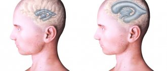
MRI images allow us to identify all pathologies that affect brain tissue. I determine affected areas by changes in color, echogenicity of individual areas of the cortex or other organ structures. Using the data obtained, specialists measure the area of the destroyed area and also predict the development of pathology.
Focal brain damage may result from:
- Demyelination;
- Presence of neoplasms;
- Tissue swelling;
- Circulatory disorders;
- Gliosis (replacement of functional cells with glial tissue).
Manifestations of pathology depend on the location of the lesion. Therefore, MRI diagnostics is considered the most informative method for detecting diseases of the central nervous system.
Based on the location of brain lesions, there are:
- Juxtacortical;
- Periventricular;
- Lacunar.
Juxtacortical lesions of nervous tissue are characteristic of multiple sclerosis. In this case, they are located as close as possible to the cerebral cortex. When describing an MRI picture, experts recommend using this particular definition, since the term “subcortical” cannot fully convey the nature of the spread of the pathology - it describes any changes in the white matter up to the ventricles.
The periventricular location of the foci of destruction is diagnosed with hypoxic-ischemic damage to the brain substance. In this case, they are located near the ventricles.
Lacunar lesions are a consequence of damage to the deep arteries. They are located in the thickness of the white matter along the blood vessels. Typically their diameter varies between 1–20 mm.
Demyelination
Characterized by the presence of areas of destruction of the myelin sheath of nerve fibers. Because of this, the transmission of nerve impulses between neurons in the area of the brain is disrupted, which negatively affects the performance of the central nervous system.
Tissue destruction of this type is observed in multiple sclerosis, multifocal leukoencephalopathy, Marburg disease, acute dissimulating encephalomyelitis, Devic's disease.
In these diseases, the MRI picture is identical: the images clearly visualize single or multiple white spots, which are located in one or several parts of the brain. The size of the areas depends on the extent of the disease, as evidenced by the presence and strength of neurological abnormalities.
At the moment, there is no common understanding of perivascular spaces. Some scientists believe that they surround only the arteries, while others believe that they surround all the large blood vessels that penetrate the brain. Some describe them as a space that is located between the wall of the vessel and the nervous tissue, others - as a natural continuation of the subarachnoid and pia mater.
Pericular spaces perform several functions at once:
- Participate in the circulation of cerebrospinal fluid;
- In them, the exchange of substances between the cerebrospinal fluid and brain tissue occurs;
- They are part of the blood-brain barrier;
- They contain immunocompetent cells, that is, with the help of them, immunoregulation occurs in the tissues of the organ.
Perivascular spaces occupy a small volume, so in a healthy person they are not visible on an MRI image.
In dangerous conditions, for example, before a stroke, the patient’s ICP increases due to an increase in the volume of cerebrospinal fluid. This leads to expansion of the cavity between the brain vessels and nervous tissue. Along with this process, the echogenicity of the area increases, which appears on an MRI image as a white spot.
The disease is characterized by the loss of neurons and a decrease in the number of synaptic connections between them. This leads to a decrease in the thickness of the gray matter and pronounced atrophy of the affected areas.
Dark spots appear on MRI images, which indicate necrosis of brain cells. An accurate diagnosis is made based on the results of several examinations, that is, over time.
It is characterized by the accumulation of fluid in the cells of the head and intercellular space. Due to this, the volume of the organ increases and intracranial pressure increases.
In the affected area on the MRI image there is a light spot, which, as the process worsens, increases and gradually covers the entire organ.
Functions of Brain Waves
The brain is an electrochemical organ. Electrical activity in the brain manifests itself in the form of brain waves. Four types of waves are recorded on the EGG:
- beta waves (the fastest oscillations with large amplitude, the frequency of which is in the range of 15–40 Hz) are generated by the awake brain, actively involved in mental activity;
- alpha waves are the opposite of beta waves, their amplitude is larger and their frequency is 9-14 Hz;
- for theta waves, the amplitude is even greater, and the frequency is 5-8 Hz, they are generated by the brain of a person who is almost asleep;
- delta waves have a maximum amplitude and frequency of 1.5-4 Hz.
If the frequency of theta waves on the ECG drops to zero, this means that brain death has occurred. Deep, dreamless sleep is characterized by a theta wave frequency of 2-3 Hz. When a person lies down in bed and reads for a few minutes before going to sleep, he or she is in a “low beta” state. The moment we put down a book, turn off the light and close our eyes, the brain waves successively pass through the stages of beta, alpha, theta, and ultimately delta.
The four types of brain vibrations are common to all people, regardless of gender, age, nationality, culture and nationality. The results of EEG studies show that although one frequency always dominates in brain oscillations, the remaining three, depending on the level of activity of the person, are also always present.