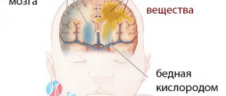
Status epilepticus is a condition in which epileptic seizures are repeated or continuous over a fairly long period of time (about 30 minutes). The patient does not have time to recover from the previous seizure before the next one overtakes him. The patient's consciousness is unclear, breathing is difficult, and signs of a coma are observed.
Causes of status epilepticus
The action of medications taken by a patient with epilepsy is aimed at inhibiting seizures. If the patient independently refuses medications prescribed for treatment, then this action can trigger the occurrence of ES.
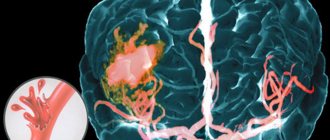
Epistatus can also occur, for example, with brain pathologies:
- Malignant neoplasms;
- Withdrawal syndrome;
- Infections, intoxications, hematomas and encephalopathy;
- Peripheral circulation disorders.
Status epilepticus can also occur in patients with diabetes. Diabetes is scary because of its complications, including ES.
Types and stages of status epilepticus
The variability and diversity of types of epileptic seizures is the main basis for the formation and identification of forms of epileptic seizures, characterized by general signs of the clinical picture of the disease. They are divided into two groups: non-convulsive and convulsive. Classifying the types of ES, we can distinguish:
- Generalized non-convulsive seizure. In this case, a short-term loss of consciousness is observed. The patient seems to freeze with interrupted activities (eating, talking, writing...), or thinking about something. In this case, the eyes are focused on one point, the face becomes pale, and the connection with the outside world is interrupted. The absence seizure stops as suddenly as it started.
- Incompletely generalized status epilepticus is characterized by muscle cramps, the patient completely loses consciousness, cardiovascular activity is disrupted, and breathing becomes unstable.
- Tonic SE is typical for children of different ages with rare and severe forms of epilepsy.
- Clonic ES, accompanied by high fever and convulsions in children and infants.
- Myoclonic ES is expressed in episodic twitching of muscle tissue.
Epileptic seizures are characterized by short duration. Usually a seizure lasts several seconds, or several tens of seconds, less often - a minute. After status epilepticus, relief occurs spontaneously, without outside intervention. Therefore, they are called self-limited epileptic seizures. Serial attacks following one after another are often encountered.
In medicine, each stage of epistatus, which is a complication of epileptic syndrome, has its own name:
- The duration of the pre-status stage can last from 1 to 10 minutes;
- The initial stage is characterized by the duration of the attack from 10 to 30 minutes;
- Expanded - from 30 to 60 minutes;
- The stage, which lasts more than an hour, is called refractory.
Epistatus is a state of a person when he does not regain consciousness during alternating epileptic seizures. Before one attack has finished, the next one begins. The second variant of epistatus is no less dangerous, and is a seizure lasting more than 30 minutes.
General part (a little theory)
Status epilepticus (or epistatus) is a syndrome that can occur in various diseases of the brain (hereinafter referred to as “brain” for brevity) and systemic diseases of the body as a whole. Here are examples of reasons that can cause epistatus:
— epilepsy (as a rule, epilepsy is caused by violation of the regularity of taking antiepileptic drugs);
- traumatic brain injury;
- tumor or other mass formation of the brain;
- inflammatory diseases of the brain and its membranes;
- acute cerebrovascular accident;
— cicatricial adhesive disorders of cerebral liquor dynamics;
— dysmetabolic conditions (alcohol withdrawal, diabetes, porphyria, acute adrenal or thyroid insufficiency, uremia, eclampsia, acute hypoglycemia, etc.);
- poisoning;
- general infections, especially with severe intoxication and hyperthermia.
Epistatus is based on continuous (or intermittent, but often repeated) paroxysmal collective electrical activity of brain neurons. Depending on the degree of involvement of different parts of the brain in this activity (in other words, depending on the degree of generalization of seizures), the nature of the epistatus may be different. Clinically, it is possible to distinguish at least the following four types of epistatus, and the first two most often end up in psychoreanimatology units:
- status of fully generalized (“full-scale”, grand mal) seizures - regularly recurring seizures with tonic and clonic phases and complete loss of consciousness;
- status of incompletely generalized convulsive seizures - regularly recurring seizures with atypical muscle activity (for example, isolated contractions of individual muscle groups, only tonic or only clonic seizures) and complete loss of consciousness;
- the status of focal (or Jacksonian) seizures with isolated ongoing convulsions in a certain muscle group (face, one limb, hemi-type convulsions) can occur without turning off consciousness, but during generalization consciousness can be turned off;
- status of non-convulsive seizures (sometimes called “absence status”, petit mal) - regularly recurring seizures without muscle activity, but with a complete loss of consciousness.
According to ICD-10, status epilepticus is classified as follows:
G41 Status epilepticus
G41.0 Status epilepticus grand mal (convulsive seizures)
Excluded: partial continuous epilepsy [Kozhevnikova] (G40.5)
G41.1 Status epilepticus petit mal (minor seizures)
G41.2 Complex partial status epilepticus
G41.8 Other specified status epilepticus
G41.9 Status epilepticus, unspecified.
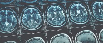
If you record an EEG during epistatus, you can see hypersynchronous high-amplitude fluctuations in brain biopotentials characteristic of epileptic seizures. The pathophysiological basis of all types of epistatus is basically the same; the difference lies only in the completeness of the involvement of the brain in epiactivity. Generalized, convulsive forms of status indicate only the involvement in the epiactivity of the cortex of the premotor areas of the brain, which are responsible for muscle movements. Manifestations of paroxysmal activity in other areas of the brain are simply not visible to us without an EEG.
An important feature that distinguishes epistatus from other paroxysmal disorders of the brain is that in the pauses between seizures the patient’s condition does not normalize, consciousness is not restored, and disturbances in the functioning of organs and systems progressively accumulate. Epistatus with pronounced muscle activity is especially dangerous: epileptic tonic-clonic convulsions of the respiratory muscles, aspiration of saliva and blood from the oral cavity, as well as post-ictal delays and arrhythmias of breathing lead to hypoxia and acidosis; the cardiovascular system experiences extreme stress due to enormous muscular work; hypoxia increases cerebral edema; acidosis increases hemodynamic and microcirculation disorders; secondly, the conditions for brain function are increasingly deteriorating. This vicious circle underlies thanatogenesis in epistatus. Mortality from epistatus in the 60s. XX century (when treated with chloral hydrate enemas, IV magnesium sulfate, etc.) was 16–33%, in the 80s. - 10–12%; currently, with proper treatment, it does not exceed 5–6% (unless, of course, the epistatus is based on some incurable condition incompatible with life).
Serial epileptic seizures differ from epileptic status only in that in the pauses between seizures (or their series) the patient’s condition is relatively normalized, consciousness is restored to one degree or another, and no progressive disruption of the functioning of organs and systems is observed. Serial seizures, however, can transform into epistatus, and the line between them may not always be clearly defined.
Psychiatric resuscitators most often have to deal with epistatus developing in psychiatric hospital patients. From admission, patients with epileptic status are rarely admitted to a psychiatric hospital, since, according to existing rules, with frequent intractable epileptic seizures, the patient must be hospitalized in the hospital closest to his location. A mental hospital is rarely such a “closest hospital.” An ambulance’s attempt to transport a patient with epistatus to a mental hospital only on the basis that he “is registered” or “has been treated there before” is a gross mistake in providing care at the prehospital stage.
Symptoms of status epilepticus
Symptoms of epistatus are expressed in circulatory disorders, disturbances of consciousness (the person “switches off”), and disruption of the respiratory system. Symptoms of status epilepticus are a consequence of previous seizures from which the patient does not recover.
Epistatus can be characterized by a frequency of attacks up to 20 per hour. The patient does not regain consciousness at the onset of the subsequent seizure; his condition can be described as numbness bordering on coma.
The comatose state worsens in direct proportion to the duration. Tonic spasms affect the muscles of the back, arms and legs. High blood pressure suddenly drops. Increased reflexivity is also unexpectedly replaced by a complete lack of reaction.
Respiratory and circulatory disorders become obvious. When the seizures disappear, epileptic prostration occurs.
The duration of the epistatus is at least 30 minutes. Usually, as expected, we draw a line between this condition and episodic seizures with partial restoration of physiology and consciousness (full or partial).
There are two phases that characterize epileptic convulsive status with the following features:
- Compensatory changes in the circulatory and metabolic system, expressed in high blood pressure, vomiting and nausea, uncontrolled urination and defecation.
- Coming in about half an hour/hour, it is a maladaptation of compensatory changes, which is expressed in acute renal failure (and liver failure), a sharp decrease in pressure, disruption of the respiratory system, and arrhythmia.
Epistatus, the course of which is not accompanied by convulsions, is characterized by complete immobility of the patient and a feeling of detachment. Usually the patient lies with his mouth open, his blank gaze does not express anything.
Since the time of M. Bourneville [1], status epilepticus (SE) has traditionally been regarded as a severe life-threatening condition. For a long time, the definition of SE by L. Clarc and T. Proute [2] remained unshakable: status epilepticus - a condition in which attacks occur with such frequency that coma and exhaustion between attacks become permanent. Since E.S. traditionally considered as a convulsive state, it was divided into primary and secondary generalized, focal (partial) and unilateral, and according to the characteristics of convulsive manifestations into the status of tonic-clonic, tonic, clonic and myoclonic seizures. Further progress in the development of the problem of ES was associated with the development of basic sciences and the introduction of their results into clinical practice. The emergence and application in clinical practice of methods for recording electrical potentials of the brain—electroencephalography (EEG)—was fundamental. This made it possible to move on to the clinical and electrographic definition of epileptic seizures and expand the understanding of the manifestations of epileptic seizures.
Back in 1974, we [3] put forward a subsequently confirmed assumption that there are as many forms of SE as there are types of epileptic seizures. Moreover, non-convulsive forms of epileptic encephalopathy have been discovered, in which progressive neurological manifestations and mental disorders, often severe, are a consequence of persistent epileptiform activity. One of the most unusual forms is electrical ES of slow-wave sleep.
In the process of studying ES, the problem of its differential diagnosis arose with cases of depression of consciousness (stupor, coma) or psychosis, when paroxysmal, including epileptiform, phenomena, and sometimes mild convulsive manifestations (with stupor and coma) can be recorded on the EEG.
All of the above served as the basis for the serious work of the Commission on the classification of E.S. International League Against Epilepsy to develop a definition and modern classification of epilepsy [4], which we are considering here with some abbreviations.
In November 2015, the said commission proposed a new definition and classification of E.S. The following definition was formulated: SE is the result of either a failure of the mechanisms responsible for the cessation of seizures, or the initiation of mechanisms that lead to abnormally prolonged seizures - after 30 minutes (t1). This is a condition that can have long-term consequences (beyond t2), including damage and death of neurons, alteration of the nervous network, depending on the type and duration of seizures.
Note that it took the League more than 40 years to define ES according to a fundamentally different, namely qualitative criterion, which was proposed by us in a discussion at the European Congress on Epilepsy in Warsaw back in 1998, but, unfortunately, was preserved only in the Russian-language literature: “SE is a qualitatively different condition from an epileptic seizure, characterized by deep depression of the antiepileptic defense system: a) with the ability to only actively suppress each epileptic seizure, but not prevent the next one; b) subsequent total failure of the antiepileptic defense system with the cessation of each seizure only in a passive way, namely due to the depletion of energy resources [5].”
From the above definition of the League it is clear that it applies time criteria, namely 30 minutes. But this can hardly be considered successful, since it is necessary to start treatment for E.S. as early as possible. The League now quite reasonably proposes different time criteria for different seizures, providing a different time frame for each patient, depending primarily on the type of seizure he is experiencing. This is reflected in the corresponding time parameters.
It is proposed to classify ES using the following 4 blocks: 1. Semiology, 2. Etiology, 3. EEG correlates, 4. Age. Ideally, each patient should be considered in each of the 4 blocks. However, it should be kept in mind that as with other acute neurological conditions, the semiology (symptoms and signs) and EEG correlates of SE are very dynamic and can change over a short period of time. Thus, during repeated neurological examinations and repeated EEG recordings during ES, changes may occur that require a different interpretation than the initial one. For example, SE may begin with focal motor symptoms, including bilateral convulsive SE (A.1.b) and may manifest several hours later as non-convulsive SE (NES) with coma and minimal motor phenomena resembling so-called “mild status” (B. 1). In addition, periodic lateralized changes may initially appear on the EEG, and upon repeated examination, a bilateral synchronized type of electrical activity is detected. Nevertheless, let's look at each of the blocks.
Block 1:
Semiology
This block includes the clinical manifestations of ES that form the basis of the classification. The two main taxonomic criteria are: 1. The presence or absence of predominant motor symptoms; 2. The degree (qualitative and quantitative) of impairment of consciousness. Forms with a predominance of motor symptoms and impaired consciousness can be combined with the concept of convulsive E.S. Moreover, the term convulsive in this case is fundamental, reflecting the usual language of the doctor. In this regard, the fundamental term is the term “status epilepticus”. In the 19th century, in French medicine, the term “état de mal” corresponded to it. The generally accepted term “convulsive” in the context of SE refers to “episodes of excessive pathological muscle contractions, usually bilateral, which may be prolonged or intermittent.”
Block 1 also includes the classification of ES:
A. SE with predominance of motor symptoms
A.1. Convulsive E.S. (SES, synonym: tonic-clonic ES). A.1.a. Generalized convulsive. A.1.b. The focal onset progresses to bilateral convulsive ES. A.1.c. It is unknown whether it is focal or generalized.
A.2. Myoclonic E.S. (predominance of epileptic myoclonic twitches). A.2.a. With a coma. A.2.b. No coma.
A.3. Focal motor. A.3.a. Recurrent focal motor seizures (Jacksonian). A.3.b. Epilepsy partialis continua (ERS)1. A.3.c. Adversive status. A.3.d. Oculoclonic status2. A.3.e. Ictal paresis (i.e. focal inhibitory ES).
A.4. Tonic status.
A.5. Hyperkinetic E.S..
B. SE without predominance of motor symptoms
(i.e. non-convulsive ES, BES)
B.1. BES with coma (including the so-called “soft” ES).
B.2. BES without coma. B.2.a. Generalized. B.2.aa Typical absence status. B.2.ab Atypical absence status. B.2.ac Myoclonic absence status. B.2.b. Focal. B.2.ba Without disturbance of consciousness (aura continua, with vegetative, sensory, visual, olfactory, gustatory, emotional/mental/experiential or auditory symptoms). B.2.bb Aphasic status. B.2.bc With impaired consciousness. B.2.c. It is unknown whether it is focal or generalized. B.2.ca Vegetative E.S..
Block 2
: Etiology
The causes underlying SE are classified in accordance with the views of the commission of the International League Against Epilepsy dating back to 2010, but in this case, established terms are used that are used by epileptologists, emergency physicians, neurologists, pediatric neurologists, neurosurgeons, family doctors and other specialists dealing with patients with ES.
Currently undefined conditions (or "borderline syndromes")
Epileptic encephalopathy. Lateralized and generalized periodic discharges with a monotonous manifestation are not considered as involving EEG patterns. Behavioral disorders (eg, psychosis) in patients with epilepsy. Acute confusion (eg, delirium) with epileptiform EEG patterns.
The term "known" or "symptomatic" is used, according to general neurological terminology, to designate SE caused by a known disorder, which may be structural, metabolic, inflammatory, infectious, toxic or genetic. Based on their temporal relationships, a division into acute, delayed and progressive manifestations can be applied.
The term "idiopathic" or "genetic" does not apply to the underlying etiology of E.S. In idiopathic or genetic epileptic syndromes, the cause of the status is not the same as the disease itself, since certain metabolic, toxic or intrinsic factors (such as sleep deprivation) can cause epilepsy in these syndromes. Therefore, the term "idiopathic" or "genetic" is not used here. SE in patients with juvenile myoclonic epilepsy (which itself is “idiopathic” or “genetic”) may be symptomatic as a result of treatment with an inappropriate antiepileptic drug (AED), abrupt drug withdrawal, or drug toxicity.
The term "unknown" or "cryptogenic" (Greek κρύπτος, hidden or unknown, τò γένος, family, class, generation, descent) is used in its strictly original meaning: unknown cause. The suggestion that this is a "supposed" symptomatic or genetic cause is inappropriate. The term "unknown" or corresponding translations in different languages may be used synonymously and in accordance with the 2010 proposal.
Known (i.e. symptomatic). Acute (for example, stroke, intoxication, malaria, encephalitis, etc.). Remote (for example, post-traumatic, post-encephalitic, post-stroke, etc.). Progressive (for example, brain tumor, Lafora disease and other PME, dementia). ES in certain electroclinical syndromes. Unknown (i.e. cryptogenic) ES in its various forms has many causes. Attached to this section is a list that will be updated periodically.
Block 3
: Electrographic correlates
None of the EEG patterns of any type of ES are specific. Epileptiform discharges are considered a characteristic feature, but with increasing duration of SE, changes in the EEG and rhythmic non-epileptiform changes may predominate. Similar EEG patterns, such as triphasic waves, can be recorded in different pathological conditions, leading to considerable confusion in the literature. Despite the fact that the EEG is overloaded with motor and muscle artifacts and thus limits its clinical value, it is indispensable in the diagnosis of BES, since clinical signs (if any) are often blurred and nonspecific.
Currently, there are no specific EEG criteria for E.S. But based on the large amount of data existing in this area, we propose the following terminology to describe EEG patterns in SE: 1. Localization: generalized (including bilateral synchronous patterns), lateralized, independent bilateral, multifocal. 2. Pattern name: periodic discharges, rhythmic delta activity or spike-wave/sharp wave, and subtypes. 3. Morphology: acuity, number of phases (for example, three-phase morphology), absolute and relative amplitude, polarity. 4. Temporal characteristics: prevalence, frequency, duration, daily pattern and index duration, onset (sudden or gradual) and dynamics (evolving, fluctuating or constant). 5. Modulation: stimulus-induced or spontaneous. 6. Effect of intervention (medicines) on EEG.
Block 4: Age of patients
In this case, the following division of ages and their definition is used: 1. Newborn (from 0 to 30 days). 2. Infancy (from 1 month to 2 years). 3. Childhood (over 2 years old and up to 12 years old). 4. Adolescence and adulthood (over 12 years and up to 59 years). 5. Elderly (60 years and older).
ES in newborns can be blurred and difficult to recognize. Some forms of SE are considered an integral part of the electroclinical syndrome; others may occur in patients with a specific electroclinical syndrome, or when triggers or precipitating causes are present, such as sleep deprivation, poisoning, or inappropriate medications. Examples are the use of phenytoin for some forms of progressive myoclonic epilepsy, carbamazepine for juvenile myoclonic epilepsy or absence epilepsy.
Considering the age aspect of SE, it is necessary to note that it is correct to talk about the predominant manifestation of certain forms of SE in certain age groups, which can also occur in other age groups.
SE, which occurs in newborns and infancy, is the debut of epileptic syndromes. Tonic status (for example, Ohtahara syndrome or West syndrome). Myoclonic status in Dravet syndrome. Focal status. Febrile status.
ES, which occurs mainly in childhood and adolescence. Vegetative E.S. with early onset of benign childhood occipital epilepsy (Panayotopoulos syndrome). BES for specific childhood epileptic syndromes and etiologies (for example, ring 20 syndrome and other karyotypic disorders, Angelman syndrome, epilepsy with myoclonic-atonic seizures, other childhood myoclonic encephalopathies. Tonic status in Lennox-Gastaut syndrome. Myoclonic status in progressive myoclonic epilepsy. Electrical status epilepticus during slow-wave sleep (EES). Aphasic status in Landau-Kleffner syndrome.
ES, which occurs predominantly in adolescence and adulthood. Myoclonic status in juvenile myoclonic epilepsy. Absence status in juvenile absence epilepsy. Myoclonic status in Down syndrome.
ES, which occurs mainly in old age. Myoclonic status in Alzheimer's disease. Nonconvulsive status epilepticus in Creutzfeldt-Jakob disease. De novo
(or recurrent) absence status in elderly and especially senile age.
Undoubtedly, the Commission of the International League Against Epilepsy has done a great job, including taking into account the dependence of the type of epileptic seizures and forms of epilepsy on age. It is important to note that the commission gave a fundamentally age-based classification of epilepsy, adapted for the diagnosis of epilepsy. The following considerations should be made as comments. The proposed classification has serious scientific significance and is the basis for further development of the problem. However, due to its bulkiness, it is not entirely convenient for use by a practicing physician. This requires a reservation and reference to a less detailed, but more convenient classification by E. Trinka et al. [6]. There are three appendices to the classification: 1) a list of neurological diseases in which ES can occur, grouped into 17 headings - about 115 items in total; 2) “a list of specific connections in which ES is an integral part of the syndrome, entity or symptom with strict clinical involvement (the list can be finalized)”: absence status in the syndrome of the 20th ring chromosome, Angelman syndrome and absence status of epilepsy3 .
Appendix 3 presents all previous definitions and classifications of ES. It would be advisable to indicate before Appendix 1 that ES can occur with any brain damage and therefore even the listing of 115 diseases cannot be exhaustive. Another necessary addition: ES in some cases can occur with extracerebral intoxication (1) as a de novo
.
Now about the definition of E.S. For a long time, the League tried to base the definition of ES on the time factor, proposing various time criteria - from 5 to 30 minutes, but this turned out to be unsuccessful. Now another criterion has been proposed: SE is a different qualitative state, characterized by insufficiency of the mechanisms responsible for the termination of an epileptic attack or the initiation of mechanisms that lead to an abnormally prolonged attack. And here is the definition of ES that we proposed in the discussion at the above-mentioned European Congress on Epilepsy in Warsaw (1998). This definition has been preserved, unfortunately, only in Russian-language literature [2]. We defined SE as a qualitatively different state from an epileptic attack, characterized by the following features: a) deep depression of the antiepileptic defense system, preserving the possibility of only actively suppressing each epileptic seizure, but not preventing the next one; b) total failure of the antiepileptic defense system with the cessation of each seizure only in a passive way, namely due to the depletion of energy resources. We also recently presented detailed data on the systemic mechanisms of antiepileptic defense in a lecture that was given in London at the 6th London-Innsbruck Colloquium on SE and acute epileptic seizures [7], to which there is also no reference.
As for the temporary criterion of SE, as indicated above, the League did not abandon it, but proposed differentiated criteria for the three main forms of SE: tonic-clonic, focal status with impaired consciousness and absence status. It is proposed to introduce two indicators: tT - the time during which seizures are likely to be prolonged to their constant activity and t2 - the time when seizures can cause long-term consequences (neuronal damage, neuronal death, damage to the neuronal network and functional deficit). This is a fundamentally new and very important approach to assessing ES and its possible consequences.
There is no reference to A.Ya. Kozhevnikova.
There is no synonym for "epileptic nystagmus".
Here is a link to the article by P. Gentone et al. “Absence status epilepsy: delineation of a distinct idiopathic generalized epilepsy syndrome.Epilepsia. 2008;49:642−649.
Emergency care for status epilepticus
First aid for status epilepticus, before the arrival of doctors, is the need to protect the patient from receiving mechanical injuries. There is no need to crowd around the patient, blocking free access to clean air.
Our recommendations:
- Place the patient on a non-traumatic surface with something soft (jacket, sweater) under his head;
- To avoid choking on saliva, carefully turn your head to the side;
- Remove the tie, belt, unbutton the collar so that nothing interferes with the patient’s ability to breathe freely;
- Remove all sharp and traumatic objects located nearby;
- If your teeth are clenched, there is no need to unclench them;
- If your mouth is open, place any soft cloth between your teeth.
You should not place sharp, metal or other objects between your teeth that could cause injury to an unconscious person.
Emergency care for status epilepticus should be provided very carefully. You should not hold the patient too tightly so as not to damage his bones (in this condition the likelihood of fractures is very high).
Complications of status epilepticus
Epistatus is characterized by irreparable consequences. Statistics. With symptomatic ES, mortality is 30–50%. With ES in patients with epilepsy - 5%.
If ES lasts more than an hour, then patients will face serious consequences:
- Diffuse saturation of brain tissue with fluid, called cerebral edema and accompanied by oxygen starvation;
- Critically low blood pressure;
- Excessive levels of lactic acid, called lactic acidosis;
- Violation of water-salt balance;
- For children, characteristic signs are delays in the development and formation of the psyche, which can lead to mental retardation.
Non-convulsive epistatus is considered less dangerous compared to generalized epistatus. Nevertheless, complications of status epilepticus often find expression in disturbances of perception, attitude, thinking, memory, and understanding.
NSICU.RU neurosurgical intensive care unit website of the intensive care unit of the N.N. Research Institute Burdenko
Introduction
Status epilepticus is a serious complication of the postoperative period in patients with neurosurgical pathology, significantly worsening the prognosis of the underlying disease and increasing the risk of an unfavorable outcome. Brain tissue damaged during neurosurgical intervention or severe traumatic brain injury can cause the formation of a focus of pathological electrical activity of brain cells, such as usually manifested by various types of convulsive seizures. The frequency of this complication during neurosurgical interventions reaches 5% - 20%, depending on the location of the tumor (7,15,16). The leading ones in this regard are cortical or meningeal lesions, and the main manifestation of this condition is partial or generalized epileptic seizures. However, in a certain situation, pathological electrical activity of brain cells can occur without characteristic clinical manifestations, but is realized by a non-convulsive attack or even non-convulsive status epilepticus (45,50,53,71). An asymptomatic (non-convulsive) epileptic attack, and in particular status epilepticus, can cause an increase in general cerebral and focal neurological symptoms, up to the development of a coma. In such a clinical situation, identifying the cause of the coma is difficult. Delayed administration of anticonvulsants is also likely, which can lead to the formation of a persistent epileptogenic focus and severe secondary brain damage as a result of neuronal death. The concept of nonconvulsive status epilepticus (NSE) was first introduced by Celesia in 1976 (17). The purpose of this work is to summarize the main clinical characteristics of BES and attract the attention of clinicians to the serious problem of diagnosis and treatment of BES.
Definition
The most accurate definition of BES, proposed by the Epilepsy Reseach Foundation Workshop, includes the following provisions: Clinically detectable changes in the level of consciousness or other equivalents: changes in the position of the eyeballs, nystagmus, autonomic manifestations (hyperhidrosis, tachycardia, changes in skin color). The presence of characteristic spike manifestations detected during EEG monitoring. The positive effect of anticonvulsant therapy in the form of normalization of the EEG and the disappearance of clinical symptoms. BES has attracted increasing attention in recent years, since some of its aspects have been poorly developed, in particular in various acute cerebral lesions, when BES significantly worsens forecast. In such a situation, based on the clinical picture of the disease, it is usually difficult to suspect BES: EEG data are crucial for diagnosis. Frequency of occurrence. The frequency of occurrence of BES varies according to various authors and ranges from 7.5 to 65 and even 89% in the studied series of patients (7,25,38,44,45,59,61,67,70). This depends to a large extent on the diagnostic methods used and on the patient population. Among patients with acute cerebral injuries, the incidence of BES is much higher compared to patients with other pathologies. In patients with brain tumors, after neurosurgical interventions, BES occurs in 20% of cases of all status epilepticus (7). In patients in a septic state who are in coma, non-convulsive seizures were detected in 10% of cases (6). In victims with severe TBI, BES was detected in 3% of patients (8). With subarachnoid hemorrhage, BES was diagnosed in 8% of cases (9,20). The development of BES after subarachnoid hemorrhage in most cases led to an unfavorable outcome (7,20). With parenchymal hemorrhage, the development of BES was combined with an increase in displacement of the midline structures and clinical deterioration (70). The main reason for unfavorable patient outcomes is late diagnosis and initiation of therapy for BES caused by acute cerebral damage. The pattern of prognosis and outcomes for BES is the same as for convulsive ES: the longer the status, the worse the prognosis. J. Yong et al. (71) based on multivariate regression analysis, they found that the development of BES in acute brain injury increases the risk of death by 46%. ###Etiology The cause of BES is a number of very different clinical conditions, manipulations and the use of pharmacological drugs: surgery of skull base tumors, pneumaticcephaly (7), the use of third generation cephalosporins (1,28,48), theophylline (55), lithium salts ( 41), phosphamide (42), tiagabine (33,36,43), carbamazepine (50), traumatic brain injury (8), chronic hemo- (32) and peritoneal dialysis (18), Creutzfeldt-Jakob disease (19 ), spontaneous subarachnoid and intraventricular hemorrhages [25,45], sclerosing encephalitis [10], abnormalities of brain development [1], herpetic encephalitis [29], necrotizing leukoencephalitis [26], consequences of electroconvulsive therapy [57] and temporal lobectomy [16, 22], and finally, withdrawal of benzodiazepines [56]. Perhaps the list of these reasons for the development of NES is not even complete, but even in its current form it is impressive and suggests the nonspecificity of NES.
Pathogenesis
Epileptiform activity leads to an unconscious state as a result of excessive excitation of cerebral activity, corresponding to an increased concentration of excitatory neurotransmitters (glutamate, aspartate) and a decrease in the content of inhibitory neurotransmitters (GABA) in the brain. Local cortical hyperperfusion was detected in 78% of patients in BES according to perfusion CT ( PCT) (30) and this was consistent with transient clinical symptoms and EEG changes, but was most likely a consequence of BES rather than its underlying cause.
Clinic
Clinical manifestations of BES include depression of consciousness, the degree of which may vary, agitation, abnormal movements of the eyeballs (including eyeball deviations and nystagmus), aphasia, and pretentious posturing of the limbs. Transient severe anterograde amnesia has been described in patients with BES after temporal lobectomy (22). In a patient with frontal BES, clinical symptoms began with somatic hallucinations (66). Patients with BES demonstrated a wide variety of clinical symptoms, ranging from subtle memory loss and unusual behavior to acute psychosis and coma (40,62). This diversity has determined the lack of a generally accepted classification of clinical manifestations of nonconvulsive epilepsy at present (37).
Classification of BES
BES is divided (52-58,64,81) into: Generalized status:
- A). Typical absence seizure;
- B). Atypical absence syndrome;
- IN). Late absence status
Partial status:
- A). Simple partial;
- B). Complex partial;
- IN). Silent (hidden) non-convulsive status.
Below we provide a more detailed clinical description of the various variants of BES.
Typical absence status is characterized by varying degrees of impairment of consciousness, decreased spontaneous activity, slowed speech, hallucinations, and rhythmic twitching of the eyelids. It usually occurs suddenly and lasts from several minutes to weeks. Typical absence status can be started or completed with the help of generalized convulsive status, provoked by tachypnea, hyperventilation, and fever. A non-seizure EEG appears to be normal baseline activity. An EEG study during BES reveals a 3 Hz spike - wave activity, which begins to gradually slow down over time. Atypical absence status. Additional clinical manifestations of this variant of BES are primarily eyelid fluttering and grimacing. Patients often feel their thoughts slow down. A risk factor for the development of this variant of BES is Lennox-Gastaut syndrome (pharmaco-resistant symptomatic generalized epilepsy) (51). The provoking factor is the prescription of carbamazepine, phenytoin, vigabatrin for idiopathic generalized epilepsy. Sudden EEG is characterized by continued generalized slowing. An EEG study during BES reveals a 3 Hz spike-wave activity. Late absence status. This variant of generalized BES is characterized by prolonged episodes of confusion in elderly patients, ranging from mild amnesia to stupor. The typical age of manifestation of this variant of BES is 6-7 decades of life. In this case, mental illnesses are often misdiagnosed. Interestingly, 50% of patients with this variant of BES had idiopathic generalized epilepsy in adolescence. The provoking factor may be intoxication with psychotropic drugs. An EEG study during BES reveals frequent, irregularly shaped 0.5 – 4 Hz spike wave activity (51). Simple partial status. This variant of BES proceeds without motor manifestations in only 5–10% of cases. At the same time, consciousness is preserved; sometimes patients’ complaints are difficult to confirm with an objective examination. There may be auditory, aphasic, sensory, gustatory, olfactory, mental, autonomic, visual symptoms and altered behavior. This variant of BES can develop against the background of long-standing focal epilepsy. An EEG study during BES reveals regional spikes and spike-wave complexes, with a mesiotemporal focus, often negative (51). Complex partial status. This variant of BES is characterized by depression of consciousness, oral and/or manual automatisms. A common clinical manifestation is a frozen gaze with an aura. Clinical symptoms fluctuate with a gradual increase in focal symptoms. Temporal and frontal attacks often occur. An EEG study during BES reveals regional spikes, spike-waves, rhythmic delta activity, often bilateral patterns (40,51). A silent (hidden) non-convulsive status develops in patients in a coma. This variant is characterized by “hidden” motor manifestations (twitching of the eyelids, deviation of the eyeballs). The EEG shows continued electrographic seizure activity. It is characteristic that pathological patterns in this form of BES are not sensitive to photostimulation.
Diagnosis of BES
The main diagnostic criteria for BES are various options for decreased levels of wakefulness and disturbances of consciousness, accompanied by epileptiform changes on the EEG. According to a study by Al-Mefty et al., (7) which included 7 patients operated on for tumors of the base of the skull and in the postoperative period in a non-convulsive state, the main clinical manifestation of this condition was coma. Upon examination, there were no focal neurological symptoms and data from additional methods (CT, MRI, MR angiography, TCD, laboratory diagnostic methods) did not explain the causes of the coma. And only video-EEG monitoring carried out within 24 hours revealed pathological seizure activity in the form of 1-3 Hz slow wave activity. The EEG picture in BES against the background of acute cerebral lesions looks like this: epileptiform discharges are superimposed on a deformed and normal slow-motion background. As for the EEG epileptiform pattern, it is represented by virtually unilateral hemispheric discharges, which can be accentuated according to the lesion and have the character of acute-slow wave complexes, with a frequency of approximately 1-1.5 Hz. (11,40,44). Previously, a similar EEG pattern was described as hemispheric lateralized epileptiform disorders (PLEDS), but was regarded as a phenomenon and not a manifestation of BES. However, in 12-24% of patients, generalized discharges may also occur, which is prognostically unfavorable. Therefore, the diagnosis of BES requires a high degree of clinical alertness, especially in patients with a decreased level of wakefulness in the neurointensive care unit and can only be confirmed using EEG monitoring.
Differential diagnosis
Differential diagnosis includes encephalitis, migraine with aura, post-traumatic amnesia, post-attack confusion, mental illness, intoxication with various substances, and global transient amnesia (40,44).
Therapy
Therapy for BES is not fundamentally different from the treatment of convulsive status epilepticus (48,51). Initial therapy for BES is preferable with intravenous lorazepam at a dose of 0.1 mg/kg. You can start with low doses of lorazepam - 4 mg and repeat this dose if the status continues according to EEG monitoring. A single 4 mg dose of lorazepam was effective in 80% of patients with status epilepticus. If IV lorazepam is not available, IV diazepam 10 mg should be given instead, followed immediately by phenytoin 18 mg/kg or an equivalent dose of fosphenytoin. Phenytoin should be administered as a continuous infusion at a rate of 50 mg/min. Treatment of resistant generalized convulsive and non-convulsive status epilepticus is recommended to begin immediately with an infusion of midazolam (0.2 mg/kg IV bolus, followed by 0.1-0.4 mg /kg/hour IV) or propofol (2 - 3 mg/kg IV bolus, followed by 1 - 2 mg/kg, and then 5 - 10 mg/kg/hour IV) or thiopental (3 - 5 mg/kg bolus, then further bolus of 1 - 2 mg/kg every 2 - 3 minutes until attacks stop, then continued infusion at a rate of 3 - 7 mg/kg/hour).
The effectiveness of therapy is confirmed by EEG monitoring; burst suppression is required to be achieved, which must be maintained for 24 hours. Therapy for refractory complex partial status epilepticus includes: Pentobarbital: at the beginning, an IV bolus of 20 mg/kg or 50 mg /min; Valproic acid: IV bolus of 25 – 45 mg/kg IV or at a rate of 6 mg/kg/min; Levetiracetam: IV bolus 1000 – 3000 mg over 15 minutes. Currently, due to the lack of comparative studies, it is not entirely clear which of these drugs is more effective.
Conclusion
Thus, BES is a serious, life-threatening condition that can lead to coma and death. In acute brain lesions, BES complicates the underlying disease quite often on average in 20% of patients, which significantly worsens the prognosis of the disease. Due to diagnostic difficulties - the lack of specific clinical manifestations - BES often remains unrecognized and, accordingly, its therapy is ineffective. The only reliable method for diagnosing latent epileptiform activity is continuous EEG monitoring.
Literature
- Karlov V.A., Ovnatanov B.S. Mediobasal epileptic foci and absence activity in the EEG. J. Neuropathol and Psychiat 1987; 6: 805–811.
- Karlov V.A., Gnezditsky V.V. Prefrontal cortex and epileptogenesis. Eastern Conference "Epilepsy and Neurophysiology", 3rd. Ukraine, Gurzuf 2001;18.
- Karlov V.A. Absence. Journal of Neurology and Psychiatry 2005; 3: 55-60.
- Karlov V.A. Epileptic encephalopathy. Journal of Neurology and Psychiatry 2006; 2: 4-9.
- Abend NS, Dlugos DJ Nonconvulsive status epilepticus in a pediatric intensive care unit.// Pediatr. Neurol. 2007 V. 37 p. 165 – 170.
- Alejandro A. Rabinstein., Continuous Electroencephalography in the Medical ICU. Neurocrit Care 2009 11:445-446
- Al-Mefty O., Wrubel D., Haddad N. Postoperative nonconvulsive encephalopathic status: identification of syndrome responsible for dekayed progressive deterioration of neurological status after skull base surgery. // J. Neurosurg. 2009. PMID: 19326988 (electronic publication).
- Amantini A., Fossi S., Grippo A., et al. Continuous EEG – SEP monitoring in severe brain injury. // Neurophysiol. Clin. 2009. V. 39 p. 85 – 93.
- Andrew S, Little, M, D, et al. Nonconvulsive status epilepticus in patients suffering spontaneous subarachnoid hemorrhage, J Neurosurg 106: 805-811, 2007
- Aydin OF, Senbil N., Gurer YK Nonconvulsive status epilepticus on EEG in a case with subacute sclerosing panencephalitis.// J. Child. Neurol/ 2006. V. 21 p. 256 – 260.
- Bearden S., Eisenschenk S., Uthman B. Diagnosis of nonconvulsive status epilepticus (NCSE) in adult with altered mental status: clinic-EEG 6. Blitcshteyn
- Beltran S., Jacobs T. An excitatory path to unconsciousness: Nonconvulsive status epilepticus.// Int. Anesth. Clin. 2008. V. 46 p. 159 – 170.
- Brenner RP Is it status? // Epilepsia. 2002. V. 43 Suppl. 3. P. 103 – 113.
- Brenner RP EEG in convulsive and nonconvulsive status epilepticus.// J. Clin. Neurophysiol. 2004. V. 21 p. 319 – 331.
- Broggi G. (Ed) Craniopharyngioma. Surgical Management of Craniopharyngiomas from 1976 to 1992. Problems and Results. Springer. Milano etc. 1995. p. 73 – 86.
- Burneo JG, Steven D., McLachlan RS Nonconvulsive status epilepticus after temporal lobectomy.// Epilepsia. 2006. V. 46 p. 1325 – 1327.
- Celesia GG Modern concepts of status epilepticus.// JAMA. 1976. V. 235 p. 1571 – 1574.
- Chow KM, Wang AY, Hui AC, et al. Nonconvulsive status epilepticus in peritoneal dialysis patients.// Am. J. Kidney Dis. 2001. V. 38 p. 400 – 405.
- Cohen D., Kutluay E., Edwards J., et al. Sporadic Creutzfeldt-Jakob disease presented with nonconvulsive status epilepticus.// Epilepsy Behav. 2004. V. 5 p. 792 – 796.
- Dennis LJ, Hirsch LJ. Mayer S.A. Nonconvulsive status epilepticus after subarachnoid hemorrhage. Neurosurgery. 2002 Nov; 51(5): 1136-43.
- Dennis LJ, Chaassen J, Hirsch J, et al. Nonconvulsive status epilepticus after subarachnoid hemorrhage. // Neurosurgery. 2002. V. 51 p. 1136 – 1144.
- Dietl T., Urbach H., Helmstaedter C., et al. Persistent severe amnesia due to seizure recurrence after unilateral temporal lobectomy.// Epilepsy Behav. 2004. V. 5 p. 394 – 400.
- Drislane FW Presentation, evaluation, and treatment of nonconvulsive status epilepticus.// Epilepsy Behav. 2000. V. 1 p. 301 – 314.
- Engel J. A proposed diagnostic scheme for people with epileptic seizures and epilepsy. Report of the ILAE Task Force on Classification and Terminology. Epilepsy 2001; 42: 801–804.
- Fernandez-Torre JL, Arce F., Martinez-Martinez M., et al. Necrotizing leukoencephalopathy associated with nonconvulsive status epilepticus and periodic short-interval diffuse disscharges: a clinicopathological study.// Clin. EEG. Neurosci. 2006. V. 37 p. 50 – 53.
- Fernandez-Torre JL, Aqiore Z., Puchades R., et al. Nonconvulsive status epilepticus causing prolonged stupor after intraventricular hemorrhage: report a case.// Clin. EEG Neurosci. 2007. V. 38 p. 57 – 60.
- Frank W. Evaluation and treatment of non-convulsive status epilepticus. Epilepsy and Behavior; 2000. 1, 301-314
- Grill MF, Maganti R. Cephalosporin-induced neurotoxocity: clinical manifestations, potential pathogenic mechanisms, and the role of EEG monitoring. // Ann. Pharmacother. 2008. V. 42 p. 1843 – 1850.
- Gunduz A., Beskardes A.F., Kutlu A., et al. Herpes encephalitis as a cause of nonconvulsive status epilepticus.// Epileptic Disord. 2006. V. 8 p. 57 – 60.
- Hauf M, Slotboom J. Cortical regional hyperperfusion in nonconvulsive status epilepticus measured by dynamic brain perfusion CT. AJNR Am J Neuroradiol, 2009 Apr; 30(4):693-8.
- Hirsch J., et al. Nonconvulsive status epilepticus in children: clinical and EEG characteristics. In: Epilepsy. Columbia Univ. NY. 2006. p. 1504 – 1509.
- Iftikhar S., Dahbour S., Nauman S. Nonconvulsive status epilepticus: high incidence in dialysis-dependent patients.// Hemodial. Int. 2007. V. 11 p. 392 – 397.
- Imperiale D., Pignatta P., Cerrato P., et al. Nonconvulsive status epilepticus due to a de novo contralateral focus during tiagabine adjunctive therapy.// Seizure. 2003. V. 12 p. 319 – 322.
- Inoue Y., Fujiwara T., Matsuda K., et al. Ring chromosome 20 and nonconvulsive epilepticus. A new epileptic syndrome.// Brain. 1997. V. 120 p. 939 – 953.
- Jagoda A. Nonconvulsive seizures.// Emerg. Med. Clin. N. Am. 1994. V. 12 p. 963 – 971.
- Jette N., Claassen J., Emerson RG, Hirsch LJ Frequency and predictors of nonconvulsive seizures during continuous EEG monitoring in critically ill children.// Arch. Neurol. 2006. V. 63 p. 1750 – 1755.
- Jordan KG Convulsive and nonconvulsive status epilepticus in the intensive care unit and emergency department. In: Miller D., Raps E., eds. Critical Care Neurology. Oxford: Heinemann, 2000: 121-47.
- Jordan KJ, Hirsh LJ Nonconvulsive status epilepticus (NCSE) treat to burst-suppression: pro and con. Epilepsy 2006; 47: Suppl 1: 41–45.
- Kaplan PW Prognosis in nonconvulsive status epilepticus.// Epileptic Disord. 2000. V. 2 p. 185 – 194.
- Kaplan PW The clinical features, diagnosis, and prognosis of nonconvulsive status epilepticus.// Neurologist. 2005. V. 11 p. 348 – 361.
- Kaplan PW, Birbeck G. Lithium-induced confusional states: nonconvulsive status epilepticus or triphasic encephalopathy? // Epilepsia. 2006. V. 47 p. 2071 – 2074.
- Kilickap S., Cakar M., Jneil IK, et al. Nonconvulsive status epilepticus due to ifosfamide.// Ann. Pharmacother. 2006. V. 40 p. 332 – 335.
- Koepp MJ, Edwards M, Collins J, et al. Status epilepticus and tiagabine therapy revisited. // Epilepsia. 2005. V. 46 p. 1625 – 1632.
- Korff CM, Nordli DRJr. Diagnosis and management of nonconvulsive status epilepticus in children. // Neurology. 2007. V. 3 p. 505 – 516.
- Little AS, Kerrigan JF, McDougall CG, et al. Nonconvulsive status epilepticus in patients suffering spontaneous subarachnoid hemorrhage.// J. Neurosurg. 2007. V. 106 p. 805 – 811.
- Livingston S., Torres L., Pauli LL, Rider RV Petit mal status. Results of long-term follow up study of 117 patients JAMA 1965; 94: 113–118.
- Lowenstain DH The management of refractory status epilepticus: an update. Epilepsia 2006:47, suppl. 1:35-40
- Maganti R., Jolin D., Rishi D., et al. Nonconvulsive status epilepticus due to cefepime in a patient normal renal function.// Epilepsy behave. 2006. V. 8 p. 312 – 314.
- Maganti R., Gerber P., Drees C., et al. Nonconvulsive status epilepticus.// Epilepsy Behav. 2008. V. 12 p. 572 – 586.
- Marini C., Parmeggiani L., Masi G., et al. Nonconvulsive status epilepticus precipitated by carbamazepine presenting as dissociative and affective disorders in adolescents.// J. Child. Neurol. 2005. V. 20 p. 693 – 696.
- Meirkord H, Boon P, et al. EFNS guideline on the management of status epilepticus in adults. European Journal of Neurology 2009; 17: 348-55.
- Murthy JM Nonconvulsive status epilepticus: An under diagnosed and potentially treatable condition.// Neurol. India. 2003. V. 51 p. 453 – 454.
- Narayamam JT, Murthy JM Nonconvulsive status epilepticus in a neurological intensive care unit: profile in a developing country.// Epilepsia. 2007. V. 48 p. 900 – 906.
- Niedermeyer E., Ribeiro M. Considerations of nonconvulsive status epilepticus.// Clin. Electroencephalogr. 2000. V. 31 p. 192 – 195.
- Nobutoki T., Nakahashi JY, Ihara T. // No To Hattatsu. 2008. V. 40 p. 328 – 332.
- Olues MJ, Golding A., Kaplan PW Nonconwulsive status epilepticus resulting from benzodiazepine withdrawal.// Ann. Intern. Med. 2003. V. 139 p. 956 – 958.
- Povlsen UJ, Wildschiodtz G., Hogenhaven H., et al. Nonconvulsive status epilepticus after electroconvulsive therapy.// J. ECT. 2003. V. 19 p. 164 – 169.
- Primavera A., Cocito L., Audenio D. Nonconvulsive status epilepticus during cephalosporine theraphy.// Neuropsychobiology. 2004. V. 49 p. 218 – 222.
- Primavera A., Bo G.-P., Venturi S. Aphasic status epilepticus. Eur Neurol 1988; 28: 255–257.
- Riggio S. Nonconvulsive status epilepticus: clinical features and diagnostic challenges.// Psych. Clin. N. Am. 2005. V. 28 p. 653 – 664.
- Rosenow F., Hamer HM, Knake S. The epidemiology of convulsive and nonconvulsive status epilepticus.// Epilepsia. 2007. V. 48 p. 82 – 84.
- Ruegg S., Dichter MA Diagnosis and treatment of nonconvulsive status epilepticus in an intensive care unit setting.// Curr. Treat. Opinions Neurol. 2003. V. 5 p. 93 – 110.
- Rüegg S. Non-convulsive status epilepticus in adults – an overview.// J. Neurol. Neurosurg. Psych. 2008. V. 29 p. 545 – 555.
- Rossetti AO, Browfield EB Levetiracetami in the treatment of status epilepticus in adults: a study 13 episodes. Eur Neurol 2005; 54: 34-38.
- Shorvon S. Status epilepticus: clinical features in children and adults and treatment. UK: Cambridge University Press 1994.
- Takaya S. Frontal nonconvulsive status epilepticus manifesting somatic hallucinations. J Neurol Sci. 2005 Jul 15;234(1-2):25-9.
- Tassinary C., Rubboli J., Volpi L. et al. Encephalopathy with electrical status epilepticus during slow sleep or ESES syndromes. Clin Neurophys 2000; 111: Suppl 2: 94–102.
- Tay KH, Hirsch LJ, Leany L. et al. Nonconvulsive status epilepticus in children: clinical and EEG characteristics. Epilepsy 2006; 47: 504–509.
- Towne AR, Waterhouse EJ, Boggs JG, Garnett LK, Smith JR, DeLornzo RJ Prevalence of nonconvulsive status epilepticus in comatose patient // Neurology 2000; 54: 340-5.
- Vespa PM, Ophelan K, Shah M et al. Acute seizures of the intracerebral hemorrhage. A factory progressive midline shift and outcome. Neurology 2003; 60: 1441–1446.
- Young JB, Jordan KJ, Doid JS An assessment of nonconvulsive seizures in the intersive care unit using continuous EEG monitoring: investigation of variable associated with mortality: Neurology 1996; 47: 83-89.
Relief of status epilepticus. Drugs
We usually relieve status epilepticus using a number of measures. At the very first stage, we provide the patient with the opportunity to breathe freely. This is followed by oxygen treatment - oxygen therapy.
We administer Diazepam intravenously, not exceeding the daily dose, which is 40 mg. A serious side effect of this medication is insufficient pulmonary ventilation.
Our doctors carry out further treatment of status epilepticus using Depakine, Diazepam, Feniton and other drugs, the choice of which depends on the stage of SE. Like all medications, medications administered to a patient for ES have side effects.
The most common of which are:
- A sharp decrease in potassium in the body;
- Sclerosis of veins;
- Acute toxic hepatitis;
- A sharp decrease in blood pressure, accompanied by dizziness, drowsiness, and often visual disturbances.
If the stage is advanced, then we treat ES using the drugs Phenobarbital, Lorazepam and others.
The measures carried out by our doctors during the refractory stage of epistatus boil down to intubation, artificial ventilation of the lungs, and correction of the water-salt balance of the body.
In case of critical condition of the patient, we provide barbiturate anesthesia. Over 20 seconds, the doctor administers 100 to 250 mg of sodium thiopental as an intravenous injection. The duration of anesthesia can range from 12 hours to a whole day.
We administer Dexamethasone and Mannitol by injection to prevent brain swelling. Our doctors use drugs such as Magnesia and others with similar effects to restore metabolism and proper circulation of cerebrospinal fluid.
Epilepsy and anesthesia
The prevalence of epilepsy, the most common neurological disorder, is 0.5–1% of the population. Although traditional AEDs (anti-epileptic drugs) still play a significant role, newer agents introduced over the last 20 years are now widely used. Anesthesiologists often deal with patients with epilepsy during elective and emergency procedures, and also deal with seizures and status epilepticus in the intensive care unit. The subject of this review is the perioperative management of epilepsy, the effects of AEDs and their interactions with anesthetics, potential adverse effects of anesthetics, and the management of seizures and refractory status epilepticus in the ICU. Relevant literature was selected by searching PubMed using the terms “epilepsy”, “status epilepticus” in combination with the names of anesthetics.
Epilepsy is a tendency to repeated spontaneous seizures. The prevalence of this most common neurological disorder is 0.5–1% of the population. A higher frequency is typical for young and old people, as well as for patients with structural or behavioral abnormalities of the brain. The International League Against Epilepsy (ILAE) classifies seizures as focal, or partial, originating in one hemisphere, and generalized, in which electrical seizure activity is initiated in both hemispheres. Lamotrigine and carbamazepine are considered the drugs of choice for focal epilepsy, while valproate is probably most effective for primary generalized seizures. If the initially prescribed AED leads to the development of undesirable effects, a trial of monotherapy with an alternative drug is prescribed. On the other hand, if seizures do not stop with usually adequate doses, a combination of drugs may be necessary.
Over the past 20 years, new generation AEDs have been introduced into practice. Many of them are newly obtained through drug development programs, others are modifications of previously known molecules and are characterized by improved pharmacokinetics. Newer AEDs tend to be less likely to cause side effects and unwanted interactions. Many anesthetics affect seizure activity in both patients with epilepsy and in patients without a history of seizures. In patients taking AEDs, it is important to consider drug interactions and maintenance doses of AEDs during the perioperative fasting period.
Patients with epilepsy often require anesthesia for elective and emergency procedures. Rational perioperative administration of AEDs to control seizures is vital for them. Anesthesiologists should be aware of the pharmacological properties of commonly used AEDs. A patient with epilepsy may require anesthesia in the treatment of status epilepticus - to maintain airway patency or induce general anesthesia for refractory status epilepticus. The purpose of the article is to review the modern treatment of epilepsy, the mechanism of action of AEDs, the action of AEDs during anesthesia and the effect of anesthesia on the course of epilepsy in adults. The use of anesthetics in the management of refractory status epilepticus is also discussed.
Mechanism of action of AEDs
Simply put, seizures can be thought of as the result of an imbalance between excitatory and inhibitory neuronal activity. This leads to hypersynchronous excitation of a large number of cortical neurons. The mechanisms of anticonvulsant activity of traditional AEDs are as follows:
- reduction of incoming voltage-dependent current (Na+,Ca2+);
- increased inhibitory neurotransmitter activity (GABA);
- decrease in excitatory neurotransmitter activity (glutamate, aspartate).
The effects are summarized in table. 1. In addition, new AEDs have an original mechanism of action. New binding sites include synaptic vesicle protein (levetiracetam), GABA receptor steroid sites (ganaxolone), and voltage-dependent potassium channels (retigabine).
| Table 1. Basic mechanisms of action of commonly used AEDs | ||
| Mechanism of action | A drug | |
| Increased GABA activity | ||
| Increasing the frequency of opening of chlorine channels | Benzodiazepines (bind to BZ2 receptors); tiagabine (prevents reuptake); gabapentin (prevents reuptake) | |
| Increasing the average duration of opening of chlorine channels | Barbiturates | |
| Blockade of GABA transaminase (blockade of GABA catabolism within a neuron) | Vigabatrin | |
| Glutamate antagonist | Topiramate | |
| Reduction of incoming voltage-dependent positive currents | Phenytoin (sodium channels); carbamazepine (sodium channels); ethosuximide (calcium channels); | |
| Increase in output voltage-dependent positive currents | Sodium valproate (potassium channels) | |
| Pleiotropic areas of action | Sodium valproate (1, 2, 3, 4), lamotrigine (2, 3), topiramate (1, 2, 3) | |
Effect of AEDs on anesthesia
There are important pharmacokinetic and pharmacodynamic interactions between AEDs and drugs used in anesthesia. This affects both the effectiveness of the drugs and the risk of seizure activity during surgery.
Induction and inhibition of cytochrome P450 isoenzymes in hepatic metabolism represents the most important mechanism of drug interactions involving AEDs. Many older AEDs, such as carbamazepine, phenytoin, phenobarbital and primidone, have potent enzyme-inducing properties. This results in decreased plasma concentrations of many medications, including immunosuppressants, antibiotics, and cardiovascular drugs, especially amiodarone, beta blockers (propranolol, metoprolol), and calcium channel blockers (nifedipine, felodipine, nimodipine, and verapamil).
In patients taking warfarin, initiation and discontinuation of enzyme-inducing AEDs requires careful monitoring of the INR (international normalized ratio, INR). Oxcarbazepine and eslicarbazepine are weaker inducers of microsomal enzymes compared to carbamazepine, but this effect may be clinically significant. Topiramate is also a dose-dependent microsomal inducer. Valproate is an inhibitor of the microsomal system and may reduce the clearance of many medications, including other AEDs. Gabapentin, lamotrigine, levetiracetam, tiagabine and vigabatrin do not induce liver enzymes. Macrolide antibiotics, especially erythromycin, are potent inhibitors of CYP3A4, which is involved in the metabolism of carbamazepine, and can lead to carbamazepine toxicity. The use of carbapenems can lead to a significant decrease in serum concentrations of valproate.
The influence of anesthetics on the course of epilepsy
Many drugs have anticonvulsant or anticonvulsant properties, which may influence the choice of anesthetic.
Inhalational anesthetics
Nitrous oxide causes seizures in animals (cats), but this has not been replicated in humans. In mice, withdrawal seizures were observed after short exposures to nitrous oxide. Electrocorticographic monitoring during surgery for epilepsy while inhaling nitrous oxide showed suppression of epileptiform activity, which resumed when inhalation was stopped. Myoclonus has been observed in volunteers exposed to hyperbaric nitrous oxide (1.5 atm) and when nitrous oxide was combined with isoflurane or halothane. There are numerous reports of seizure-like activity provoked by sevoflurane, especially in children and when high concentrations are used in combination with hypocapnia. At high concentrations, enflurane exhibits periods of suppression with paroxysmal epileptimorphic bursts in cats and rats. There are numerous reports of seizure activity in humans following enflurane anesthesia. Isoflurane has well-documented anticonvulsant properties. Both isoflurane and desflurane can be used for refractory status epilepticus, described in the next section.
Opioids
Meperidine is the opioid most closely associated with myoclonus and tonic-clonic seizures. However, generalized seizures have been reported with low to moderate doses of fentanyl, alfentanil, sufentanil, and morphine, especially when administered intrathecally. No anticonvulsant properties of fentanyl and its analogues have been demonstrated.
Opioids are used to enhance EEG activity in patients with focal epilepsy. Both remifentanil and alfentanil are used to induce spike activity in epileptogenic zones during epilepsy surgery, with alfentanil being the more potent activator. The addition of alfentanil to propofol anesthesia for ECT also increases the duration of seizures.
Intravenous anesthetics
The role of barbiturates (thiopental, methohexital and pentobarbital) in the treatment of refractory status epilepticus is well established. All anesthetics have been reported to produce excitatory activity (myoclonus, opisthotonus and, rarely, generalized convulsions upon induction of anesthesia). This is most commonly observed with etomidate, followed by thiopental, methohexital and propofol. Etomidate, compared with thiopental, has been shown to increase seizure duration during ECT. In higher doses, all of these drugs act as anticonvulsants. The property of ketamine, a non-competitive glutamate antagonist, to act on NMDA receptors may be useful in the management of epilepsy refractory to other drugs. Low doses of ketamine, like other anesthetics, can produce a proconvulsant effect, but in doses that cause sedation or anesthesia, the drug exhibits anticonvulsant properties.
Benzodiazepines
All benzodiazepines have powerful anticonvulsant properties. Diazepam, midazolam, and lorazepam are widely used to abort epilepsy (see below).
Local anesthetics
Local anesthetics readily cross the blood-brain barrier, causing sedation and analgesia and, in higher doses, generalized convulsions. High blood levels may result from inadvertent intravenous injection or rapid systemic absorption from well-vascularized areas. Intravenous lidocaine has been used to treat ES in several small studies, mostly in children. These reports did not indicate any serious adverse effects, but the effectiveness and role of lidocaine in the management of epilepsy is currently unclear.
Neuromuscular conduction blockers
None of the muscle relaxants exhibits either proconvulsant or anticonvulsant properties. Laudanosine, the primary metabolite of atracurium, produces electroencephalographic and clinical signs of seizure activity in animals. This phenomenon has not been reproduced in humans but should be considered in the management of patients with significantly prolonged laudanosine half-life (liver failure). Succinylcholine causes EEG activation associated with increased cerebral blood flow; this effect is partially neutralized by the preliminary administration of a non-polarizing relaxant. It is not associated with seizure activity.
Anticholinergic and anticholinesterase drugs
Increased concentrations of acetylcholine when prescribed atropine or scopolamine can cause central cholinergic blockade (central cholinergic syndrome). It manifests itself as agitation with convulsions, hallucinations and anxiety or stupor, coma and apnea. The most effective treatment is physostigmine. Glycopyrrolate does not penetrate the blood-brain barrier and does not cause such symptoms.
Perioperative prescription of AEDs
When managing patients with well-controlled epilepsy, it is extremely important to avoid perioperative interruption of anticonvulsant therapy. Patients should be advised to take the morning dose of medications on the day of surgery and resume regular dosing as soon as possible after surgery. If one dose is missed (for example, in "one day surgery"), it should be taken as soon as possible after surgery. If many doses are likely to be missed, parenteral AEDs should be prescribed if possible. Oral forms of phenytoin, sodium valproate and levetiracetam are available (IV doses equivalent to oral), and carbamazepine is available in suppository form. If the patient is taking medications that are not available in parenteral forms, a neurologist should be consulted regarding an alternative to cover the perioperative period.
Routine perioperative monitoring of concentrations is usually not required since anesthetics do not have a significant effect on the pharmacokinetics of AEDs. However, prolonged ICU stay, changes in serum pH and albumin levels, and use of drugs that interact with AEDs may affect serum concentrations of anticonvulsants. Of all the commonly used AEDs, phenytoin poses the greatest challenges in this regard due to its unique pharmacokinetics. In ICU patients, serum phenytoin concentrations should be measured daily to adjust dosages.
Status epilepticus
An emergency condition such as ES occurs quite often. The traditional definition of SE is a convulsive seizure that lasts or recurs without restoration of consciousness for more than 30 minutes - it is more useful in epidemiological terms. In clinical practice, most seizures stop within 2-3 minutes; In the case of a seizure lasting more than 5 minutes, there is little chance of spontaneous cessation of seizures, and emergency administration of anticonvulsants should be started.
Physiological changes in SE
During the first stage of convulsive status epilepticus (SES), there is an increase in cerebral metabolism, increased blood flow, and increased concentrations of glucose and lactate. It is associated with massive catecholamine release, increased cardiac output, hypertension, tachycardia, and increased central venous pressure. These compensatory mechanisms prevent brain damage during the first 30-60 minutes. After this time, if seizure control is not achieved, compensatory mechanisms begin to break down and brain damage may occur. Cerebral autoregulation becomes incompetent, leading to hypoxia, hypoglycemia, increased intracranial pressure and cerebral edema. The result is hyponatremia, potassium imbalance, and metabolic acidosis, leading to sweating coagulopathy, rhabdomyolysis, and multiorgan failure. These changes are presented in Figure 1. It should be noted that these changes develop more quickly with SES, but can also occur with non-convulsive status epilepticus (NSE).
Figure 1. Physiological changes during long-term SE. PED, periodic epileptic seizure; CBF, cerebral blood flow. 1, loss of cerebral oxygen tension reactivity; 2, discrepancy between increased utilization of oxygen and glucose and decreased cerebral blood flow; 3, depletion of glucose and glycogen; 4, drop in cerebral energy status.
Stages of GSES (generalized convulsive status epilepticus) and drug treatment
Intervention is required in all cases where a seizure lasts more than 2 minutes longer than the patient's usual seizures. Typically, this means giving an anticonvulsant for seizures lasting 5 minutes. First-line drugs are benzodiazepines. There is evidence that the longer the seizure continues, the less effective treatment becomes. This is due to the impaired localization of GABAergic receptors on the neuronal membrane caused by seizures. Therefore, a benzodiazepine should be started as soon as it is determined that the seizure is not stopping spontaneously. Patients who have experienced one episode of SES, especially those with structural brain abnormalities or learning disabilities, should be prescribed benzodiazepines to prevent refractory SES. Traditionally, rectal diazepam has been used, but buccal or nasal midazolam appears to be equally effective and more acceptable in adult and pediatric patients.
Urgent laboratory evaluation should include arterial blood gases, glucose, renal and hepatic function tests, calcium, magnesium, complete blood count with platelets, coagulation, and AED levels. Consider storing blood and urine samples for subsequent studies, including toxicology, if the cause is unclear. A chest x-ray may be performed to rule out aspiration pneumonia. Other studies may be based on possible etiology (brain imaging, lumbar puncture). When managing SES, respiratory depression often occurs, associated with the sedative effect of anticonvulsants and requiring intubation.
The cause of SES should be determined and treated if possible. Seizures may be associated with alcohol withdrawal and metabolic disturbances, including hypoglycemia and hyponatremia. In patients with epilepsy, the development of SES may be triggered by poor adherence to treatment with a rapid decrease in serum levels of AEDs. SES may be a manifestation of infectious and inflammatory diseases of the brain, which may negatively affect the prognosis of these conditions. Failure to treat the underlying conditions is a common cause of seizures refractory to anticonvulsants.
Premonitory stage (in out-of-hospital conditions or in the first 5 minutes)
Early stage (first 5-10 min)
Initial seizure management is supportive—airway protection, oxygen administration, assessment of cardiorespiratory function, and establishment of venous access. If hypoglycemic seizures are suspected, 50 ml of 50% glucose should be administered immediately. If a malnutrition or alcohol dependence is suspected, a large dose of thiamine (250 mg) should be administered with glucose.
Benzodiazepines are used as first line in the early stages of SES. Although all benzodiazepines have an affinity for the same GABAergic receptor subunits, their pharmacokinetics differ. Lorazepam has demonstrated higher rates of successful seizure control than phenytoin, phenobarbital, and phenytoin with diazepam and is the treatment of choice. If lorazepam is not available, diazepam can be used, but the risk of seizure recurrence is higher due to its rapid redistribution. If venous access is delayed, subsequent doses of rectal diazepam or buccal (nasal) midazolam may be administered. An alternative may be intramuscular midazolam. A study is currently being conducted comparing IM midazolam with the gold standard for the management of early SES, IV lorazepam.
Developed SES (5-30 min)
Currently, 4 drugs are considered as options in the treatment of established SES: phenytoin (or its prodrug, fosphenytoin), valproate, phenobarbital and levetiracetam.
There is little data on their relative effectiveness, and adequate comparative studies are urgently needed.
Phenytoin is probably the most widely used drug in the UK for continued treatment of SES after benzodiazepine administration. It is insoluble in water, and IV forms are highly alkaline. Therefore, phenytoin should only be administered through a large-bore peripheral cannula or central line, as drug extravasation may result in widespread tissue necrosis. Heart rate and blood pressure should also be monitored, as hypotension and bradycardia are possible, especially in the elderly. Fosphenytoin is rapidly converted to phenytoin after IV administration. It can be given more quickly intravenously or intramuscularly and usually causes fewer local complications. However, fosphenytoin is significantly more expensive than phenytoin and is not widely available in the UK.
Phenobarbital has been used as an anticonvulsant for a century and remains the most widely used AED in the world. IV phenobarbital is an alternative to phenytoin as a second-line drug for the treatment of epilepsy. High doses are often required, which carries the risk of sedation. The drug is used infrequently due to concerns about respiratory depression with the benzodiazepine already administered.
An intravenous form of sodium valproate has been available since the late 1990s. and is increasingly used to treat established ES. In a randomized comparative trial of IV valproate and phenytoin as first-line treatment for SES in 68 patients, valproate demonstrated a greater seizure freedom rate (66% vs 42%, P.0.05), compared with 79% when comparing these drugs as second line. (vs. 25% phenytoin, which showed ineffectiveness, P,0.005). Participants in this study were not receiving benzodiazepines as first-line medications, which would be the acceptable standard. Therefore, the relative effectiveness data from this study should be interpreted with caution. Moreover, another study comparing these two drugs as initial treatment for epileptic seizures and acute recurrent seizures found no significant difference in efficacy, but valproate appeared to be better tolerated. Acute encephalopathy and hyperammonemia remain potentially serious but fortunately rare complications of valproate therapy.
Several small SES studies (case series) have reported the effectiveness of the newer AED levetiracetam. It is characterized by favorable pharmacokinetic properties, the absence of clinically significant interactions and sedative properties. Its effectiveness as a second-line drug for SES needs clarification. Prospective studies are lacking, but a retrospective analysis of 187 cases of SES treated with levetiracetam, phenytoin, or valproate as second-line agents was recently published. The authors reported that levetiracetam therapy was more likely to fail (48.3%) than valproate therapy (25.4%) (odds ratio 2.69, 95% confidence interval: 1.19–6.08). Phenytoin was not significantly different from the other two drugs (41.4%).
For other AEDs, effective control of SES has been reported with topiramate and lacosamide in a small retrospective case series. Their role in the management of ES remains uncertain.
Refractory status (30-60 min)
Refractory SES (RES), in which the SE continues despite the administration of two AEDs (eg, benzodiazepine and phenytoin), is associated with a high risk of complications, including tachyarrhythmias, pulmonary edema, pyrexia, rhabdomyolysis, and aspiration pneumonia. RES has a high mortality rate, and less than a third of patients recover to premorbid levels.
In patients not responding to other measures, general anesthesia should be initiated and maintained with midazolam, propofolom, or barbiturates (thiopental or pentobarbital). High-dose propofol infusion should be considered with caution due to the risk of propofol infusion syndrome and for this reason it is not recommended in children. An EEG is necessary to titrate doses and to ascertain electrographic signs of cessation of seizures. Maximum therapy should continue until 12-24 hours after the last clinical and electrographic signs of seizures, after which the dose should be reduced. If seizures recur, therapy should be resumed or reconsidered.
Both propofol and thiopental are effective in the treatment of RES. If one of these drugs is ineffective, another can be successfully used. Thiopental provides a lower rate of seizure failure and “breakthrough” seizures, but leads to prolonged recovery, prolongation of ventilation and hospital stay. There are growing concerns about long-term use of high doses of propofol (risk of propofol infusion syndrome). Cardiovascular collapse and mortality have been reported in patients without a history of cardiac disease.
Ketamine may be effective in patients refractory to other drugs. There is also experimental evidence that ketamine may reduce neuronal loss associated with SE. However, this potential benefit must be weighed against the risk of ketamine-related neurotoxicity observed in some cases.
In patients unresponsive to intravenous anesthetics, inhalational anesthesia (isoflurane and desflurane) has been shown to effectively suppress seizure activity on the EEG. In a series of 7 patients, isoflurane concentrations of 1.2-5% were used for an average of 19 days, with occasional recurrence of seizure activity after discontinuation of inhalation. The authors consider this measure as a tool for controlling seizures until regular treatment is established. However, the administration of volatile anesthetics is fraught with technical difficulties.
Nonconvulsive status epilepticus
The term BSES applies to the detection of electrographic seizure patterns on the EEG without obvious clinical phenomena characterizing seizures. In the ICU setting, such patients are usually unconscious. These may be cases of advanced SES, when motor activity weakens over time. Various acute neurological conditions (encephalitis, stroke, trauma and post-circulatory arrest) may also present with coma with electrographic evidence of seizures on the EEG. Detection of BSES usually indicates a worse prognosis for the underlying neurological disease. However, in some of these patients, after treatment with AEDs, the level of consciousness increases. Thus, treatment with AEDs is urgently required for all patients when ES is detected by EEG. The diagnosis of BSES based on EEG, however, is not straightforward, and it may be difficult to identify EEG patterns that require treatment.
In the literature, the term BSEP is also used to describe absence or complex partial ES. Absence status occurs in various idiopathic and symptomatic generalized forms of epilepsy. Complex partial seizures are probably not always recognized and may be more common in older people. Typical manifestations of impaired consciousness with automatisms are not always present. This should be taken into account in the differential diagnosis of all cases of confusion, especially if there is a history of epilepsy or structural abnormalities of the brain. Absence and complex partial statuses are not associated with as severe brain damage as generalized clonic-tonic seizures. The risk of aggressive intravenous therapy is therefore considered unjustified. Adjustment of already prescribed anticonvulsant therapy and the use of oral benzodiazepines is often sufficient to improve the level of consciousness. EEG diagnostics may require the administration of rapid-acting IV AEDs, such as diazepam, at the time of EEG recording to observe the clinical and EEG response.
Non-epileptic seizures (psychogenic non-epileptic seizures)
It should be noted that in specialized centers the number of psychogenic pseudostatuses may exceed the number of true ES. Psychogenic nonepileptic seizures may also occur in the perioperative period. There are many clinical signs that help differentiate them from epileptic seizures. Close clinical observation is key.
conclusions
Anesthesiologists often encounter epilepsy in the perioperative period. They may also be involved in airway management and general anesthesia in the treatment of status epilepticus. Knowledge of the pharmacology of AEDs and their potential interactions with drugs used in anesthesia is important for the adequate management of patients with epilepsy. Although some anesthetics may precipitate seizures, recovery from anesthesia may be accompanied by tremors and myoclonus, which do not indicate epilepsy. Patients with epilepsy may experience “breakthrough” seizures in the perioperative period, but psychogenic non-epileptic seizures may also occur under these conditions. Status epilepticus is a common neurological emergency that requires urgent intervention. Loss of response to benzodiazepines is a key feature of advanced SES, and prompt anticonvulsant treatment along with resuscitation is necessary. Second-line drugs include phenytoin (fosphenytoin) and valproate, and the effectiveness of new drugs such as levetiracetam and lacosamide has not yet been definitively established. For refractory ES, general anesthesia with midazolam, propofol, or thiopental is currently an acceptable option. Opioids should be avoided. Clinical seizure manifestations may become less obvious over time, and electrographic monitoring is essential to ensure seizure control is achieved. Nonconvulsive status epilepticus should be considered in the differential diagnosis of unconsciousness if its cause is unclear.