Good afternoon, dear readers. Today we will study the midbrain pathways. Before you begin today's topic, be sure to read my tutorials on the spinal cord, medulla oblongata, and pons.
If you follow this order and study the parts of the central nervous system using my articles, you will not have to learn the pathways separately later - you will already know them, because in each new lesson I make connections with the structures from previous lessons.
What is a pathway? A conduction path is a sequence of neurons connected to each other to conduct an impulse in a strictly defined direction. For example, the Gowers pathway, which mediates unconscious proprioceptive sensitivity, consists of two neurons:
- The body of the first neuron is located in the dorsal ganglion, and the axon extends into the muscles/joint capsules/tendons;
- The body of the second neuron is located in the dorsal horn of the spinal cord, its axons extend into the white matter of the spinal cord; then passing through the medulla oblongata and the pons, they enter the cerebellum;
- The body of the third neuron is located in the cortex of the cerebellar vermis.
When we talk about the body and process of a neuron, we understand that in reality we are talking not about one, but about a huge number of neuron bodies (their cluster is called the nucleus) and entire bundles of neuron processes. At the level of each part of the central nervous system there are its own nuclei and its own bundles of fibers. For example, in the last lesson we already studied the nuclei of the midbrain. We will now study the arrangement of the fiber bundles passing through the midbrain. In anatomical textbooks, bundles of fibers that form pathways are called pathways, tracts, or simply pathways.
Midbrain pathways
Like the pathways of other underlying parts of the central nervous system, the pathways of the midbrain are divided into:
- Sensitive (conduct information from receptors, for example, rods/cones, to the central nervous system);
- Motor (conduct information to executive organs located outside the central nervous system, for example, to skeletal muscles);
- Associative (connect areas of the same part of the central nervous system with each other).
However, there is another group of unusual pathways in the midbrain that is associated with the presence of the substantia nigra. The substantia nigra, as you remember from the last guide, synthesizes dopamine and transmits it to the overlying structures of the brain. We will definitely analyze these pathways, but we will start with the more familiar and already known to us motor and sensory pathways of the midbrain.
Motor pathways of the midbrain
Red nuclear spinal tract
When you look at a section of the midbrain, you can't help but notice large, high-contrast structures called red nuclei. The red nuclei are one of the key components of the extrapyramidal motor system - that is, a system of unconscious movements that do not require careful processing in the cerebral cortex.
From the red nuclei down to the spinal cord and, through it, to the skeletal muscles, there is a path called the red nucleus-spinal tract (tractus rubrospinalis). There are also names:
- Monakovsky bundle;
- Rubrospinal tract -
- these are equivalent names for the same conducting path.
We make sure to note the intersection of the rubrospinal fibers, which is located here, at the level of the midbrain. The point of intersection of two rubrospinal tracts has its own name - the anterior tegmental decussation (decussatio tegmenti anterior). According to the author, this anatomical formation is called the cross of Trout.
It looks like a ladder, because in previous lessons I designated the rubrospinal tract that way. I try to keep the same conductive paths in the same color scheme so that it is easier for us to collect them.
Roof-spinal tract
This is another pathway of the extrapyramidal system. The roof-spinal tract (tractus tectospinalis) begins from the sensory nuclei located in the superior and inferior colliculi. As you remember, the subcortical centers of vision and hearing lie there, respectively.
In our case, the term “subcortical” means that there will be fast, immediate processing of visual and auditory signals. After this processing, a switch will be made to the motor neurons of the roof-spinal tract.
Imagine that a metal tray falls out of your field of vision in the room where you are now. You will suddenly hear a sudden, sharp crash. Your body's first reaction will be an involuntary contraction of the skeletal muscles - you will flinch or even jump. It is this reaction that is provided by the roof-spinal tract.
The situation is similar with vision - if some object flashes in your field of vision, you involuntarily turn your head in its direction. If the object's movement is too close to you, you are likely to close your eyes for a split second and cover your face with your hands.
It is these reactions that are subcortical. Let's draw our tectospinal tract, not forgetting to draw the first neurons. Of course, I made a big mistake by not drawing them in the last lesson about the nuclei of the midbrain:
As you can see, the roof-spinal tract also forms a decussation. This section of intersection is called the posterior tegmental decussation (decussatio tegmenti posterior), and according to the author, the decussation of Meynert.
Medial longitudinal fasciculus
The processes of the neuron bodies that form the nuclei of Cajal and Darshkevich are connected into bundles called medial longitudinal bundles (fasciculus longitudinalis medialis). As you remember, this pathway anastomoses with the nuclei of the cranial nerves responsible for eye movement, as well as with the motor fibers of the spinal cord. As a result, the medial longitudinal fasciculus is responsible for combined movements of the head and eyes.
A violation of this function is very similar to what a cheating student looks like, turning his head towards the teacher and his eyes towards his knees and the gadget with cheat sheets.
Frontopontine tract
We will now move to the cerebral peduncles, which are essentially raised, shaped areas of white matter. The fibers of the frontopontine tract (tractus frontopontinus) will be located most medially here. As you remember, in the basilar part of the pons we observed many small nuclei scattered over a large area, which were called the proper nuclei of the pons. The axons of neurons that were grouped into these nuclei formed the middle cerebellar peduncles and were directed, accordingly, to the cerebellum.
It turns out that these axons and nuclei receive information from neurons in the cerebral cortex. The set of fibers that connect the frontal lobe cortex and the pontine nuclei is called the frontopontine tract.
Unlike the tracts we previously studied, the frontopontine tracts do not begin from the nuclei of the midbrain, but pass through it in transit. These pathways are located medial to all other tracts of the cerebral peduncles:
Occipitotemporopontine tract
The occipitotemporopontine tract (tractus occipitotemporopontinus) is completely similar to the frontopontine tract, with the exception that in this case the bodies of the first neurons are not in the frontal gyrus, but in the temporal and occipital gyri. In addition, the bundles of this pathway pass not in the medial, but in the most lateral part of the cerebral peduncles. This is where the differences end:
Corticospinal tract
Finally, we get to the corticospinal tracts (tractus corticospinalis), which in the underlying structures transform into the well-known pyramids.
The corticospinal tracts begin from the cells of the cerebral cortex. These cells lie in the 5th layer of the cortex and are called Betz pyramidal cells. The adjective “pyramidal” is used here because of some similarity between these neurons and pyramids. This is clearly visible when examining microscopic images of Betz pyramidal cells.
The corticospinal tract is a very important evolutionary adaptation. It is the pyramidal system (sometimes the pathways starting from the pyramidal cells are called the pyramidal system) that allows us to make meaningful movements with a preliminary deep, high-quality cortical analysis. For example, when I first tried to tie a surgical knot (it was a Dzhanelidze knot), I had to think for a very long time before each movement. This is where my pyramid system worked at full capacity!
But then, after several thousand repetitions, tying this knot no longer required such a long and thorough analysis on the part of the pyramidal system, and the extrapyramidal system partially came into play.
The fibers of the corticospinal tract, starting from the Betz cells, descend down, passing in transit through the underlying structures of the brain and spinal cord. As you remember, at the level of the medulla oblongata, these tracts cross, after which 80% of the fibers end up on the opposite side. But at the level of the midbrain there are no crossovers, so we simply paint over the part of the cerebral peduncle in which the corticospinal tract lies:
Note 1: the presence of transverse stripes in my drawing is simply the need to somehow visually separate the pathways of the cerebral peduncles from each other, despite the fact that, according to anatomical tradition, all motor pathways should be red, and all sensory pathways should be blue.
Note 2: Topographically, my drawing is not entirely correct because the corticospinal tracts should occupy the largest space of the cerebral peduncle of all the other tracts.
Corticonuclear pathway
The corticonuclear tract (tractus corticonuclearis) is another tract that forms the pyramidal system. However, unlike the corticospinal tract, which innervates the skeletal muscles of the trunk and limbs, the corticonuclear tract innervates the skeletal muscles of the face - chewing and facial muscles, as well as the muscles of the neck.
Sensory pathways of the midbrain
Our midbrain looks too red, doesn't it? A little blue—the traditional color of sensitive pathways—would definitely go a long way here.
Spinothalamic tract
Well known to us from previous lessons, the spinothalamic tract (tractus spinothalamicus) is responsible for transmitting tactile, temperature and pain sensations from skin receptors. At the level of the mesencephalon, the spinothalamic tract does not switch to any nuclei, does not begin and does not end, so we mark the fiber bundle as a horizontal slice:
Medial loop
The medial lemniscus (lemniscus medialis) is the fibers of the pathways of conscious proprioceptive sensitivity. At the level of the spinal cord, these pathways are called the Gaul and Burdach pathways. Further, crossing at the level of the myelencephalon, these paths are connected into one tract, called the medial lemniscus. In the form of a medial loop, these fibers will continue to rise to the thalamus, where they switch to neurons heading to the cerebral cortex.
At the level of the midbrain, no switching occurs; we simply see bundles of fibers:
Lateral loop
The lateral loop (lemniscus lateralis) is the fibers of the pathway that connects the organ of hearing and parts of the central nervous system. At the level of the midbrain, part of the fibers of this pathway are located in the subcortical centers, that is, the lower colliculi of the quadrigeminal plate, and the rest goes directly to the cerebral hemispheres, more precisely, to the cortex of the superior temporal gyrus.
In reality, the lateral loop is not that wide relative to the medial loop, I just had to “flatten” it a little due to lack of space inside the design. In atlases, the lateral loop is depicted as
Routes of dopamine transport. Nigrostriatal pathway
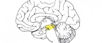
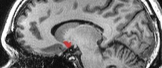
The mesencephalon contains an anatomical formation called the substantia nigra. Neurons of the substantia nigra synthesize dopamine, a hormone and neurotransmitter that transmits impulses in pathways responsible, among other things, for the emergence of a feeling of satisfaction from work and motivation to action. Dopamine is also one of the mediators of the extrapyramidal system.
The disruption of dopamine synthesis in the substantia nigra is undoubtedly associated with a terrible neurological suffering - Parkinson's disease. Another disabling disease, schizophrenia, is also believed to be associated with dopamine metabolism.
In order to deliver dopamine to the overlying structures of the cerebral hemispheres, neurons of the substantia nigra send their processes to the striatum - we will definitely analyze this anatomical formation later. And now we must remember that a significant part of the processes of neurons in the substantia nigra goes upward, to the cerebral hemispheres and carries dopamine there. Three pathways are responsible for this, the most important of which is the nigrostriatal pathway (tractus nigrostriatalis). I schematically marked the course of the nigrostriatal pathway in our illustration:
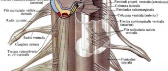
1. Anterior cord, funiculus ventralis (anterior)
, includes the following pathways:
1.1.
Anterior corticospinal (pyramidal) tract, tractus corticospinalis (pyramidalis) ventralis (anterior) - motor tract.
It contains processes of giant pyramidal neurons (gigantopyramidal neurons). The bundle of nerve fibers forming this path lies near the anterior median fissure, occupying the anteromedial sections of the anterior cord. This path transmits control signals from the nerve centers of the cerebral cortex to the anterior horns of the spinal cord. 1.2.
The reticular-spinal tract, tractus reticulospinalis , conducts control signals from the reticular formation of the brain to the motor nuclei of the anterior horn of the spinal cord.
It is located in the central part of the anterior funiculus, lateral to the corticospinal tract. 1.3.
The anterior spinothalamic tract, tractus spinothalamicus ventralis (anterior) , is located somewhat anterior to the reticular-spinal tract.
This tract carries information related to tactile sensation (touch and pressure). 1.4.
The tectospinal tract, tractus tectospinalis , connects the subcortical centers of vision (superior colliculi of the midbrain roof) and hearing (inferior colliculi) with the motor nuclei of the anterior horns of the spinal cord.
It is located medial to the anterior corticospinal (pyramidal) tract. The bundle of these fibers is directly adjacent to the anterior median fissure. This tract transmits control signals intended to carry out reflexive protective motor reactions in response to visual and auditory influences of the environment. 1.5.
The posterior longitudinal fasciculus, fasciculus longitudinalis dorsalis (posterior) , is located between the anterior corticospinal (pyramidal) tract in front and the anterior gray commissure in the back.
This bundle extends from the brain stem to the upper segments of the spinal cord. The fibers of this bundle conduct control signals that coordinate the functions of the muscles of the eyeball and neck muscles. 1.6.
The vestibulospinal tract, tractus vestibulospinalis , is located on the border of the anterior cord with the lateral cord.
This pathway is located in the superficial layers of the white matter of the anterior funiculus of the spinal cord, immediately adjacent to its anterior lateral sulcus. The fibers of this pathway go from the vestibular nuclei of the VIII pair of cranial nerves, located in the medulla oblongata, to the motor cells of the anterior horns of the spinal cord. 2. The lateral cord, funiculus lateralis
, of the spinal cord contains the following pathways:
2.7.
The posterior spinocerebellar tract, tractus spinocerebellaris dorsalis (posterior), (Flexig's bundle) , conducts afferent information (proprioceptive sensitivity).
It occupies the posterolateral sections of the lateral funiculus near the posterior lateral sulcus. On the medial side, the bundle of fibers of this pathway is adjacent to the lateral corticospinal (pyramidal) tract, the red nuclear spinal cord and the lateral spinothalamic tract. Anteriorly, the posterior spinocerebellar tract is in contact with the anterior tract of the same name. P.E. Flechsig, Paul Emil Flechsig, 1847-1929, was a German anatomist and neurologist. 2.8.
The anterior spinocerebellar tract, tractus spinocerebellaris ventralis (anterior), (Gowers' bundle) , also carrying proprioceptive information to the cerebellum, is located in the anterolateral sections of the lateral cord.
Anteriorly, it adjoins the anterior lateral groove of the spinal cord and borders the olivospinal tract. On the medial side, the anterior spinocerebellar tract is adjacent to the lateral spinothalamic and spinocerebellar tracts. V.R. Gowers, William Richard Gowers, 1845-1915, British neurologist. 2.9.
The lateral spinothalamic tract, tractus splnothalamicus lateralis , is localized in the anterior sections of the lateral cord, between the anterior and posterior spinocerebellar tracts on the lateral side and the red-nucleus-spinal and vestibulospinal tracts on the medial side.
This pathway carries information related to pain and temperature sensitivity. The descending fiber systems of the lateral cord include the lateral corticospinal (pyramidal) and extrapyramidal red-nuclear-spinal cord pathways. 2.10.
The lateral corticospinal (pyramidal) tract, tractus corticospinalis (pyramidalis) lateralis , conducts control signals from the cerebral cortex to the anterior horns of the spinal cord.
The bundle of fibers of this pathway, which are processes of giant pyramidal neurons, lies medial to the posterior spinocerebellar tract and occupies a significant part of the area of the lateral cord, especially in the upper segments of the spinal cord. In front of this path is the red nucleus-spinal tract. In the lower segments, it occupies less and less area in sections. 2.11.
The red nucleus-spinal tract, tractus rubrospinalis , is located anterior to the lateral corticospinal (pyramidal) tract.
Adjacent to it on the lateral side in a narrow area are the posterior spinocerebellar tract (its anterior sections) and the lateral spinothalamic tract. The red nucleus spinal tract is a conductor of control signals to the anterior horns of the spinal cord for automatic (subconscious) control of tone and movements carried out through skeletal muscles. In the lateral cords of the spinal cord there are also bundles of nerve fibers that form other pathways (for example, spinal tegmental, olivospinal, etc.). 3. The posterior cord, funiculus dorsalis (posterior)
, at the level of the cervical and upper thoracic segments of the spinal cord, is divided into two bundles by the posterior intermediate groove.
3.12.
The medial bundle, or thin bundle (Gaull's bundle), fasciculus graciiis , is directly adjacent to the posterior longitudinal groove.
F. Goll, Friedrich Goll, 1829-1903, Swiss anatomist. 3.13.
Wedge-shaped bundle (Burdach's bundle), fasciculus cuneatus , is adjacent on the medial side to the posterior horn lateral to the medial bundle. K.F. Burdach, Karl Friedrich Burdach, 1776-1847, German anatomist and physiologist. The thin bundle consists of longer conductors running from the lower torso and lower extremities of the corresponding side to the medulla oblongata. It includes fibers that form part of the dorsal roots of the 19 lower segments of the spinal cord and occupy its more medial part in the posterior cord. Due to the entry into the 12 upper segments of the spinal cord of fibers belonging to neurons innervating the upper limbs and upper body, a wedge-shaped bundle is formed, occupying a lateral position in the posterior cord of the spinal cord. The thin and wedge-shaped fascicles are conductors of information caused by proprioceptive sensitivity (articular-muscular sense). These bundles carry information to the cerebral cortex about the position of the body and its parts in space. In different parts of the spinal cord, the ratios of areas (on horizontal sections) occupied by gray and white matter are not the same. Thus, in the lower segments, in particular in the area of the lumbar thickening, the gray matter occupies the majority of the section. Changes in the quantitative relationships of gray and white matter are explained by the fact that in the lower parts of the spinal cord the number of fibers of the descending tracts coming from the brain is significantly reduced, and the ascending tracts are just beginning to form. The number of fibers forming the ascending tracts gradually increases from the lower to the upper segments. In transverse sections of the middle thoracic and upper cervical segments of the spinal cord, the area of white matter is larger. In the area of the cervical and lumbar thickenings, the area occupied by gray matter is greater than in other parts of the spinal cord.
See: Neurology: Dictionary, Neurology: Internet Resources.
| LIBRARY = LIBRARY Human Physiology = Human Physiology, Human Anatomy = Human Anatomy, Human Biochemistry = Human Biochemistry, Human Psychology = Human Psychology, Medicine = Medicine, Mathematics = Mathematics, Chemistry = Chemistry, Physics = Physics, General Scientific Literature = General Science Lexis. Click here to access any source in the site's library! Click here and receive access to the any reference of the library! “I AM LEARNED. . . N E D O U C H A ?” T E S T V A S H E G O I N T E L L E C T A Premise : The effectiveness of the development of any branch of knowledge is determined by the degree of compliance with the methodology of knowledge - the knowable entity. |
Error? Click here and fix it! Search on the site E-mail of the author (author)
Conducting pathways of the midbrain and underlying structures
Let's note the connection of the midbrain pathways with the pathways of the structures we previously studied, so that we can form a complete concept of the pathways of the central nervous system. In this collection, I will “build” only those pathways that are not directly connected to the cranial nerves, because we will examine the cranial nerves in a separate series of articles.
Red nuclear spinal tract
This path starts from the red nuclei of the mesencephalon, and, going down, first passes through the bridge. Once again we pay attention to the Trout cross and connect the midbrain and the pons:
We can also complete the whole picture using diagrams of the medulla oblongata and spinal cord:
In this diagram, I did not draw the body of the second neuron, which is located in the ventral horns of the spinal cord. We will draw the pathways in more detail in a special lesson, but for now we will remember all the parts of the central nervous system in which we can see the rubrospinal tract.
Do not forget that the rubrospinal tract is one of the main components of the extrapyramidal system, that is, the system of unconscious movements.
Roof-spinal tract
The roof-spinal cord, also known as the tectospinal tract, begins, according to its name, from the roof of the spinal cord, crosses there (Meynert's cross) and then follows all the way to the spinal cord. Let us note the connection between the mesencephalon and the bridge along the roof-spinal tract:
This figure may give the impression that the pons contains nuclei of the tectospinal tract due to the too wide section of the fascicles. Of course, this is just a flaw in my drawing, because the bodies of the second neurons of the tectospinal tract are located in the ventral horns of the spinal cord.
We include the previously studied parts of the central nervous system into our diagram and obtain an almost complete tectospinal tract (with the exception of the body of the second neuron):
Corticospinal (pyramidal) tract
Unlike the previous two tracts, we do not see the entire corticospinal tract. A significant part of it passes through the internal capsule of the cerebral hemispheres, and it ends in the cerebral cortex. However, the corticospinal tract runs like a red thread (ba-dum-shh) through all the parts of the central nervous system we have studied.
Therefore, we can draw an approximate diagram of the lower part of this path, not forgetting that it is repainted at the level of the myelencephalon, and in the spinal cord it is completely divided into two bundles - anterior and lateral.
Spinothalamic tract
We have now moved on to the sensitive pathways. As you remember, the spinal cord had two spinothalamic tracts - lateral and anterior (as well as the pyramidal tracts). Both spinothalamic tracts collected information from the sensations perceived by the skin and transmitted it upward. The spinothalamic fibers transit through the mylencephalon, pons and mesencephalon, heading to their final stop - the thalamus.
This allows us to roughly draw the course of the spinothalamic tracts on our diagram:
Medial loop
In fact, the medial lemniscus is part of the deep conscious sensitivity pathway. The word “deep” in this case means “articular-muscular”, that is, the receptors of this pathway are located in the muscles, joints, fascia and ligaments. Deep sensitivity is contrasted with superficial sensitivity, the receptors of which are located in the skin and carry an impulse along the spinothalamic tract we just discussed.
The full name of this pathway is “ganglio-bulbar-thalamo-cortical pathway.” The previous one, the spinothalamic tract, is completely called the ganglio-dorsal-thalamo-cortical tract. I specifically emphasize this because many students get confused when they call the medial lemniscus the spinothalamic tract. It is easy to confuse these pathways, because both go to the thalamus, and after the thalamus - to the cerebral cortex.
Their morphological difference lies in the fact that the body of the second neuron of the spinothalamic tract is located in the spinal cord, hence the “ganglio-spinal-thalamo-cortical tract.” The body of the second neuron in the tract, a portion of which is called the medial lemniscus, is located in the medulla oblongata, also known as the bulbus, hence the gangliobulbothalamocortical tract. I will draw the pathways with neuron bodies in other lessons, but for now let’s just note the connection of the mesencephalon with the underlying structures along the ganglio-bulbar-thalamo-cortical pathway, which for the most part is called the medial lemniscus:
Latin terms from this article:
- Tractus rubrospinalis;
- Decussatio tegmenti anterior;
- Tractus tectospinalis;
- Decussatio tegmenti posterior;
- Fasciculus longitudinalis medialis;
- Tractus frontopontinus;
- Tractus occipitotemporopontinus;
- Tractus corticospinalis;
- Tractus corticonuclearis;
- Tractus spinothalamicus;
- Lemniscus medialis;
- Lemniscus lateralis;
- Tractus nigrostriatalis.