1.What are extrapyramidal disorders?
Extrapyramidal disorders
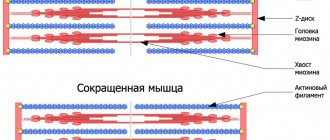
represent disturbances in muscle tone, which affects motor activity.
Movements can be obsessive, uncontrollable, or, conversely, impossible (although previously they did not present any difficulties). The severity of these disorders varies from small tics and paresis to constant trembling or obsessive voluntary contraction of a muscle group.
A must read! Help with treatment and hospitalization!
ES functions
The pyramidal system is responsible for conscious movements in the human body. For example, in order for a person to be able to bring a spoon or fork to his mouth while eating, he needs to think about it in advance. What processes are considered unconscious and do not require the participation of the cerebral cortex to run? You can get the answer only by studying in detail the functions of the extrapyramidal structure. So, she is responsible for:
- regulatory processes of tone in muscle muscles. There is a muscle group that does not relax even during a period of rest. However, a person never thinks twice before “preparing” them for physical activity; this is what ES does;
- protective reflexes of the body. For example, when there is a loud clap or sound, a person involuntarily closes his eyes or flinches;
- maintaining balance. In this case, when a person slips on ice: the tilt of the body changes, and the hands are involved. This is all done on an unconscious level with the participation of the ES;
- skills that are acquired throughout the life cycle and are reinforced. In this case, they talk about the “Caesar” syndrome, which performed several actions at the same time. A person can work calmly and at the same time talk on the phone. All this was made possible with the help of a unique extrapyramidal complex.
If one of the areas of this structure is damaged, a person develops serious impairments in coordination, etc. To understand these processes in more detail, let us consider its structure in detail.
2. Causes of the disease
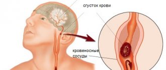
The cause of these disorders is associated with damage to the extrapyramidal system of the brain and neurotransmitter imbalance. The extrapyramidal part of the brain ensures control of posture, smoothness of movements, and their compliance with the intended action. Accuracy, speed and coordination of different muscle groups are also controlled by this system.
Quite often, extrapyramidal disorders occur as a side effect of taking antipsychotic drugs.
. Drug-induced extrapyramidal disorders can also be observed when taking antidepressants, calcium antagonists, antiarrhythmic drugs and drugs prescribed for Parkinson's disease. Side effects of these drugs can occur in the first days of treatment or as a consequence of prolonged regular use (respectively, “early” and “late” drug disorders). Late extrapyramidal disorders can develop even after discontinuation of the drug and be irreversible. This risk must be taken into account when including these drugs in a therapeutic regimen.
Extrapyramidal disorders significantly reduce the quality of life of patients, severely limiting social activity. The psychological status is characterized by anxiety, feelings of inferiority, cognitive impairment, isolation, loss of interest in the outside world and serious feelings of loneliness.
Visit our Neurology page
General concept of ES
So, the pyramidal system is responsible for all conscious movements (walking, speaking, hand movements, etc.).
However, deep in the human brain there is a special extrapyramidal system that is responsible for all our new skills and capabilities. Scientists divided its formation (evolution) into two separate periods:
- non-ostrianar;
- palleostriatal.
The first arose much earlier than the palleostriatal one; together they complement each other. Due to it, processes of slowing down motor activity occur, which in turn are triggered by the second system.
ES originates in the brain (in the area of the pons and protongus) and is directed to the spinal cord. It is considered one of the first to be responsible for human motor activity.
3.Types of extrapyramidal disorders
Ametosis
This type of disorder most often manifests itself in the hands and facial muscles. Characterized by slow wriggling movements of the fingers, which look worm-like and devoid of bones. Twitching of the lips and tongue, curvature and asymmetry may be observed on the face. The facial muscles alternately tense and relax. Such disorders can be the result of birth trauma, encephalitis, syphilis and traumatic brain injuries.
Chorea
This type of disorder is manifested by erratic, irregular movements of the entire body. At the same time, the muscles of the trunk and limbs are characterized by a decrease in tone.
Torsion spasm
A combination of dystonia of the trunk muscles with spasms, up to complete freezing of the entire body. This type of disorder begins with the neck muscles, which involuntarily turn the head to the side. This type of torsion torticollis can develop, involving other muscle groups. In some cases, a “writer’s cramp” is observed - while writing or even when trying to give the fingers a “writing” position, a spasm of the hand occurs due to hypertonicity in the fingers.
Teak
Involuntary repeated contractions of certain muscles (usually the face or neck). This disorder can range from eyelid twitching to obsessive winking, head tilting, and shoulder twitching. As a rule, stressful situations and anxiety intensify the manifestation of this type of extrapyramidal disorder.
Hemiballism
Sweeping unilateral movements of the limbs are observed, reminiscent of throwing up or an attempt to make a grasping movement. This type of obsessive movements most often develops against the background of an infectious brain lesion (tuberculosis, syphilis, encephalitis). It can also occur with severe vascular disorders and metastasis to the brain.
Tremor
Hand tremors, head tremor. When trying to make a precise movement, the amplitude and frequency of movements increase with increasing concentration on the object. In some forms (Parkinson's disease), a “rest tremor” is observed - trembling occurs in a static position, but does not appear during movement.
Facial hemispasm
Spasm of half the face, including the tongue, eyes and neck. This type may be accompanied by sounds such as laughter, crying, or screaming.
The listed types of extrapyramidal disorders are most often combined with each other in different combinations
and are included in the symptom complex of serious diseases of hereditary or acquired origin.
Severe metabolic and cerebral circulation disorders, trauma, and neuroinfections lead to muscle spasms and dystonia. Any changes in tone and loss of control over movements can be a manifestation of severe brain disorders and require immediate contact with a neurologist.
About our clinic Chistye Prudy metro station Medintercom page!
Conducting pathways of the pyramidal and extrapyramidal systems. The limbic system and its connections
PYRAMIDAL PATHWAYS. Main motor
, or the pyramidal corticospinal tract is a system of nerve fibers through which voluntary motor impulses from giant pyramidal neurocytes (Betz pyramidal cells), located in the cortex of the precentral gyrus (5th layer) and the pericentral lobule, are sent to the motor nuclei of the cranial nerves and to the anterior horns of the spinal cord, and from them to the skeletal muscles.
Depending on the direction and location of the fibers, the pyramidal tract is divided into three parts: the corticonuclear tract, going to the nuclei of the cranial nerves; lateral and anterior corticospinal tracts leading to the nuclei of the anterior horns of the spinal cord. The corticonuclear tract
is a bundle of axons of giant pyramidal cells of the precentral gyrus.
This pathway begins in the lower third of the precentral gyrus and passes through the genu of the internal capsule, the base of the cerebral peduncle. The fibers of the cortical-nuclear tract pass to the opposite side to the motor nuclei of the cranial nerves, where they end at synapses on their neurons. The axons of the motor neurons of these nuclei leave the brain as part of the corresponding cranial nerves and are directed to the skeletal muscles of the head and neck. The lateral and anterior corticospinal (pyramidal) tracts begin from the giant pyramidal neurocytes of the precentral gyrus. The fibers of this pathway are directed to the internal capsule, pass through the anterior part of its posterior peduncle, then through the base of the cerebral peduncle and pons, and pass into the medulla oblongata, forming its pyramids. At the border of the medulla oblongata and the spinal cord, part of the fibers of the corticospinal cord passes to the opposite side, continues into the lateral cord of the spinal cord (lateral corticospinal tract) and ends in the anterior horns of the spinal cord with synapses on their motor cells. Thus, the lateral corticospinal tract lies in the lateral cord, consists of neurites of the cells of the cortex of the opposite hemisphere and gradually becomes thinner, since in each segment of the spinal cord part of its fibers ends on the cells of the anterior horns. This pathway carries voluntary motor impulses, stimulating and inhibitory, from the cortex. The fibers of the corticospinal tract that do not pass to the opposite side at the border of the medulla oblongata with the spinal cord descend down as part of the anterior cord of the spinal cord, forming the anterior corticospinal tract . These fibers pass segment by segment to the opposite side through the white commissure of the spinal cord and end with synapses on motor neurocytes (motoneurons) of the anterior horns of the opposite side of the spinal cord. The axons of the anterior horn cells emerge from the spinal cord as part of the anterior root and innervate skeletal muscles. So, all pyramid paths are crossed. It is interesting that the corticospinal tracts terminate on motor neurons of the spinal cord only in humans and primates, while in subprimates, and sometimes in primates, an interneuron is included between them. When the pyramidal tracts are damaged, the reflex mechanisms of the spinal cord are disinhibited, an increase in spinal cord reflexes and muscle tone is observed, protective reflexes are revealed, as well as reflexes that are normally observed only in infants. Damage to the pyramidal tract leads to the development of central paralysis or paresis. EXTRAPYRAMIDAL PATHWAYS . Extrapyramidal pathways in mammals and humans are the morphological basis through which unconditioned reflexes are carried out, regulating the tone of skeletal muscles and carrying out their involuntary automatic innervation. When these pathways are damaged, various types of hyperkinesis and akinesis occur. Extrapyramidal pathways are phylogenetically older than pyramidal ones. They have many connections with the cells and nuclei of the brain stem and with the cerebral cortex, which controls and controls the extrapyramidal system. In this regard, the general beginning of the extrapyramidal tracts can be considered the cerebral cortex, and the place where they end is the nuclei of the brain stem and the anterior horns of the spinal cord. The influence of the cerebral cortex is carried out through a number of formations: the cerebellum, red nuclei, reticular formation, connected to the thalamus and striatum through the vestibular nuclei. The extrapyramidal tracts are divided into three parts: cortical, striopallidal and truncospinal tracts. The cortical extrapyramidal tracts are composed of nerve fibers going from the cells of the cortical motor centers to the formations of the extrapyramidal system. Here the following pathways can be distinguished: corticothalamic, cortico-hypothalamic, corticopontine, cortico-red nuclear and corticotegmental. For example, the cerebral cortex controls the functions of the cerebellum, which is involved in the coordination of movements, through the bridge along the cortico-pontine-cerebellar pathway. There are two of them: fronto-pontocerebellar and occipitotemporal-pontocerebellar. They pass from the cerebral cortex to the pons own nuclei and from them to the cerebellar cortex of the opposite side. Striopallidal pathways are represented by neurites of cells located in the subcortical basal ganglia (in the striatum - in the caudate nucleus, globus pallidus, putamen); these pathways go to the nuclei of the thalamus, hypothalamus, red nucleus, and substantia nigra. Anatomically, these neurites form three main efferent bundles: the lenticular loop, the lenticular bundle and the subthalamic bundle. Truncospinal tracts are formed by nerve conductors running from the nuclei of the midbrain, diencephalon and medulla oblongata to the motor nuclei of the spinal cord and cranial nerves, as part of the following anatomically separate motor tracts. One of the functions of the red nucleus is to maintain muscle tone, which is necessary to involuntarily keep the body in balance. From the red nucleus, nerve impulses are sent to the motor nuclei of the anterior horns of the spinal cord (red nuclear spinal tract). It begins from the midbrain (from the red nucleus), descends along the lateral funiculus of the opposite side of the spinal cord and ends on the motor neurons of the anterior horns. This pathway carries involuntary motor impulses and is important for extrapyramidal movement. In the coordination of human movements in case of imbalance, an important role is played by the vestibulospinal tract , which lies between the anterior and lateral cords and connects the vestibular nuclei of the rhomboid fossa with the anterior horns of the spinal cord. The first neuron of this pathway lies in the nuclei of the 8th pair of cranial nerves. These nuclei are connected to the cerebellum and, through the posterior longitudinal fasciculus, to the motor nuclei of the 3rd, 4th and 6th pairs of cranial nerves. This ensures that the position of the eyeball is maintained during movements of the head and neck. The axons of the second neurons of the vestibulospinal tract descend down as part of the anterior funiculus of the spinal cord and end at synapses on the motor cells of the anterior horns of the spinal cord. Neurons of the reticular formation provide communication between the vestibulospinal tract and the basal ganglia. The tegmental-spinal tract begins from the nuclei of the tegmentum of the quadrigeminal and ends at the cells of the anterior horns of the cervical segments, establishes connections between the extrapyramidal system, as well as the subcortical centers of vision and hearing with the cervical muscles; thus it is associated with auditory and visual perceptions. The olivospinal tract is present in the cervical segments of the spinal cord; starts from the neurons of the olive and ends on the cells of the anterior horns. The spinal reticular tract runs from the reticular formation of the brain stem to the motor neurons of the spinal cord and has a great influence on the functions of the spinal cord. The posterior longitudinal fasciculus begins from the Darkshevich nucleus and ends segment by segment at the motor neurons of the spinal cord. It has connections with all nuclei of the oculomotor nerves and the vestibular nerve. Ensures simultaneous rotation of the eyeballs and head, consistency of movements of the eyeballs. This beam is also called the Schutz beam. The medial longitudinal fasciculus lies in the anterior cord and consists of both descending and ascending fibers;
originates and ends on the nuclei of the brain stem and on the cells of the anterior horns; innervates the muscles of the neck. The bundle is a very ancient system of fibers, which in lower vertebrates serves as the most important association pathway of the brain. All these pathways functionally unite the body into a single whole and ensure the consistency of its actions. Limbic System Connections
The limbic system is connected with the higher centers of the cerebral cortex, as well as with the more primitive centers of the brainstem. It not only allows emotions to influence the body, but also regulates emotional reactions.
There is danger!
The consequences of each specific syndrome may differ from each other, but there are also similar indicators:
- depression;
- sleep disturbance;
- body aches;
- skeletal muscle necrosis;
- vein thrombosis;
- renal failure;
- liver failure;
- breathing problems;
- relapse of psychiatric illness;
- arrhythmias, myocardial infarction.
Extrapyramidal disorders are a complex system of syndromes of a neurological nature that affect not only a person’s health status, but also his adaptation in society. Adequate treatment may be the only way for a person to live and work normally.
Relationships between the structures of the extrapyramidal system
Until now, the interaction processes remain incompletely studied. The ES is directly connected to the thalamus, reticulatory nuclei, pons, cerebellum, etc. For full functioning, gamma motor neutrons from the spinal cord are added to all of these structures.
This system is closely related to the pyramidal structure. Thanks to this interaction, a person organizes all movements that are provoked by parts of the pyramidal system. The processes of the red nucleus of the extrapyramidal system form the so-called rubrospinal tract. It is responsible for the motor processes of the human upper limbs.
The vestibular region of the ES is closely connected with the area of the inner ear, cerebellum and some parts of the spinal cord. Thanks to this, a person makes movements with his neck, torso, head and limbs. In addition, the relationship with various structures of the brain provides the functions of blinking, turning the head and controlling muscle contractions. When one of the processes is disrupted, a person experiences various complications.