Localization
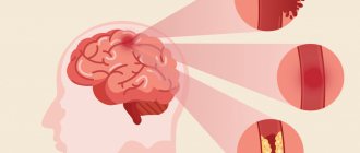
Color agnosia occurs in most cases with lesions in the left occipital lobe that extend to the temporal or parietal lobe. There are known cases of color agnosia with bilateral occipital lesions. Functional and localization studies indicate that areas V4, V8 of the visual cortex and the lingual gyrus are involved in color processing. Thus, studies of single neurons have shown a possible role in the development of this syndrome of the V4 region, which is responsible for color perception. This area contains many cells that are sensitive to the shape and color of visual stimuli. It is assumed that the color properties of the stimulus can be integrated with the system that describes contours and shapes, and their connection with the development of color agnosia can be destroyed. However, it is unclear under what conditions lesions of area V4 lead to the development of visual object agnosia and under what conditions to color agnosia. Probably, color agnosia occurs when a lesion spreads from the medial occipitotemporal region to various parts of the temporal and parietal lobes involved in the process of recognizing a visual stimulus based on the properties of shape, contour and spatial relationships, which in certain specific areas are integrated with the results of color processing information. This assumption requires additional research. Modern studies report cases of covert processing of information about colors, when subjects with color agnosia successfully coped with implicit color recognition tasks. This led the authors to propose that color agnosia arises from a failure to access and retrieve chromatic information. This study supports the involvement of multiple neural mechanisms and different neuroanatomical regions in color processing.
Agnosia - what is it, causes, types, symptoms, treatment, prognosis
Agnosia is a consequence of certain disorders that are often observed in the brain. Perception is distorted, which becomes a symptom of agnosia, which comes in several types. Its causes lie in diseases that require treatment.
A person perceives the world through the senses. The eyes see, the ears hear, the tongue tastes, the nose smells, etc. These analyzers can work fully. However, perceptual error can occur at the level of information processing in the brain. A person perceives everything that surrounds him distortedly. This may be agnosia, which will be discussed in the online magazine psytheater.com.
Symptoms of agnosia
Symptoms of agnosia are recognized by the manifestations that occur. Depending on the area of brain damage, certain types of agnosia arise, which are considered.
For example, damage to the occipital-parietal lobe leads to the fact that a person is able to see an object, but cannot name it.
And damage to the temporal region leads to the inability to perceive familiar speech (it is heard as a set of sounds).
- A person is not able to navigate in space and on a map.
- A person cannot recognize objects by touch.
- A person does not perceive the presence of defects in himself, although they undoubtedly exist.
- A person is indifferent to the fact that he has defects.
- A person cannot recognize sounds, their nature, source of origin, etc.
- A person incorrectly perceives his own body, which seems to him to be of a different shape or with a different quantity.
- A person does not recognize familiar faces, but remembers information about these people.
- A person does not see the picture as a whole, although he recognizes individual objects in it.
- A person sees only half of space.
go to top
Causes of agnosia
The question naturally arises as to what causes agnosia. No person would want to face such a disease. Avoiding factors can help achieve this desire.
The main cause of agnosia is damage to the parts of the brain that are responsible for processing information received through analyzers. Factors that lead to damage are:
- Chronic circulatory disorder in the brain (stroke), which leads to dementia.
- Alzheimer's disease, which is accompanied by the accumulation of amyloid, a protein that should normally break down.
- Inflammatory processes in the brain: encephalitis.
- Parkinson's disease.
- Tumors in the brain.
- Consequence of traumatic brain injury.
go to top
Treatment of agnosia
Agnosia occurs quite rarely, but it significantly prevents an ordinary person, who has all his senses and sensitivity, from living fully.
Efficiency and quality of life decrease because a person cannot adequately and fully perceive the world around him.
Treatment is prescribed after diagnosing agnosia, where the cause and location of the pathology are first identified.
Complaints are collected (when the disease appeared, how quickly symptoms develop, what preceded agnosia), after which the following measures are prescribed:
- Neurological examination to identify changes and pathologies in tissues. The sharpness of the analyzer is checked.
- Examination by a neuropsychologist who conducts tests and surveys.
- CT and MRI to identify damaged areas in the brain.
- Consultation with a psychiatrist, otolaryngologist, cardiologist, ophthalmologist as necessary.
There are no specific treatments for agnosia. Doctors focus all their efforts on eliminating the cause that provoked damage to the brain, after which the individual is sent for additional corrective work as rehabilitation:
- Speech therapy sessions, especially for auditory agnosia.
- Occupational therapy.
- Psychotherapeutic sessions.
- Working with teachers.
Treatment of the underlying disease is prescribed individually. Here, tumors can be removed from the brain, blood pressure can be monitored, and medications can be prescribed to improve blood circulation in the brain and medications to eliminate neuropsychological pathologies.
The rehabilitation period can last about 3 months. In case of serious damage to a part of the brain, treatment may take 10 months or more.
go to top
Autotopagnosia
And this is the second type of disorder related to somatoagnosia. A person’s presence can be recognized by his ignoring half of his body. And many patients simply do not recognize some of its individual parts or incorrectly assess their position in space.
This type of pathology includes:
- Hemisomatoagnosia. A person ignores half of the body, but its functions are partially preserved. The patient simply does not use them. He may “forget” that he has a left arm, leg, etc.
- Somatoparagnosia. The patient perceives the affected part of the body as foreign. He may seriously believe that there is someone else next to him, and it is he who owns his second leg, arm, etc. In severe cases, people feel the “separateness” of the body, as if it were sawn into two parts.
- Somatic allosthesia. The patient feels an increase in the number of his limbs. He may think that he has two or three left hands, for example.
- Autotopagnosia of posture. A person is not able to determine exactly what position his body parts are in. He really doesn't know whether his hand is down or up, whether he's sitting or lying down.
- Disorientation. With this pathology, the patient does not know such concepts as “right” and “left”. This is usually caused by damage to the left parietal lobe.
- Finger agnosia. Specific pathology. If it is present, a person is not able to show on his hand the same finger that the doctor shows on his hand.
Apractoagnosia is also a disorder of this type. This is a special case. People with this disorder have difficulty performing spatially oriented movements. It is difficult for them to make the bed (they place the bedspread crosswise rather than lengthwise), find their way into a room or ward, get their foot in a pant leg, or put on a T-shirt with the right side.
As in any other case, when diagnosing somatoagnosia, neuropsychological methods, samples and tests are used.
Treatment of agnosia
There is no specific therapy for this pathological condition. Basic treatment of agnosia is aimed at treating conditions that have led to damage to the cortical and subcortical structures of the brain. In each individual case, the therapeutic tactics are determined by the severity of the manifestations, the severity of the condition and the location of the pathological changes, the course of the disease and the presence of complications.
To develop a plan for adequate therapeutic care, it is necessary to conduct a full diagnostic examination:
— conducting a complete examination of the patient, collecting anamnesis, determining the presence of hereditary diseases;
— diagnostic manipulations aimed at identifying the tumor process, the presence of trauma, the presence of vascular accidents;
— consultations with specialists of a narrow profile (ophthalmologist, otolaryngologist, cardiologist, psychiatrist) to exclude other possible causes of this symptomatology;
— conducting diagnostic tests that reveal the degree of change in perception;
— carrying out diagnostic procedures aimed at identifying areas of damage to the brain cortex (CT, MRI).
To directly correct the manifestations of agnosia, it is necessary to work with a neuropsychologist, speech therapist, and the use of occupational therapy.
The recovery period is about three months. The severe course of the disease leading to agnosia and its complications can increase the duration of therapeutic procedures to up to a year. If necessary, treatment is repeated, but when the cause is eliminated and agnosia is completely corrected, relapses often do not occur.
According to the latest statistics, with proper and timely diagnosis of the underlying disease and its manifestations, adequate and complete therapy and corrective measures, carried out fully, will lead to a complete restoration of the analyzers’ functioning.
If you do not consult a doctor in a timely manner, ignore prescribed recommendations or do not implement them in full, or use self-medication, the prognosis may be unfavorable, and the risk of developing irreversible processes in the structure of the brain cortex may increase. The unfavorable outcome of the disease may be influenced by the patient’s age, nature and severity of the disease.
The impact of agnosia on the patient’s quality of life depends on the type of this pathology, for example, simultaneous agnosia or spatial perception disorder significantly worsens the patient’s quality of life, reduces work activity, and impairs communication skills. Whereas, tonal or finger agnosia occurs almost unnoticeably.
Primary prevention of agnosia comes down to the prevention of major diseases, the manifestations of which may be agnosia - maintaining a healthy lifestyle, a nutritious, healthy diet, prevention of stressful conditions. If the first signs of pathology occur, you should immediately contact a specialist.
Symptoms
Symptoms of the disease depend on the damage to one or another analyzer and can serve as a diagnostic criterion for differentiating damage to parts of the brain. Depending on their manifestation, I distinguish the following types of agnosia:
- Prosopagnosia
is a violation of memorization and subsequent recognition of faces with preservation of mnemonic functions relating to objects. In severe cases, patients cannot recognize their own reflection in the mirror; - Lissauer object agnosia
is a violation of the recognition of individual objects; - Color agnosia
- the inability to determine the color belonging to certain objects, as well as to select identical colors or their shades; - Simultaneous agnosia
- limitation of the visual field to only one visible object or the ability to perceive only one object, regardless of its shape, color and size; - The weakness of optical concepts is the inability to create a description of any characteristics of a visible object;
- Balint's syndrome
is the inability to direct gaze towards an object and then fixate it, while maintaining the ability to move the eyeballs; - One-sided
spatial agnosia - loss of one side of space from the visual field; - Violation of stereoscopic vision
- the impossibility of comparing a single whole of visual images entering each eye; - Deep agnosia
is the inability to correctly determine the location of objects located in front of the patient; - Violation of topographic orientation
- loss of orientation in places familiar to the patient, while maintaining other cognitive functions; - Auditory speech agnosia
- recognition of speech as a set of unrelated sounds; - Simple auditory agnosia
- the inability to recognize individual sounds; - Tonal agnosia
- the inability to recognize timbre, as well as the emotional coloring of speech, while maintaining other functions of the auditory analyzer; - Autotopagnosia hemicarpus
- complete ignorance or denial of the presence of the left half of the body with complete or partial preservation of its functioning; - Somatic aloesthesia
is a feeling of the presence of static or dynamic extra limbs; - Autotopagnosia of posture
is the inability to determine the position of the body or its individual parts in space; - Pretman syndrome
(finger agnosia) - the inability to correctly point to the finger shown by the interlocutor; - Violation of “right-left” orientation - the inability to point to the right or left parts of the body.
Kinds
The disease has three main types: visual agnosia, auditory agnosia and tactile agnosia. In addition, there are several less common types of the disease (spatial agnosia and other perceptual disorders).
In visual agnosia, the lesions are localized in the occipital lobe of the brain. This type is characterized by the patient's inability to recognize objects and images, despite the fact that he retains sufficient visual acuity for this. Visual agnosia can be expressed in different ways and manifest itself in the form of the following disorders:
- object agnosia (damage to the convexital surface of the left part of the occipital region): inability to recognize various objects, in which the patient can only describe individual features of an object, but cannot say what kind of object is in front of him;
- color agnosia (damage to the occipital region of the left dominant hemisphere): inability to classify colors, recognize identical colors and shades, correlate a specific color with a specific object;
- visual agnosia, manifested in weakness of optical representations (bilateral damage to the occipital-parietal region): inability to imagine any object and characterize it (name size, color, shape, etc.);
- agnosia for faces, or prosopagnosia (damage to the lower occipital region of the right hemisphere): a violation of the process of recognizing faces while maintaining the ability to distinguish between objects and images, which in especially severe cases can be characterized by the patient’s inability to recognize his own face in the mirror;
- simultaneous agnosia (damage to the anterior part of the dominant occipital lobe): a sharp decrease in the number of simultaneously perceived objects, in which the patient is often able to see only one object;
- Balint's syndrome, or visual agnosia caused by optomotor disorders (bilateral damage to the occipital-parietal region): inability to direct the gaze in the right direction, focus it on a specific object, which can be especially pronounced when reading - the patient cannot read normally, since It is very difficult for him to switch from one word to another.
Auditory agnosia occurs when the temporal cortex of the right hemisphere is damaged. This type is characterized by the patient’s inability to recognize sounds and speech, while the function of the auditory analyzer is not impaired. The following disorders are distinguished in the category of auditory agnosia:
- simple auditory agnosia, in which the patient cannot recognize simple, familiar sounds (the sound of rain, rustling paper, knocking, creaking doors, etc.);
- auditory-verbal agnosia - the inability to distinguish speech (for a person suffering from this type of auditory agnosia, native speech is presented as a set of unfamiliar sounds);
- tonal auditory agnosia - the patient cannot perceive the tone, timbre, or emotional coloring of speech, but at the same time he retains the ability to normally perceive words and correctly recognize grammatical structures.
With tactile agnosia, the patient is unable to identify objects by touch. One type of tactile agnosia is the patient’s inability to recognize parts of his own body and evaluate their location relative to each other. This type of tactile agnosia is called somatoagnosia. Tactile agnosia, in which the process of recognizing objects through touch is disrupted, is called astereognosia.
There are also spatial agnosias, which are expressed as a violation of the identification of various parameters of space. With lesions of the left hemisphere, it manifests itself in the form of impaired stereoscopic vision; with lesions of the middle parts of the parieto-occipital region, the disease can be expressed as the patient’s inability to correctly localize objects in three spatial coordinates, especially in depth, as well as to recognize parameters further or closer.
There are also types of agnosia such as unilateral spatial agnosia - the inability to recognize one of the halves of space (usually the left), and spatial agnosia, expressed in a violation of topographic orientation, in which the patient may not recognize familiar places, but he does not have any memory impairment .
One of the rarest types of agnosia is impaired perception of time and motion - a condition in which a person is unable to judge the speed of time and perceive the movement of objects. The last disorder (inability to perceive moving objects) is called akinetopsia.
Pathogenesis
The cerebral cortex has three main groups of associative fields that provide multi-level analysis of information entering the brain. Primary fields are connected to peripheral receptors and receive afferent impulses coming from them. Secondary association areas of the cortex are responsible for analyzing and summarizing information coming from primary fields.
Next, the information is transferred to tertiary fields, where higher synthesis and development of behavioral tasks are carried out. Dysfunction of secondary fields leads to disruption of this chain, which is clinically manifested by the loss of the ability to recognize external stimuli and perceive holistic images. In this case, the function of the analyzers (auditory, visual, etc.) is not impaired.
Treatment
Initially, treatment is aimed at eliminating the underlying disease, for example, antibiotic therapy for brain abscesses, surgery, radiation therapy for brain tumors, etc.
Rehabilitation through speech therapy, correction of disorders by speech pathologists, psychological support, consultations with a neuropsychologist who can help adapt to the characteristics and manifestations of the disease can be effective.
The doctors
specialization: Neuropsychologist / Neurosurgeon / Speech therapist / Psychologist
Chesnova Tatyana Andreevna
10 reviewsSign up
Find a doctor and make an appointment
Medicines
Cavinton Picamilon Fezam Lutsetam
Conservative drug treatment is carried out using drugs that improve metabolism and cerebral blood flow. These include:
- Cavinton is a psychostimulant nootropic drug that helps reduce the severity of neurological and mental disorders. Intended for intravenous infusion at a rate of 80 drops/min, 20 mg per day.
- Picamilon is a nootropic drug that dilates blood vessels in the brain with additional tranquilizing, psychostimulating, antiplatelet and antioxidant effects. The standard daily dose of 60-150 mg can be divided into 2-3 doses, the duration of treatment is 1-2 months, it is recommended to repeat the course six months later.
- Trental is a vasodilating agent that can reduce viscosity and improve the rheological properties of blood, as well as promote microcirculation in areas of impaired blood flow. Usually doctors prescribe taking 1-2 tablets 3 times a day, maximum 15 tablets.
- Phezam is a drug that improves metabolic and microcirculatory processes in the brain. Recommended daily dose: take 1-2 caps. 3 times a day for 1-3 months, repeat the course of treatment 2-3 times a year.
- Lucetam is a nootropic drug that can increase concentration and improve cognitive function. Typically, the daily dose is 2.4-4.8 g/day, divided into 2-3 doses, but it is best that the treatment regimen is selected by the attending physician, depending on the characteristics and severity of the disorders.
Patients are most often recommended individual psychotherapy sessions, sessions with speech pathologists and speech therapists.
Diagnostic methods
The Clinical Institute of the Brain has all the conditions for a full diagnosis of patients with agnosia. At the initial stage, it is important to determine exactly what difficulties the patient has with the perception of the environment. For this purpose, there are special tests, during which the patient is asked to describe objects, sounds, and point to parts of the body. After determining the type of disorder, it is important to conduct brain imaging using computed tomography or magnetic resonance imaging. This research method will detect neoplasms, heart attacks, vascular damage and other anomalies that can cause agnosia.
Types of agnosia
The described disorder is characterized by three main types: tactile, visual and auditory perceptual disturbances. In addition, we can distinguish a number of less common forms of the disease in question (for example, spatial agnosia).
Visual agnosia is characterized by the presence of a lesion in the occipital region of the brain. This form of the disease manifests itself in the inability of patients to recognize images and objects while maintaining visual acuity. The type of pathology in question can manifest itself in different ways. The following forms of visual agnosia are distinguished: object, color, visual, simultaneous agnosia, prosopagnosia and Balint's syndrome.
Auditory perceptual dysfunctions arise due to damage to the temporal cortex of the right hemisphere. This type of agnosia is represented by the inability of individuals to recognize speech and sounds against the background of normal functioning of the auditory analyzer. Auditory agnosia, in turn, is divided into simple auditory perception disorder, auditory speech and tonal auditory agnosia.
A simple disorder of auditory perception is characterized by the inability of people to recognize simple, previously familiar sounds, such as the sound of rain, the rustling of the sea, knocking, a doorbell, creaking, etc.
Auditory speech agnosia is the inability to recognize speech. To a person suffering from the described form of agnosia, native speech seems to be a set of unfamiliar sounds.
Tonal hearing disorder is characterized by the inability to perceive the emotional coloring, tone, and timbre of speech while maintaining the ability to adequately perceive words and correctly distinguish grammatical structures.
Tactile agnosia is the inability to identify objects or things by touch. The following types of agnosia are distinguished: somatoagnosia, astereognosia and disturbance of spatial perception. The patient's inability to recognize parts of his own body and evaluate their location relative to each other is called somatoagnosia. A disorder of tactile perception, in which the process of recognizing objects and things through touch is called astereognosia.
There are also disturbances in spatial perception, expressed in the form of incorrect identification of space parameters. Damages to the middle areas of the occipital-parietal region are revealed in the inability to measure quantities closer or further away, as well as to correctly place objects in three-dimensional space, especially in depth; damage to the left hemisphere entails spatial agnosia, manifested by impaired stereoscopic vision. In addition, there are such types of agnosia as a unilateral violation of spatial perception and a perceptual disorder consisting in the inability to topographically navigate the terrain. Unilateral spatial agnosia is the inability to recognize one half of space. Violation of topographic orientation is expressed in the inability to recognize familiar places against the background of intact memory function.
One of the rarest types of agnosia is dysfunction of the perception of movement and time. This disease manifests itself in a violation of the correct understanding of the movement of objects and an adequate assessment of the speed of the passage of time. The inability to perceive objects in motion is called akinetopsia.
Treatment of the disease
In medicine, there is no single protocol for the treatment of agnosia, since it depends on the root cause of the disease, its type and degree of neglect. In parallel with the treatment of the main illness, work is carried out with a psychiatrist, speech therapist, and neuropsychologist. This is necessary in order to help a person adapt to life in the presence of such pathologies. There are cases when treatment brought instant results and when it lasted for many years. The effect depends on how quickly the patient sought professional help. The most commonly used drugs are the following groups: vascular drugs, neuroprotectors and nootropics, B vitamins.
Symptoms of agnosia
Damage to the cerebral cortex, which is responsible for the analysis and synthesis of information, gives rise to agnosia. Therefore, the symptoms will depend on the location of the affected area of the brain. For example, as a result of damage to the left zone of the occipital region, object agnosia arises, which consists of the patient losing data about the object and its purpose. In other words, an individual suffering from this perceptual disorder sees an object and can describe it, but is unable to name it or talk about its purpose. When the temporal region is damaged, an auditory-verbal perception disorder occurs: the patient perceives the speaker’s speech as if it were an ordinary set of sounds; he is unable to perceive the meaning of phrases and distinguish individual words. Statistics confirm that the disorder in question is quite rare.
The causes of agnosia are as follows: dysfunctions of the temporal and parietal areas of the brain, where data on the use of familiar objects are stored (more often it occurs suddenly after a stroke, heart attack or head injury, when the cortex and nearby subcortical formations of the brain are affected, and damage to the cortex can cause a tumor process ). In addition, the pathology in question may arise as a result of degeneration of areas of the brain that are responsible for the integration of perception, memory and identification processes.
Thus, the main causes of agnosia are damage to the parietal and occipital areas of the cerebral cortex, which occur, in addition to the above pathologies, with the following ailments:
- chronic circulatory disorder in the brain, which later develops into dementia;
- inflammatory processes of the brain (for example, encephalitis);
- Alzheimer's disease, which is associated with the accumulation of amyloid in the brain (a specific protein that normally breaks down quickly in the brain);
- Parkinson's disease, characterized by the occurrence of progressive muscle stiffness, tremors and a number of neuropsychological disorders, including apraxia.
There are different types of perceptual dysfunction depending on the location of the affected area in the brain. For example, if the parieto-occipital zone is damaged, a violation of topographic orientation occurs; if the right subdominant part of the parietal lobe is damaged, anosognosia occurs, which is the absence in patients of a critical assessment of their own illness or defect. For example, people suffering from this form of dysfunction consider themselves completely healthy even against the background of immobility on one side of the body (state of paralysis).
Many people far from medicine wonder about agnosia, what is it, what are the symptoms of this disease, how do they manifest themselves?
The following manifestations and symptoms of agnosia can be distinguished:
- violation of spatial orientation and the ability to “read” on the map, that is, to understand the location of cities, regions and other places on the map;
- a disorder in the ability to recognize objects by touch (sick people find it difficult to determine the texture, configuration and shape of an object;
- denial of the fact of having a physical defect or illness (for example, blindness, deafness), despite the indisputability of the existing defects;
- indifference to the existing defect (a person may be little worried about sudden deafness, blindness or other defects;
- impairment of the ability to recognize sounds (the patient is not able to distinguish the nature of the sound, understand where it is coming from, for example, when he hears a bell in his own house or the voice of a relative;
— dysfunction of perception of one’s own body (people are not able to correctly determine the number of their limbs or their length);
- a disorder in the ability to recognize the faces of friends, along with this, patients are able to name their approximate age or gender;
- impaired recognition of complex visual images, while patients retain the ability to recognize individual components of these images, for example, an individual, looking at an image, recognizes a jug on the table, but is not able to understand that the presence of a jug, glasses, plates, food on the table, shows that the picture shows a feast;
- ignoring part of the visible space (for example, a patient, while eating food, eats food only from the right side of the plate).
Anosognosia
This is one of the types of somatoagnosia - a pathology in which a person does not recognize parts of his own body. This type of disease includes the following manifestations:
- Anosognosia hemiplegia. The person does not realize that he has unilateral paresis or paralysis, or denies it.
- Anosognosia of blindness. The patient denies or is unaware that he is not sighted. He perceives confabulatory images as real.
- Anosognosia aphasia. With this disorder, a person does not notice his speech errors at all, even if he speaks as unintelligibly as possible.
In simple terms, the term “anosognosia” refers to a lack of awareness of one’s illnesses and illnesses. A person not only denies them out of principle - he really sincerely believes that he is healthy.
Classification of impaired perception
Depending on the location, the types of agnosia differ.
Agnosia comes in three main types, each of which in turn is divided into subspecies.
Visual agnosia
With this type of disorder, brain damage is localized in the occipital region. With visual agnosia, the patient cannot recognize an object or picture. The patient does not have visual impairment.
This type of agnosia manifests itself in different ways:
- Subject. With damage to the surface of the brain, which is adjacent to the left side of the occipital bone: objects are unrecognizable (the patient describes it in general terms).
- Colored. With lesions in the occipital region of the left hemisphere, which is considered dominant: the patient has difficulty distinguishing colors and shades, cannot match an object and a color.
- Visual, manifested in optical weakness (optical spatial). If the occipital and parietal region is affected on both sides: the patient cannot imagine the object and describe it.
- Face perception disorder. With lesions of the lower region of the back of the head of the right hemisphere: the patient cannot recognize faces, even his own reflection, but distinguishes between objects and pictures.
- Simultaneous. With lesions of the anterior part of the dominant occipital lobe: the patient cannot simultaneously perceive several objects. Autotopagnosia is characterized by the inability to recognize one's body.
- Balint's syndrome. It is noted with bilateral damage to the occipital-parietal region: the patient cannot look in the right direction, focus his gaze, and cannot read normally.
Auditory agnosia
With this type of disorder, damage occurs to the cortex in the temporal region of the right hemisphere.
A patient with auditory agnosia cannot recognize speech or sounds, but a disorder of the auditory analyzer is not diagnosed.
This type of pathology has such disorders that characterize each subtype of auditory agnosia, such as:
- Simple. The patient does not recognize simple sounds.
- Hearing-speech. The patient is unable to distinguish speech.
- Tonal. The patient does not grasp tone, timbre, or emotionality, but at the same time perceives phrases and words normally.
Disorders of tactile and spatial sensations
Tactile agnosia is characterized by the fact that the patient cannot recognize an object by touch. It happens that the patient cannot even recognize a part of his own body and evaluate its location (somatoagnosia). With astereognosia, the patient cannot recognize an object using touch.
Spatial agnosia:
- damage to the left cerebral hemisphere: impairment of spatial vision (optical-spatial), in this case there is often an intersection with visual agnosia;
- damage to the middle part of the parietal and occipital region: the patient cannot localize an object in space.
There is also a spatial neurological disorder:
- which is expressed in the violation of one of the halves of the space;
- which is expressed in a violation of topographic orientation.
A rare type of disorder is considered when the patient does not perceive time frames and movements (akinetopsia).
Visual object agnosia in brain lesions (review)
G.V. Tikhomirov, I.O. Konstantinova, M.M. Tsirkova, N.A. Bulanov, V.N. Grigorieva
Key words : visual agnosia; theory of two streams of visual information; striate and extrastriate cortex; object agnosia; method of mapping the lesion.
Violations of visual gnosis are one of the possible causes of disability in patients with brain damage, but their prevalence and clinical significance in the neurological clinic are underestimated. The review provides an idea of visual object agnosia as a manifestation of brain pathology.
Modern views on the neuroanatomical and neurophysiological foundations of visual object gnosis are presented. Clinical variants of visual object agnosia, their morphological substrates, features of neuropsychological diagnostics and basic approaches to the rehabilitation of patients are described.
The unique capabilities of computer technologies for implementing the principles of physical measurements, digital mapping and controlled optimization in the diagnostic and rehabilitation process and, in particular, in visual object agnosia are presented.
The need to develop standardized valid methods for diagnosing visual object agnosia to improve ways of their correction in neurological practice is emphasized.
Introduction
Visual gnosis (recognition, recognition) - the ability to recognize the object seen, i.e. understand the meaning of a previously familiar visual stimulus when perceiving it as a whole or individual parts [1, 2]. Accordingly, visual agnosia is understood as the inability of a person to recognize an object or part of an object using vision alone while maintaining elementary visual functions (visual acuity and visual fields, sensitivity to spatial contrast, color vision), speech, memory and the ability to recognize objects by sound or tactile characteristics [2–6].
Visual agnosia is a monomodal disorder, therefore, clinical cases in which patients, along with a violation of visual gnosis, have signs of a disorder of the gnostic functions of other modalities, according to a number of experts, should not be regarded as cases of visual agnosia itself [3]. Visual agnosia can be observed in the clinic of many diseases and brain injuries, but they still remain one of the least studied disorders in neurology [7]. Meanwhile, the relevance of timely diagnosis of visual gnosis disorders is determined by their negative impact on the quality of life of patients and the need for early correction [8].
Currently, most authors identify such types of visual agnosia as visual agnosia of objects and shapes, facial agnosia, topographic agnosia, and letter agnosia [2, 8]. The least studied is visual agnosia of objects and forms (object agnosia), which is understood as the inability to visually recognize complex objects or drawings and differentiate between classes of stimuli, despite the intactness of basic visual functions [9]. Patients with object visual agnosia do not recognize previously familiar objects and are unable to learn to recognize new objects based on appearance alone [10]. In addition, such patients have reduced control over the correct recognition of an object [2]. Object visual agnosia may be accompanied by impaired recognition of familiar faces (prosopagnosia), and less commonly, letters and words (“pure” alexia without agraphia) [3, 11].
Classification of visual object agnosia
Object agnosia is divided into apperceptive and associative forms [5, 10, 12, 13]. Apperceptive object agnosia is manifested by the inability to copy an object, as well as to find similarities/differences between objects [13–15], while associative agnosia of objects is characterized by a violation of the identification of the latter due to the loss of knowledge about their meaning: the patient is able to draw an object, describe its parts and find similarities between different objects, but is not able to recognize the object that was just presented and sketched [14, 15].
Apperceptive visual agnosia. It is divided into form agnosia, transformational agnosia and integrative agnosia [5, 16, 17].
With apperceptive shape agnosia, patients cannot recognize simple geometric shapes and, therefore, such elementary properties of objects as curvature and volume. They make mistakes in tests for recognizing and comparing objects, and are also unable to draw or copy an object they see [3]. Recognition is aided by tracing the outline of an object with one’s hands and feeling it, whereby visual perception is converted into kinesthetic perception [10, 12]. A number of authors believe that this disorder is not purely agnostic and is more correctly designated as “pseudoagnosia” [12].
A patient with apperceptive integrative visual agnosia is not able to put the details of an object into a single whole and therefore cannot recognize an object and distinguish between correct and incorrect images of real objects, although he can perceive their individual elements and copy images in parts [10, 12, 16, 18].
Apperceptive transformational agnosia is the inability to recognize three-dimensional objects in cases where the patient looks at them from unusual points of view and must perform a “mental rotation” on them [10, 12, 19]. Some authors attribute such a deficit to spatial agnosia. Other researchers believe that the term “spatial agnosia” in this context can create confusion, since it gives the specialist the impression that transformational agnosia is based on errors in spatial information processing, and not on impairments in the ability to perceive the same object from different angles of view [12 ].
Associative object agnosia . It is characterized by the fact that the patient is not able to identify an object and determine its semantic category, although he can analyze the structure of the object [18]. Some authors question the possibility of recognizing this disorder as agnosia itself, since its mechanisms are closely related to selective impairment of visual memory. It is believed that in patients with associative object agnosia, not only previously acquired knowledge about objects suffers, but also the ability to acquire new visual experience [10, 15]. JJ Barton [10] proposed to distinguish two variants of the associative form of visual agnosia, depending on whether the patient has impaired access to stored traces of the visual image of an object (semantic access agnosia) or whether the visual representation of the object in memory itself is lost (complete semantic agnosia).
Along with the two described forms of associative visual object agnosia, the existence of category-specific agnosia is discussed - pathological conditions in which patients are not able to recognize stimuli related to specific specific categories, in particular to a group of living or inanimate objects [10].
Neurophysiological and morphological basis of visual object gnosis
Explanations of the nature of visual agnosia are in most cases based on ideas about the normal processes of visual image formation, which are conventionally divided into three levels [4]. At the first (lowest) of them, the visual stimulus is processed and information about the simplest physical properties of the object is analyzed. The anatomical basis of these processes are the structures of the eye, the corresponding pathways, visual subcortical centers and the primary (striate) visual cortex. The second (middle) level includes the synthesis of visual information about the properties of an object, which is carried out with the participation of the extrastriate cortex and at the psychological level correlates with the formation of images [20–22]. At the third (highest) level, a synthesis of multimodal information occurs, which is necessary to endow the image of an object with a certain meaning; the neurophysiological basis of such synthesis is the activity of multimodal associative areas of the cortex [4].
To understand the nature of object gnosis, the hypothesis of Leslie Ungerleider and Mortimer Mishkin about two streams of extrastriate visual impulses, one of which follows from the primary visual cortex to the parietal cortex and is associated with the processing of information about the position and direction of movement of an object (the “where?” path) is important. , and the second goes to the temporal cortex and is related to image recognition and categorization (the “what” path?). Later, it was suggested that these streams have slightly different functional differences: with the participation of the ventral pathway, information necessary for the perception of stimuli and awareness of the surrounding world (vision for recognition) is processed, and with the participation of the dorsal pathway, information important for the control and programming of actions is processed ( vision for action) [5, 22–26]. The ventral pathway involves the medial occipitotemporal structures, and the dorsal pathway involves the lateral occipito-parietal structures [12, 25].
Currently, the idea of the isolation of visual information flows in the extrastriate cortex has begun to be challenged, as evidence has emerged that both recognition of objects and actions with them can be impaired regardless of the location of the lesion in the structures of the ventral or dorsal pathway [5, 27, 28] . In this regard, a modified model of the above-described two-stream theory has been proposed, according to which parallel and hierarchical coding of object information in the dorsal and ventral systems can be constantly modified within the framework of the interaction of the two streams before convergence in the prefrontal cortex [14, 22, 28, 29].
To verify the morphological substrates of visual object gnosis disorders, a comparison of clinical data and the results of neuroimaging studies is carried out. The method of lesion mapping is becoming increasingly widespread, which consists of a statistical analysis of the dependence of the clinical picture (in particular, manifestations of visual agnosia) on the localization of lesions according to neuroimaging data [30].
Anatomical areas critical for visual object gnosis (object-sensitive) are the lateral occipital complex, posterior temporal cortex, parahippocampal place area, and fusiform face recognition area [14]. The lateral occipital complex is an area on the lateral surface of the occipital lobe [14]. It is activated when an object is presented from different points of view, but not when the size or location of the latter changes [18]. The posterior parts of the temporal cortex constitute the neurophysiological basis for linking the image of an object with its semantic meaning, and the parahippocampal “place” zone is maximally activated in response to visual stimuli in the form of buildings and topographical landmarks [14, 31]. As for the fusiform gyrus, this area is activated primarily in response to faces, not objects [14].
Visual object agnosia has been described in carbon monoxide poisoning, strokes, posterior reversible leukoencephalopathy syndrome, multiple brain metastases, herpetic encephalitis, posterior cortical atrophy, and Creutzfeldt–Jakob disease [2, 9, 12]. With local lesions of the brain, object agnosia is more often observed in cases of bilateral temporo-occipital lesions, although it is also possible with isolated lesions of the left or right hemisphere [2, 3, 12, 32].
Diagnosis of visual object agnosia
The difficulty of diagnosing visual object agnosia is determined by the fact that in case of brain lesions it is often combined with other severe neuropsychological and neurological disorders that mask a violation of gnosis [5, 33]. Another problem is the lack of standardized methods for identifying violations of gnosis [12, 34]. At the same time, timely clinical detection of object agnosia is of great importance, since it can be an early sign of neurodegenerative diseases for which neuroimaging diagnostic methods have low sensitivity [33]. Accurate determination of the nature of agnosia makes it possible to develop an individual rehabilitation strategy for the patient, which improves the prognosis of his recovery [12].
The initial stage of examination of a patient with suspected visual agnosia includes a clinical examination with assessment of basic visual functions and perception of simple visual stimuli. If they are intact, they proceed to testing the ability to recognize objects [3, 16]. To do this, the patient is shown known objects, asked to name them, and also to describe their properties. The latter is necessary for the differential diagnosis of agnosia with anomia: a patient with anomia cannot name an object, but correctly describes its purpose [2, 3]. Tasks for recognizing crossed out images and superimposed figures are widely used [1, 16].
In patients with apperceptive visual agnosia of objects, recognition of intersecting images is sharply worsened compared to separately presented images [16]. Tasks are used to distinguish between images of real and meaningless (created by adding or replacing details) objects, which present the greatest difficulties for patients with visual object apperceptive integrative agnosia [16]. To diagnose visual object apperceptive shape agnosia (accompanied by copying impairment), tasks for copying geometric figures and letters are also presented [12]. In the diagnosis of visual object apperceptive transformational agnosia, tasks are used to recognize objects that are demonstrated from unusual visual angles and require mental rotations in space for their recognition [12, 19]. Visual object associative agnosia is identified using tasks to recognize objects from various semantic categories and determine their type characteristics [12, 16].
Currently, there are few standardized tests to identify visual object agnosia, which reduces the reliability of diagnosing object agnosia [35].
Computer technologies for diagnosis and digital display of object agnosia
Traditional methods of diagnostics and rehabilitation of cognitive functions are adapted to the cognitive-affective resources of the human expert. The advantage of these methods is the active emotional and motivational component, which ensures the involvement of a person in diagnostic and rehabilitation procedures (training). However, a number of important shortcomings are also noted: a limited space of features for describing the structure of an individual cognitive system; low detection accuracy; significant distortions in assessments associated with the cognitive-affective status of the expert. Current clinical strategies lack the ability to provide objective digital mapping and controlled optimization of cognitive function.
Thanks to the development of computer technologies, virtual reality technologies and software tools, unique opportunities have opened up for objectifying diagnostics and increasing the efficiency of correction of cognitive functions [36]. Technological prerequisites have been created for the implementation of the basic principle of physical measurements in relation to the human cognitive system: comparing like with like, an object with a standard. Information objects and event contexts of a virtual computer environment can be considered as standards for measuring the properties of subjective cognitive space. In this case, the measurement procedure can be reduced to formalized estimates of errors in recognition, control or reproduction of virtual standards. The results of the measurements are digital cognitive maps, which provide an objective reflection of the cognitive system of a particular person in a wide range of parameters of the cognitive process [37–39].
Currently existing local and Internet-based software tools for cognitive diagnostics and rehabilitation (Lumosity.com, Cognifit.com, Wikium.ru, platform.apway.ru, etc.) successfully provide measurement and training of perception, memory, attention, speed , flexibility in relation to visual objects in a variety of event contexts [40–43].
Testing is carried out in relation to visual objects of different semantics (object images, geometric figures, letters, words, faces with various emotional expressions) in a limited set of event contexts. Event contexts ensure the completion of tasks of recognition, search and comparison of visual images [44–51]. Various types of recognition errors, reaction times, psychophysical detection and discrimination thresholds are measured. The Wundt technique (Fig. 1, a), the Stroop test (Fig. 1, b), and the “Gottschaldt Figures” test (Fig. 1, c) in various modifications were used as basic models for implementing the tests.
| Rice. 1. Examples of Internet implementation of basic test models (https://wikium.ru/science/techniques): a - Wundt’s technique; b — Stroop test; c — “Gottschaldt Figures” test |
In most computer diagnostic and rehabilitation simulators, tests are implemented in the form of exciting computer games. A mandatory element is feedback with a digital assessment of the trained functions and display of the training history in the form of a time diagram of assessments. Game and sports excitement motivates long-term training on cognitive simulators. However, each game actualizes many cognitive processes, making differential diagnosis and correction of the damaged cognitive module difficult.
An alternative to cognitive simulators presented in the open Internet space can be an expert system located on the ApWay Web platform (platform.apway.ru), which was developed at the Volga Research Medical University (Nizhny Novgorod). It provides the ability to digitally map cognitive functions across a broad feature space and provides a convenient interface for constructing original custom tests [52]. To date, the platform has hosted 350 scenarios that allow measurements of individual cognitive modules in three target contexts: sensorimotor activity across a wide range of visual features and objects; object search; associations of multimodal information images.
In the ApWay environment, based on the original “computer campimetry” test model [53], a unique infrastructure has been created for testing the function of identifying visual objects from the background (Fig. 2).
| Rice. 2. Options for semantics and localization of visual objects on the ApWay.ru Web platform |
Test scenarios based on this model can use visual objects with different semantics (object images, geometric shapes, letters, words) localized in different areas of the screen. The user is given the task of showing a figure on a color background, pointing to the pictogram of this figure, and hiding the figure (Fig. 3).
| Rice. 3. Samples of object images in the computer campimetry method |
The same sequence of events for different shades of the background makes it possible to construct a psychophysical color discrimination function (Fig. 4), which is a digital map of the subjective color space and displays the color discrimination features of a particular person. The opportunity opens up for instrumental diagnosis of visual object agnosia, regardless of speech and mnestic functions.
| Rice. 4. An example of a diagram of the color discrimination function based on the results of computer campimetry |
Each test on the ApWay cognitive platform can be optimized to update individual cognitive modules and provide both the construction of personal digital cognitive maps and the formation of individual cognitive training programs based on them. A significant disadvantage of this technology is the lack of a convenient user interface for feedback and remote monitoring of the rehabilitation process.
Internet platforms that provide cognitive simulators have been successfully tested for the diagnosis and rehabilitation of patients with dementia, for post-stroke rehabilitation [54], as well as for improving cognitive functions in cancer patients after chemotherapy [55].
Thus, technological prerequisites have been created for the objectification of cognitive diagnostics and rehabilitation. Template digital maps are being developed based on the basic features of information objects (spatial, temporal, quantitative, qualitative) and basic cognitive processes (feature selection, identification and classification of objects, selective attention, decision making) for major neuropsychological disorders, including visual object agnosia. The unique opportunities for personalized digitalization of the cognitive system have so far been poorly implemented. However, it should be recognized that there is active movement in this direction.
Rehabilitation of patients with visual object agnosia
Spontaneous recovery from visual object agnosia is rare, which increases the importance of special training with patients [2, 8]. Meanwhile, very little attention is paid to methods for restoring or compensating visual gnosis, significantly less, for example, than issues of correcting visual deficiency [7]. Thus, J. Heutink et al. in 2021, they found only seven works in scientific publications devoted to the rehabilitation of patients with visual object agnosia [8]. Research in this area is based only on isolated clinical observations, indicating that individual training with patients with visual agnosia can improve their ability to recognize objects and lead to some generalization of the positive effect [56].
Humphreys [16], based on the results of a 26-year observation of a patient with visual object apperceptive integrative agnosia, came to the conclusion that disorders of visual gnosis can be partially compensated over time due to the involvement of preserved structures of the dorsal pathway in recognition. It is recommended that patients be deliberately trained in the use of compensatory techniques, such as the conscious use of contextual, tactile and auditory cues and verbal descriptions of objects [2, 8].
New principles for a formalized description of individual cognitive systems may lead to a revision of the classification of cognitive disorders and the creation of fundamentally new models of cognitive diagnostics and rehabilitation [57].
Conclusion
Violations of visual object gnosis can significantly limit the life activity of patients with brain lesions, but in practice they are often underestimated by doctors. It is necessary to develop standardized methods for diagnosing visual object agnosia and improving approaches to the rehabilitation of patients with this disorder.
Research funding and conflicts of interest. The study was not funded by any sources and there are no conflicts of interest associated with this study.
- Luria A.R. Fundamentals of neuropsychology. M: Publishing; 2013.
- Zihl J. Rehabilitation of visual disorders after brain injury. Psychology Press; 2010, https://doi.org/10.4324/9780203843253.
- Cooper SA Higher visual function: hats, wives and disconnections. Pract Neurol 2012; 12(6): 349–357, https://doi.org/10.1136/practneurol-2011-000153.
- Fundamental neuroscience. Squire L., Berg D., Bloom F.E., du Lac S., Ghosh A., Spitzer N.C. (editors). Elsevier; 2012.
- Haque S., Vaphiades MS, Lueck CJ The visual agnosias and related disorders. J Neuroophthalmol 2018; 38(3): 379–392, https://doi.org/10.1097/wno.0000000000000556.
- Martinaud O. Visual agnosia and focal brain injury. Rev Neurol (Paris) 2017; 173(7–8): 451–460, https://doi.org/10.1016/j.neurol.2017.07.009.
- Hanna KL, Rowe F. Clinical versus evidence-based rehabilitation options for post-stroke visual impairment. Neuroophthalmology 2017; 41(6): 297–305, https://doi.org/10.1080/01658107.2017.1337159.
- Heutink J., Indorf DL, Cordes C. The neuropsychological rehabilitation of visual agnosia and Balint's syndrome. Neuropsychol Rehabil 2018; 1–20, https://doi.org/10.1080/09602011.2017.1422272.
- Barton JJS Objects and faces, faces and objects…. Cogn Neuropsychol 2018; 35(1–2): 90–93, https://doi.org/10.1080/02643294.2017.1414693.
- Barton JJ Disorders of higher visual processing. Handb Clin Neurol 2011; 102: 223–261, https://doi.org/10.1016/b978-0-444-52903-9.00015-7.
- Cavina-Pratesi C., Large ME, Milner AD Visual processing of words in a patient with visual form agnosia: a behavioral and fMRI study. Cortex 2015; 64: 29–46, https://doi.org/10.1016/j.cortex.2014.09.017.
- Unzueta-Arce J., García-García R., Ladera-Fernández V., Perea-Bartolomé MV, Mora-Simón S., Cacho-Gutiérrez J. Visual form-processing deficits: a global clinical classification. Neurology 2014; 29(8): 482–489, https://doi.org/10.1016/j.nrleng.2012.03.023.
- Chechlacz M., Novick A., Rotshtein P., Bickerton WL, Humphreys GW, Demeyere N. The neural substrates of drawing: a voxel-based morphometry analysis of constructional, hierarchical, and spatial representation deficits. J Cogn Neurosci 2014; 26(12): 2701–2015, https://doi.org/10.1162/jocn_a_00664.
- Baars BJ, Gage NM Fundamentals of cognitive neuroscience: a beginner's guide. Elsevier; 2013.
- Kolb B., Whishaw IQ Fundamentals of human neuropsychology. New York: Worth; 2015.
- Humphreys G. A reader in visual agnosia. Routledge; 2021, https://doi.org/10.4324/9781315668444.
- Strappini F., Pelli DG, Di Pace E., Martelli M. Agnosic vision is like peripheral vision, which is limited by crowding. Cortex 2017; 89: 135–155, https://doi.org/10.1016/j.cortex.2017.01.012.
- Ptak R., Lazeyras F., Di Pietro M., Schnider A., Simon SR Visual object agnosia is associated with a breakdown of object-selective responses in the lateral occipital cortex. Neuropsychology 2014; 60: 10–20, https://doi.org/10.1016/j.neuropsychologia.2014.05.009.
- Searle JA, Hamm JP Mental rotation: an examination of assumptions. Wiley Interdiscip Rev Cogn Sci 2017; 8(6), https://doi.org/10.1002/wcs.1443.
- Angelucci A., Roe AW, Sereno MI Controversial issues in visual cortex mapping: extrastriate cortex between areas V2 and MT in human and nonhuman primates. Vis Neurosci 2015; 32: E025, https://doi.org/10.1017/s0952523815000292.
- Kujovic M., Zilles K., Malikovic A., Schleicher A., Mohlberg H., Rottschy C., Eickhoff SB, Amunts K. Cytoarchitectonic mapping of the human dorsal extrastriate cortex. Brain Struct Function 2013; 218(1): 157–172, https://doi.org/10.1007/s00429-012-0390-9.
- Goodale MA, Milner AD Two visual pathways - where have they taken us and where will they lead in the future? Cortex 2018; 98: 283–292, https://doi.org/10.1016/j.cortex.2017.12.002.
- Merabet LB, Mayer DL, Bauer CM, Wright D., Kran BS Disentangling how the brain is “wired” in cortical (cerebral) visual impairment. Semin Pediatr Neurol 2017; 24(2): 83–91, https://doi.org/10.1016/j.spen.2017.04.005.
- Goodale MA Separate visual systems for perception and action: a framework for understanding cortical visual impairment. Dev Med Child Neurol 2013; 55(Suppl 4): 9–12, https://doi.org/10.1111/dmcn.12299.
- Goodale MA How (and why) the visual control of action differs from visual perception. Proc Biol Sci 2014; 281(1785): 20140337, https://doi.org/10.1098/rspb.2014.0337.
- Foley RT, Whitwell RL, Goodale MA The two-visual-systems hypothesis and the perspectival features of visual experience. Conscious Cogn 2015; 35: 225–233, https://doi.org/10.1016/j.concog.2015.03.005.
- Rossetti Y., Pisella L., McIntosh R.D. Rise and fall of the two visual systems theories. Ann Phys Rehabil Med 2017; 60(3): 130–140, https://doi.org/10.1016/j.rehab.2017.02.002.
- Meichtry JR, Cazzoli D., Chaves S., von Arx S., Pflugshaupt T., Kalla R., Bassetti CL, Gutbrod K., Müri RM Pure optic ataxia and visual hemiagnosia—extending the dual visual hypothesis. J Neuropsychol 2018; 12(2): 271–290, https://doi.org/10.1111/jnp.12119.
- Takahashi E., Ohki K., Kim DS Dissociation and convergence of the dorsal and ventral visual working memory streams in the human prefrontal cortex. Neuroimage 2013; 65: 488–498, https://doi.org/10.1016/j.neuroimage.2012.10.002.
- Martinaud O., Pouliquen D., Gérardin E., Loubeyre M., Hirsbein D., Hannequin D., Cohen L. Visual agnosia and posterior cerebral artery infarcts: an anatomical-clinical study. PLoS One 2012; 7(1): e30433, https://doi.org/10.1371/journal.pone.0030433.
- Ishii K., Koide R., Mamada N., Tamaoka A. Topographical disorientation in a patient with right parahippocampal infarction. Neurol Sci 2017; 38(7): 1329–1332, https://doi.org/10.1007/s10072-017-2925-6.
- Rennig J., Cornelsen S., Wilhelm H., Himmelbach M., Karnath HO Preserved expert object recognition in a case of visual hemiagnosia. J Cogn Neurosci 2018; 30(2): 131–143, https://doi.org/10.1162/jocn_a_01193.
- Cooper SA, O'Sullivan M. Here, there and everywhere: higher visual function and the dorsal visual stream. Pract Neurol 2016; 16(3): 176–183, https://doi.org/10.1136/practneurol-2015-001168.
- De Vries SM, Heutink J., Melis-Dankers BJM, Vrijling ACL, Cornelissen FW, Tucha O. Screening of visual perceptual disorders following acquired brain injury: a Delphi study. Appl Neuropsychol Adult 2018; 25(3): 197–209, https://doi.org/10.1080/23279095.2016.1275636.
- Chiu E.-C., Wu W.-C., Chou C.-X., Yu M.-Y., Hung J.-W. Test-retest reliability and minimal detectable change of the test of visual perceptual skills-third edition in patients with stroke. Arch Phys Med Rehabil 2016; 97(11): 1917–1923, https://doi.org/10.1016/j.apmr.2016.04.023.
- Velichkovsky B.M., Soloviev V.D. Computers, brain, cognition: advances in cognitive science. M: Science; 2008. 293 p.
- Morrison GE, Simone CM, Ng NF, Hardy JL Reliability and validity of the NeuroCognitive Performance Test, a web-based neuropsychological assessment. Front Psychol 2015; 6: 1652, https://doi.org/10.3389/fpsyg.2015.01652.
- Hardy JL, Nelson RA, Thomason ME, Sternberg DA, Katovich K., Farzin F., Scanlon M. Enhancing cognitive abilities with comprehensive training: a large, online, randomized, active-controlled trial. PLoS One 2015; 10(9): e0134467, https://doi.org/10.1371/journal.pone.0134467.
- Jiang T. Brainnetome: a new-ome to understand the brain and its disorders. Neuroimage 2013, 80: 263–272, https://doi.org/10.1016/j.neuroimage.2013.04.002.
- Feenstra HEM, Murre JMJ, Vermeulen IE, Kieffer JM, Schagen SB Reliability and validity of a self-administered tool for online neuropsychological testing: the Amsterdam Cognition Scan. J Clin Exp Neuropsychol 2021 40(4): 253–273, https://doi.org/10.1080/13803395.2017.1339017.
- Fliessbach K., Hoppe C., Schlegel U., Elger CE, Helmstaedter C. NeuroCogFX—a computer-based neuropsychological assessment battery for the follow-up examination of neurological patients. Fortschr Neurol Psychiatr 2006; 74(11): 643–650, https://doi.org/10.1055/s-2006-932162.
- Guimarães B., Ribeiro J., Cruz B., Ferreira A., Alves H., Cruz-Correia R., Madeira MD, Ferreira MA Performance equivalency between computer-based and traditional pen-and-paper assessment: a case study in clinical anatomy. Anat Sci Educ 2018; 11(2): 124–136, https://doi.org/10.1002/ase.1720.
- Segalowitz SJ, Mahaney P., Santesso DL, MacGregor L., Dywan J., Willer B. Retest reliability in adolescents of a computerized neuropsychological battery used to assess recovery from concussion. NeuroRehabilitation 2007; 22(3): 243–251.
- Tsotsos LE, Roggeveen AB, Sekuler AB, Vrkljan BH, Bennett PJ The effects of practice in a useful field of view task on driving performance. Journal of Vision 2010; 10(7): 152–152, https://doi.org/10.1167/10.7.152.
- Crabb DP, Fitzke FW, Hitchings RA, Viswanathan AC A practical approach to measuring the visual field component of fitness to drive. Br J Ophthalmol 2004; 88(9): 1191–1196, https://doi.org/10.1136/bjo.2003.035949.
- Edwards JD, Vance DE, Wadley VG, Cissell GM, Roenker DL, Ball KK Reliability and validity of useful field of view test scores as administered by personal computer. J Clin Exp Neuropsychol 2005; 27(5): 529–543, https://doi.org/10.1080/13803390490515432.
- Tombaugh TN TOMM, Test of Memory Malingering. North Tonawanda, NY: Multi-Health Systems; 1996.
- Korkman M., Kirk U., Kemp S. NEPSY. A developmental neuropsychological assessment. San Antonio, TX: The Psychological Corporation; 1998.
- Hooper HE Hooper Visual Organization Test (VOT) manual. Los Angeles, CA: Western Psychological Services; 1983.
- Conners CK Conners' Rating Scale manual. North Tonawanda, NY: Multi-Health Systems; 1989.
- Greenberg LM, Kindschi CL, Corman CL TOVA test of variables of attention: clinical guide. St. Paul, MN: TOVA Research Foundation; 1996.
- Polevaya S.A., Mansurova (Yachmonina) Yu.O., Vetyugov V.V., Fedotchev A.I., Parin S.B. Features of cognitive functions and their autonomic support in cases of disorders of the endogenous opioid system. In the book: XX International Scientific and Technical Conference “Neuroinformatics-2018”. M: National Research Nuclear University MEPhI; 2018; 2: 162–170.
- Polevaya S.A., Parin S.B., Stromkova E.G. Psychophysical mapping of human functional states. In the book: Experimental psychology in Russia: traditions and prospects. Ed. Barabanshchikova V.A. M: Publishing house "Institute of Psychology RAS"; 2010; c. 534–538.
- Shatil E., Mikulecká J., Bellotti F., Bureš V. Novel television-based cognitive training improves working memory and executive function. PLoS One 2014; 9(7): e101472, https://doi.org/10.1371/journal.pone.0101472.
- Bray VJ, Dhillon HM, Bell ML, Kabourakis M., Fiero MH, Yip D., Boyle F., Price MA, Vardy JL Evaluation of a web-based cognitive rehabilitation program in cancer survivors reporting cognitive symptoms after chemotherapy. J Clin Oncol 2017; 35(2): 217–225, https://doi.org/10.1200/jco.2016.67.8201.
- Behrmann M., Peterson M.A., Moscovitch M., Suzuki S. Independent representation of parts and the relations between them: evidence from integrative agnosia. J Exp Psychol Hum Percept Perform 2006; 32(5): 1169–1184, https://doi.org/10.1037/0096-1523.32.5.1169.
- Polevaya S.A., Runova E.V., Nekrasova M.M., Fedotova I.V., Bakhchina A.V., Kovalchuk A.V., Shishalov I.S., Parin S.B. Telemetric and information technologies in diagnosing the functional state of athletes. Modern technologies in medicine 2012; 4(4): 94–98.
Causes and symptoms
According to statistics, agnosia is a fairly rare disease. The main causes are head injuries, stroke, brain tumors. The type of pathology depends on the area of damage to the cerebral cortex: with lesions of the posterior parietal and anterior occipital cortex, visual agnosia develops, auditory agnosia develops in the temporal lobe of the left hemisphere, and spatial agnosia develops in the parieto-occipital lobe.
Visual agnosia is a disorder of object recognition with completely normal vision. The following varieties are distinguished:
- Simultaneous – a disorder in the ability to perceive a group of images, limiting perception to one object.
- Color vision is a violation of the ability to distinguish colors, although color vision is not impaired; this pathology should not be confused with color blindness.
- Letter - letter recognition disorder. In this case, speech is practically not affected, but the person does not distinguish between letters and is not able to read and write. Occurs when the dominant hemisphere of the occipital cortex is damaged.
- Facial – loss of the ability to distinguish the faces of familiar people; in severe cases, a person is unable to recognize himself when looking in the mirror. Occurs with lesions in the lower occipital region of the right hemisphere.
The essence of tactile agnosia is a violation of the recognition of objects by touch. There are the following types:
- Object - a violation of the ability to determine by touch the shape of an object, its size and the material from which the object is made, although a person can visually describe the object.
- Finger - a disorder due to which a person with his eyes closed is unable to independently determine which finger the doctor (or any other person) is touching.
- Somatoagnosia is a disorder of recognition of body parts and their location. Occurs when various parts of the right hemisphere are damaged.
Auditory agnosia is a disturbance in the perception of sounds and speech, sometimes a complete loss of the ability to distinguish extraneous noise from useful sound information. Often accompanied by an inability to write text from dictation or read aloud. There are the following types:
- Simple auditory agnosia is the loss of the ability to recognize the simplest sounds, for example, creaking, knocking, rustling leaves, clicking, etc.
- Hearing speech is the inability to recognize human speech, which is perceived as a set of unfamiliar sounds or noise.
- Tonal - the patient does not grasp the emotional nuance of speech, timbre of voice, tone, although he clearly perceives the meaning of what he heard.
Spatial agnosia is characterized by loss of the ability to orientate oneself in the area; the patient cannot distinguish between right and left, confuses the hands on a watch dial, confuses letters and numbers when reading. In some cases, the patient sees only part of the spatial image, for example, only the left half of the road, the other part of the road is not recognized.
General information
Gnosis translated from Greek means “knowledge.” It is a higher nervous function that ensures recognition of objects, phenomena, and one’s own body. Agnosia is a complex concept that includes all violations of the gnostic function. Gnosis disorders often accompany degenerative processes of the central nervous system and are observed in many organic brain lesions resulting from injuries, strokes, infectious and tumor diseases.
Classic agnosia is rarely diagnosed in young children, since their higher nervous activity is in the developmental stage and the differentiation of cortical centers is not complete. Gnosis disorders most often occur in children over 7 years of age and in adults. Women and men get sick equally often.
Agnosia
