The brain is the most important organ; all functions in the human body are under its control. It's hard to imagine how this is possible. Smell, vision, taste buds, hearing and many other functions are controlled by an organ weighing about 1.5 kg.
With the development of modern pharmacology, people have become accustomed to buying this or that pill based on the first symptoms and self-medicating. Fortunately, the situation in our country is changing and now not every drug can be bought at a pharmacy without a prescription, which encourages citizens to seek qualified help from a doctor. So, in order to know your body and understand what is happening to it and when you need to run to the doctor, this article was written. We will talk about the occipital lobe of the brain.
Interesting facts about the spinal cord and white matter
The white matter of the spinal cord contains a lot of interesting things and is the best conductor of nerve impulses, but the bone marrow itself is a very interesting structure that hides quite a large number of mysteries.
Here are the most interesting facts that scientists told the world about this system of the human body:
- The human spinal cord actively grows and develops from infancy to five years, after which it reaches a size of 45 centimeters.
- The older a person is, the more white matter is contained in the spinal cord, because it replaces dead nerve cells.
- The human spinal cord underwent evolutionary changes much earlier than the brain.
- The nerve centers responsible for sexual arousal are located exclusively in the spinal cord.
- Music is very beneficial for the spinal cord.
The most interesting thing is that the white matter of the spinal cord has a beige tint, and the name says something completely different. This component of the brain and spinal cord has approximately the same functions and undergoes the same morphological changes over the course of a person’s life.
- Location
- Assigned functions
- METHODS FOR STUDYING THE FUNCTIONS OF THE CEREBRAL CORTEX
- Assigned functions
- Dorsal and lateral routes
- Pathologies of the cerebral hemispheres
- Types and functions
The white matter contained in the bone marrow of animals has a completely different shape than in humans, and it also differs in different species of fauna. Scientists still have not figured out why everything turned out this way, but they can say with confidence that this structure is reliably protected from external influences by bone tissue.
The spinal cord, containing white and gray matter, is responsible for the sensuality of the entire body. If some of its parts are damaged, a person has to face various physical problems - loss of motor activity, speech, sensitivity, hair. This mechanism consists of many nerve endings, more than half of which are lost after the baby is born, and the rest can be destroyed due to the person’s lifestyle.
In the brain and spinal cord, white matter is located in certain areas, thanks to which it can transmit impulses to the central nervous system quickly and correctly. Even if you watch a person’s brain activity all day long, it is impossible to say exactly what the white matter does, because everything happens so quickly that the human eye cannot catch it.
The spinal cord, like the brain, contains white and gray matter and they are closely connected. Their work can be compared to the working mechanism of a Swiss watch and it will always be justified. The scientific facts about these structures are simply amazing. White matter consists of millions of small components that intertwine with each other, connecting into a complete, complex, functional structure. All this happens even before a person is born, and is consolidated in infancy. This structure is closely connected with the human nervous system, and if problems arise in this direction, then physical health will suffer. To prevent this from happening, a person needs to protect his back and nerves from the adverse effects of the outside world, and then he will be able to live a happy, long, healthy life.
The gray matter of the spinal cord and its white matter cannot exist separately and therefore a person must always monitor the condition of his body so that no malfunctions occur. If the diencephalon, medulla oblongata, or midbrain and white matter lose communication, the body will be at serious risk, and no one wants that.
Subcortical structures
Beneath the main cortex is a collection of neurons: the thalamus, basal ganglia, and hypothalamus.
The thalamus is necessary for connecting the senses with parts of the sensory cortex. Thanks to it, the processes of wakefulness and attention are supported.
The basal ganglia are responsible for the initiation and inhibition of coordination movements.
The hypothalamus regulates the functioning of hormones, the body's water metabolism, the distribution of fat reserves, sex hormones, and is responsible for the normalization of sleep and wakefulness.
Location
The temporal lobe is part of the telencephalon and is included in the structure of the cortex. It is located on both hemispheres of the brain on the sides below, in close contact with neighboring areas - the frontal and parietal lobes. This area of the cortex has the most pronounced boundary lines. The upper part of the temple is slightly convex, and the lower part is concave. The temporal lobe is separated from all the others by a groove called the lateral (lateral). The close location of the temporal and frontal lobes is not accidental: speech develops in parallel with thinking (frontal cortex), and these two functions are closely interconnected, since the ability to formulate and express oneself clearly (speech) is ensured by the degree of development of mental functions.
The convolutions of the temporal lobe are located parallel to the grooves that limit the area. Anatomically, there are 3 gyri: superior, middle and inferior. However, the superior cerebral fold includes 3 more small convolutions located in the sulcus itself. This group of small structures is called Heschl's convolutions. The inferior gyrus of the temple borders the transverse medullary fissure. On the lower part of the temporal lobe, in addition to the inferior gyrus, additional structures are also distinguished: the hippocampal peduncles, the lateral occipitotemporal gyrus.
Medulla
It is located on the border of the spinal cord and the bridge and is responsible for vital functions. The oblong part consists of elevations called pyramids. Its presence is characteristic only of erectus. Thanks to them, thinking appeared, the ability to understand commands, and small movements were formed.
The pyramids are no more than 3 cm long; they are flanked by olive trees and posterior pillars. They have a large number of pathways throughout the body. In the neck region, motor neurons on the right side of the brain go to the left side and vice versa. Therefore, the loss of coordination occurs on the opposite side of the problem area of the brain.
The cough, respiratory and swallowing centers are concentrated in the medulla oblongata, and it becomes clear which part of the brain is responsible for breathing. When the ambient temperature drops, skin thermoreceptors send information to the medulla oblongata, which reduces the breathing rate and increases blood pressure. The medulla oblongata forms appetite and thirst.
Suppression of the function of the medulla oblongata may be incompatible with life. There is a violation of swallowing, breathing, and heart activity.
Assigned functions
The functionality of the temporal cortex is insignificant, however, it is highly specialized. The functions of the temporal lobe of the brain are associated with the perception, analysis and synthesis of speech, the perception of auditory information, and partly gustatory and olfactory information. Also, the location of one part of the seahorse determines another function - memory, namely its mechanical component. One area has a special purpose: Wernicke's center (sensory speech area) - located on the back of the superior temporal gyrus. This zone is responsible for the perception and comprehension of oral and written speech.
What matters is the functional asymmetry of the brain, that is, the location of the dominant areas of the cortex on the surface of the brain. This specificity of the central nervous system did not bypass the temporal lobe.
Important Mydocalm: instructions for use, price and reviews
The left temporal lobe is responsible for the following functions (it should be noted that the list of tasks is based on the fact that the left hemisphere is dominant):
- Understanding audio information (music, words and speech);
- Short-term memory;
- Choice of words during a conversation;
- Synthesis of visual information with auditory information;
There is an interesting phenomenon here - synesthesia. Only 0.05% of the population has this phenomenon. The essence of the phenomenon is the ability to see the qualitative parameters of sounds in a different color spectrum. Physiologically, this is explained by the process of irradiation (spread of action potential), when the excitation of an overly irritated area of the cortex passes to the neighboring part of the brain. As a rule, famous musicians (Rimsky-Korsakov, Franz Liszt) possessed and still possess this ability. - The connection between music and emotions;
The right temporal lobe of the brain is responsible for the following functions and abilities:
- Recognition of facial expressions;
- Identification of speech intonation;
- Musical tones and rhythm;
- Memorizing and fixing visual data.
In addition to recognizing speech intonation, the non-dominant lobe also analyzes it and subsequently integrates images into the general emotional attitude towards the interlocutor. It is this part of the brain that allows a person to know whether his conversation partner is happy with him or wants to get rid of him as soon as possible.
Structure
The bulk of the brain is made up of cells called neurons. They are capable of creating electrical impulses and transmitting data. In order for neurons to function, they require neuroglia, which collectively are support cells and make up half of all cells in the central nervous system. A neuron consists of two parts:
- axons - cells that transmit impulses;
- dendrites are cells that receive impulses.
Brain structure:
- Diamond-shaped.
- Oblong.
- Rear.
- Average.
- Front.
- Finite.
- Intermediate.
The main functions of the cerebral hemispheres are the interaction between higher and lower nervous activity.
METHODS FOR STUDYING THE FUNCTIONS OF THE CEREBRAL CORTEX
A significant number of methods are used in physiology to study the activity of the cerebral cortex. Some methods can only be used in so-called acute experiments, when the animal is under anesthesia and dies after the experiments; other methods make it possible to study for a long time. To study the functions of such a complex organ as the cortex, the greatest results are obtained by methods that allow research to be carried out over several months and even years.
Rice. 2 DIAGRAM OF THE COURSE OF NERVE FIBERS IN THE LARGE HEMISPHERES OF THE BRAIN. 1 - short associative fibers; 2 - long associative fibers; 3 - commissural fibers that communicate between both hemispheres of the pulp; 4 - centrifugal fibers
Let's get acquainted with some methods for studying the activity of the cerebral cortex.
Removal of individual sections of bark.
The essence of the method is that certain areas of the animal’s cortex are surgically removed. After the wound heals, when the animal recovers, the changes that have occurred in the animal’s behavior are observed. Based on the resulting disturbances, conclusions are drawn about the functions of the remote area of the cortex.
Electrical stimulation method
This method makes it possible, after opening the skull of an experimental animal or a person during brain surgery, to apply electrical stimulation to various points of the cortex. Thus, it is possible to establish the motor zone of the cortex and study its individual areas, the irritation of which causes the contraction of certain specific muscle groups. When studying the functions of the human cortex, this method turned out to be productive, since a person, when the cortex is irritated, is able to respond and communicate to the researcher the sensations that he experiences.
Chemical irritation method
To cause chemical irritation to the cerebral cortex, some poisons are used, most often strychnine.
To study the cortex, the property of strychnine to sharply increase the excitability of the nervous system was used. A small piece of filter paper is moistened with a solution of strychnine and applied to the area of the bark being examined. The excitability of the area of the cortex to which strychnine is applied increases sharply, which is reflected in the animal’s reactions. By studying these changes and knowing where the piece of paper moistened with strychnine solution is attached, they get an idea of the functions of this area.
Study of brain currents
The study of electrical phenomena in the brain first began in our country.
Much earlier than foreign authors, these studies were carried out by V. Ya. Danilevsky, I. M. Sechenov, N. E. Vvedensky, B. F. Verigo, V. V. Pravdich-Neminsky. In 1877, V. Ya. Danilevsky first published his research, which showed the presence of rhythmic electrical oscillations in the brain. He established a connection between the activity of the brain and the electrical fluctuations it observed. Soon after the work of V. Ya. Danilevsky, I. M. Sechenov in 1882, while studying electrical phenomena in the medulla oblongata, established the rhythmic nature of these phenomena and made a number of other observations.
In 1884, N. E. Vvedensky, applying to the cerebral cortex the technique he developed of listening to electrical muscle currents through a telephone receiver, grasped the rhythmic nature of electrical phenomena.
The modern method of electroencephalography, i.e., recording the biocurrents of the brain, allows, by applying special electrodes to the cerebral cortex or scalp during the experiment, to divert the currents of the cortex and record them. A recording of brain currents is called an encephalogram. Recording action currents during work and at rest, during sleep and during various other types of activity, as well as their further comparison, make it possible to draw certain conclusions. In Fig. 125 shows the electroencephalogram of a person during rest and work.
Clinical method
It consists in studying changes in the normal activities of individual
organs and organ systems that are observed in people as a result of hemorrhages, injuries or brain tumors. If patients die, an autopsy is performed to determine which areas of the brain have undergone changes. Knowing the disturbances in the body's activity, it is possible to establish the function of the affected area of the cerebral hemispheres.
As already indicated, all these methods for studying the functions of the cerebral cortex make it possible to study only specific issues of the activity of the cerebral hemispheres. The study of the true physiology of the cerebral cortex became possible only in connection with the creation of the method of conditioned reflexes by I. P. Pavlov.
Important What is ventriculomegaly in the fetus
Article on the topic of the cerebral hemispheres
Midbrain
The midbrain is located in the stem part. It is a conductor of signals from the front to various departments. Its main function is to regulate muscle tone. It is also responsible for the transmission of tactile sensations, coordination and reflexes. The functions of parts of the human brain depend on their location. For this reason, the midbrain is responsible for the vestibular apparatus. Thanks to the midbrain, a person can simultaneously perform several functions.
In the absence of intellectual activity, brain function is disrupted. People over 70 years of age are susceptible to this. When the middle part malfunctions, coordination failures occur and visual and auditory perception shifts.
Assigned functions
The functions of the occipital lobe of the brain are associated with the analysis, perception and containment (storage) of visual information. The visual tract consists of several points:
- An eye with its retina. This paired organ is only a mechanical component of vision, performing an optical function.
- Optic nerves, through which electrical impulses directly travel at a certain frequency and carry certain information.
- The primary centers are represented by the visual thalamus and quadrigeminal.
- Subcortical and cortical centers. All of the above structures act as points of elementary perception and delivery of information. The visual cortex, in contrast, plays the role of a higher analyzer, that is, it processes the received nerve impulses into mental visual images.
It is noteworthy that the retina of the eye perceives a set of light waves, each of which has a length and consists of quanta of electromagnetic radiation. But the cortex, evolving over millions of years, “learned” to work with such signals and turn them into something more than a set of energy and impulses. Thanks to this, people have a picture of the environment and the world. Thanks to this crust, we see the elements of the universe as they appear.
The visual cortex, located on both hemispheres of the occipital lobe, provides binocular vision - the world appears three-dimensional to the human eye.
The human brain is a multifunctional structure, like each area of its cortex - therefore, the occipital lobe of the brain in a standard functional state takes little part in processing auditory and tactile signals. Under conditions of damage to neighboring areas, the degree of participation in signal analysis increases.
The visual cortex, called the association area, constantly interacts with other brain structures, forming a complete picture of the world. The occipital lobe has strong connections with the limbic system (especially the hippocampus), parietal and temporal lobes. Thus, a particular visual image may be accompanied by negative emotions, or vice versa: a long-standing visual memory evokes positive feelings.
The occipital lobe, in addition to simultaneous analysis of signals, also plays the role of an information container. However, the amount of such information is small, and most environmental data is stored in the hippocampus.
The occipital cortex is strongly associated with theories of feature integration, the essence of which is that the cortical analytical centers process individual properties of an object (color) both separately, in isolation, and in parallel.
To summarize, we can answer the question of what the occipital lobe is responsible for:
- processing visual information and integrating it into a general attitude towards the world;
- storage of visual information;
- interaction with other areas of the telencephalon and partial succession of their functions;
- binocular perception of the environment.
https://youtube.com/watch?v=c-grDqnqQgM
https://youtube.com/watch?v=c-grDqnqQgM
Scientists distinguish three main parts of the human brain: the hindbrain, the midbrain and the forebrain. All three are clearly visible already in a four-week embryo in the form of “brain bubbles.” Historically, the hindbrain and midbrain are considered more ancient. They are responsible for vital internal functions of the body: maintaining blood flow, breathing. The forebrain is responsible for human forms of communication with the outside world (thinking, memory, speech), which will interest us primarily in the light of the problems discussed in this book.
To understand why each disease affects the patient's behavior differently, you need to know the basic principles of brain organization.
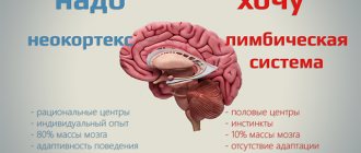
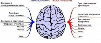
- The first principle is the division of functions into hemispheres - lateralization. The brain is physically divided into two hemispheres: left and right. Despite their external similarity and active interaction provided by a large number of special fibers, functional asymmetry in the functioning of the brain can be seen quite clearly. Some functions are better handled by the right hemisphere (for most people it is responsible for imaginative and creative work), while others are handled better by the left hemisphere (associated with abstract thinking, symbolic activity and rationality).
- The second principle is also related to the distribution of functions across different areas of the brain. Although this organ works as a single whole and many higher human functions are ensured by the coordinated work of different parts, the “division of labor” between the lobes of the cerebral cortex can be seen quite clearly.
The cerebral cortex can be divided into four lobes: occipital, parietal, temporal and frontal. In accordance with the first principle - the principle of lateralization - each lobe has its own pair.
Frontal lobes
The frontal lobes can be called the command post of the brain. Here are centers that are not so much responsible for an individual action, but rather provide such qualities as a person’s independence and initiative, his ability for critical self-assessment. Damage to the frontal lobes causes carelessness, meaningless aspirations, fickleness and a tendency to make inappropriate jokes. With the loss of motivation due to atrophy of the frontal lobes, a person becomes passive, loses interest in what is happening, and remains in bed for hours. Often others mistake this behavior for laziness, not realizing that changes in behavior are a direct consequence of the death of nerve cells in this area of the cerebral cortex
According to modern science, Alzheimer's disease, one of the most common causes of dementia, is caused by the formation of protein deposits around (and inside) neurons, which prevent these neurons from communicating with other cells and lead to their death. Since scientists have not found effective ways to prevent the formation of protein plaques, the main method of drug control against Alzheimer's disease remains the impact on the work of mediators that ensure communication between neurons. In particular, acetylcholinesterase inhibitors affect acetylcholine, and memantine drugs affect glutamate. Others mistake this behavior for laziness, not suspecting that changes in behavior are a direct consequence of the death of nerve cells in this area of the cerebral cortex.
An important function of the frontal lobes is to control and manage behavior. It is from this part of the brain that the command comes, preventing the performance of socially undesirable actions (for example, the grasping reflex or unseemly behavior towards others). When this zone is affected in dementia patients, it is as if their internal limiter is turned off, which previously prevented them from expressing obscenities and using obscene words.
The frontal lobes are responsible for voluntary actions, their organization and planning, as well as the development of skills. It is thanks to them that gradually work that initially seemed complex and difficult to complete becomes automatic and does not require much effort. If the frontal lobes are damaged, a person is doomed to do his work every time as if for the first time: for example, his ability to cook, go to the store, etc. falls apart. Another variant of disorders associated with the frontal lobes is the patient’s “fixation” on the action being performed, or perseveration. Perseveration can manifest itself both in speech (repetition of the same word or whole phrase) and in other actions (for example, aimlessly moving objects from place to place).
The dominant (usually left) frontal lobe has many areas that deal with the infectious aspects of a person's speech, attention, and abstract thinking.
Let us finally note the participation of the frontal lobes in maintaining the vertical position of the body. When they are affected, the patient develops a shallow mincing gait and a bent posture.
Temporal lobes
The temporal lobes in the upper regions process auditory sensations, turning them into sound images. Since hearing is the channel through which speech sounds are transmitted to humans, the temporal lobes (especially the dominant left) play a critical role in facilitating speech communication. It is in this part of the brain that the words addressed to a person are recognized and filled with meaning, as well as the selection of language units to express their own meanings. The non-dominant lobe (right in right-handed people) is involved in recognizing intonation patterns and facial expressions.
The anterior and medial parts of the temporal lobes are associated with the sense of smell. Today it has been proven that the appearance of problems with the sense of smell in an elderly patient may be a signal of developing, but not yet identified, Alzheimer's disease.
A small, seahorse-shaped area on the inner surface of the temporal lobes (the hippocampus) controls human long-term memory. It is the temporal lobes that store our memories. The dominant (usually left) temporal lobe deals with verbal memory and object names, the non-dominant is used for visual memory.
Simultaneous damage to both temporal lobes leads to serenity, loss of visual recognition and hypersexuality.
Parietal lobes
The functions performed by the parietal lobes differ for the dominant and non-dominant sides.
The dominant side (usually the left) is responsible for the ability to understand the structure of the whole through the correlation of its parts (their order, structure) and for our ability to put the parts together into a whole. This applies to a variety of things. For example, to read you need to be able to put letters into words and words into phrases. Same with numbers and numbers. The same lobe allows you to master the sequence of related movements necessary to achieve a certain result (a disorder of this function is called apraxia). For example, the inability to dress independently, often noted in patients with Alzheimer's disease, is not caused by impaired coordination, but by forgetting the movements necessary to achieve a specific goal.
The dominant side is also responsible for the sensation of your body: for distinguishing its right and left parts, for knowing the relationship of a separate part to the whole.
The non-dominant side (usually the right) is the center that, by combining information from the occipital lobes, provides three-dimensional perception of the world around us. Disruption of this area of the cortex leads to visual agnosia - the inability to recognize objects, faces, or the surrounding landscape. Because visual information is processed in the brain separately from information coming from other senses, the patient in some cases has the opportunity to compensate for problems in visual recognition. For example, a patient who does not recognize a loved one by sight can recognize him by his voice during a conversation. This side is also involved in the spatial orientation of the individual: the dominant parietal lobe is responsible for the internal space of the body, and the non-dominant one is responsible for recognizing objects in external space and for determining the distance to these objects and between them.
Both parietal lobes are involved in the perception of heat, cold and pain.
Occipital lobes
The occipital lobes are responsible for processing visual information. In fact, everything that we see, we do not see with our eyes, which only record the irritation of the light acting on them and translate it into electrical impulses. We “see” with the occipital lobes, which interpret signals from the eyes. Knowing this, it is necessary to distinguish between weakened visual acuity in an elderly person and problems associated with his ability to perceive objects. Visual acuity (the ability to see small objects) depends on the work of the eyes, perception is a product of the work of the occipital and parietal lobes of the brain. Information about color, shape, and motion is processed separately in the occipital lobe of the cortex before being received in the parietal lobe to be converted into a three-dimensional representation. When communicating with dementia patients, it is important to take into account that their failure to recognize surrounding objects may be caused by the inability of normal signal processing in the brain and has nothing to do with visual acuity.
Concluding a short story about the brain, it is necessary to say a few words about its blood supply, since problems in its vascular system are one of the most common (and in Russia, perhaps the most common) causes of dementia.
For neurons to function normally, they need constant energy supply, which they receive thanks to three arteries supplying blood to the brain: two internal carotid arteries and the basilar artery. They connect to each other and form an arterial (Willisian) circle, which allows you to nourish all parts of the brain. When, for some reason (for example, a stroke), the blood supply to certain parts of the brain is weakened or completely stopped, neurons die and dementia develops.
Often in science fiction novels (and in popular science publications) the work of the brain is compared to the work of a computer. This is not true for many reasons. Firstly, unlike a man-made machine, the brain was formed as a result of a natural process of self-organization and does not require any external program. Hence the radical differences in the principles of its operation from the functioning of an inorganic and non-autonomous device with an embedded program. Secondly (and for our problem this is very important), the various fragments of the nervous system are not connected in a rigid way, like computer blocks and cables stretched between them. The connection between cells is incomparably more subtle, dynamic, responding to many different factors. This is the power of our brain, allowing it to sensitively respond to the slightest failures in the system and compensate for them. And this is also its weakness, since not a single one of these failures goes away without a trace, and over time their combination reduces the potential of the system, its ability to perform compensatory processes. Then changes begin in a person’s condition (and then in his behavior), which scientists call cognitive disorders and which over time lead to a disease such as dementia.
The article uses a fragment of the book “Dementia: diagnosis, treatment, patient care and prevention”
Tags: #Dementia
Dorsal and lateral routes
Once information has passed through the primary visual cortex into the occipital lobe, the data stream that this area emits bifurcates along two different routes: ventral and dorsal. As we will see, they propagate in parallel, interacting with parts of the brain that the other route does not directly access.
Through the abdominal
The ventral pathway starts from the primary visual cortex in the occipital lobe and goes to the frontal region of the brain through the lower part of the brain, which includes the visual cortices V2 and V4, which, as indicated by their number, are responsible for processing information already processed by v1.
It is believed that neurons involved in this "assembly line" of visual information are responsible for processing the characteristics of individual elements that are viewed at any time - that is, the content of the vision. Therefore, this route is also called the "what" route.
Dorsal path
This pathway runs from the occipital lobe to the frontal cortex through networks of neurons near the top of the skull. In it, information processed by the primary visual cortex reaches the parietal lobe through visual cortices v3 and v5. This visual processing area is thought to be responsible for establishing the location and motion characteristics of what is seen; This is why the spinal track is also called "where and how".
Along with the ventral pathway, this visual processing pathway, associated with the occipital lobe, tells us about how the brain works: sometimes mental processes that seem to form a single whole and come to our consciousness as a holistic experience are actually the product of several brain pathways that operate in parallel, each focusing on a different aspect.
What fields are included?
The occipital lobe of the cerebral cortex contains:
- 17th field – accumulation of gray matter of the visual analyzer. This field is the primary zone. Consists of 300 million nerve cells.
- 18th field. It is also a nuclear cluster of the visual analyzer. According to Brodmann, this field performs the function of perceiving written speech and is a more complex secondary zone.
- 19th field. This field takes part in assessing the meaning of what is seen.
- 39 field. However, this brain area does not entirely belong to the occipital region. This field is located on the border between the parietal, temporal and occipital lobes. The angular gyrus is located here, and its list of tasks includes the integration of visual, auditory and general sensitivity of information.
Pathologies of the cerebral hemispheres
When the cortex of any lobe of the cerebral hemispheres is damaged, various neurological symptoms and syndromes occur.
The reasons for the development of such conditions are:
- head injuries;
- oncological diseases (benign and malignant brain tumors);
- atrophic diseases of the brain (Pick's disease, Alzheimer's disease);
- congenital disorders (insufficient development of nervous system structures);
- birth injuries of the skull;
- hydrocephalus;
- infectious and inflammatory processes in the membranes of the brain (meningitis, encephalitis);
- circulatory disorders in the vessels of the brain.
Disorders in the frontal cortex
When the frontal cortex is damaged, depending on the location, the following symptoms occur:
- frontal ataxia - imbalance, unsteadiness of gait;
- increased muscle tone in the limbs (passive movements are limited or difficult);
- paralysis of a limb/limbs on one side;
- tonic/clonic seizures;
- seizures (tonic-clonic or epileptic);
- speech difficulty (a person cannot find synonyms, case, time of action) - Broca's aphasia;
- symptoms of the frontal psyche (a person behaves foolishly, relaxed, rage may appear for no reason);
- “frontal signs” (the appearance of primitive reflexes, such as in a baby - proboscis, grasping, etc.);
- loss of smell on one side.
Important Nervousness
In addition to the pronounced symptoms of the frontal psyche, the patient may behave apathetically, indifferently, and not come into contact with others. In severe cases, there may be a tendency to immoral social actions: fights, rowdyism, arson.
Pathological disorders in the parietal lobe cortex
When the cortex of the parietal lobe is damaged, disturbances in sensitivity and surrounding perception occur. The following symptoms are characteristic:
- disorders of skin sensitivity;
- posturality (changes in position in space, passive movements that the patient feels, but this does not happen to him);
- lack of perception of parts of your body;
- inability or refusal to respond to stimuli in areas of superficial and deep sensitivity;
- loss of reading, writing, and counting skills;
- inability to find familiar places;
- when examining objects with closed eyes, the patient cannot recognize a familiar thing.
Pathological disorders in the temporal lobe cortex
The main manifestations of damage to the temporal lobe are:
- cortical deafness (hearing loss without injury to the ear);
- Wernicke's aphasia – loss of the ability to perceive speech, music, etc.;
- noise in ears;
- dream-like states (the patient remembers something that he has not seen or heard before, but claims that it happened to him in reality, and not in a dream);
- the occurrence of auditory hallucinations;
- short- or long-term memory loss (amnesia);
- the occurrence of moments of déjà vu;
- combined hallucinations (auditory + visual, auditory + olfactory);
- temporal lobe seizures.
Pathological disorders in the occipital lobe cortex
Damage to the cortex of this area is accompanied by problems with the visual analyzer. Conditions such as:
- cortical blindness (complete loss of vision without damage to the visual analyzer);
- loss of vision, in which the patient claims that he has not lost his sight;
- hemianopsia – loss of visual fields on one side;
- inability to remember an object, color, or person's face;
- changes in surrounding objects that seem small - visual illusions;
- visual hallucinations – flashes of light, zigzags, individual for each eye.
When the limbic system is damaged, memory loss or confusion of memories occurs, there is an inability to create and remember bright moments in life, low emotional lability, lack of smell, loss of the ability to analyze and make decisions, as well as master new skills.
The cerebral hemispheres play a huge functional role in the human body. The ability to write, read, analyze information, perceive and navigate in space, feel, hear, see, smell help the body adapt to the world around it. When certain areas of the cortex are damaged, pathological syndromes and symptoms arise, which can be used to indicate the location of the affected area.
Diencephalon
It responds to external stimuli, is located at the end of the brain stem and is covered by the cerebral hemispheres. Thanks to it, a person can navigate in space, receive visual and auditory signals. Participates in the formation of all types of feelings.
All functions of the human brain are interconnected. Without an intermediate, the functioning of the entire organism will be disrupted. Damage to part of the midbrain leads to disorientation and dementia. If the connections between the lobes of the hemispheres are disrupted, speech, vision or hearing will be disrupted.
The diencephalon is also responsible for pain sensations. A malfunction increases or decreases sensitivity. This part forces a person to show emotions and is responsible for the instinct of self-preservation.
The diencephalon controls the production of hormones, regulates water metabolism, sleep, body temperature, and sexual desire.
The pituitary gland is part of the diencephalon and is responsible for height and weight. It regulates procreation, sperm and follicle production. Provokes skin pigmentation and increased blood pressure.
Parietal
In order to understand the functions of the parietal lobes, it is important to understand that the dominant and non-dominant side will do different jobs.
The dominant parietal lobe of the brain helps to understand the structure of the whole through its parts, their structure, order. Thanks to her, we know how to put individual parts into a whole. The ability to read is very indicative of this. To read a word, you need to put the letters together, and you need to create a phrase from the words. Manipulations with numbers are also carried out.
The parietal lobe helps to link individual movements into a complete action. When this function is disrupted, apraxia is observed. Patients cannot perform basic actions, for example, they are not able to get dressed. This happens with Alzheimer's disease. A person simply forgets how to make the necessary movements.
The dominant area helps you feel your body, distinguish between the right and left sides, and relate parts and the whole. This regulation is involved in spatial orientation.
The non-dominant side (in right-handed people it is the right side) combines information that comes from the occipital lobes and allows you to perceive the world around you in three-dimensional mode. If the non-dominant parietal lobe is disrupted, visual agnosia may occur, in which a person is unable to recognize objects, landscapes, or even faces.
The parietal lobes are involved in the perception of pain, cold, and heat. Their functioning also ensures orientation in space.