General information
Vascular spasm (syn. angiospasm) is a pathological transient narrowing of the lumen of large/small arteries and capillaries due to prolonged intense/excessive contraction of the muscles of the vascular wall, causing disturbances in blood circulation/tissue metabolism.
Angiospasm can be considered as a variant of vascular crisis (acute vascular dystonia). Normally, due to their elasticity, blood vessels ensure uninterrupted and adequate blood flow to various organs, which ensures their functioning. With vascular spasm of varying degrees/localization, the parameters of local/systemic hemodynamics are correspondingly disrupted.
Maintaining vascular tone is carried out due to the tension of the vascular muscle layer, and the contractile activity of the muscle layer is directly regulated by nerve impulses from the brain arriving through sympathetic nerve fibers. It is the myogenic reactions of blood vessels that support circulatory homeostasis . In addition to muscle regulation of tone, its maintenance is carried out due to metabolic, humoral-hormonal and neurogenic mechanisms of vascular regulation.
Developing arterial insufficiency due to spasm of the arterial network leads to tissue ischemia in the area of the spasmodic artery and the development of hypoxia of various organs and tissues, which is manifested by disturbances in their functions. Previously, vasospasm was classified as a disease of “old age,” but currently there is a clear trend towards rejuvenation of vascular disorders. The reason is the high pace of life with frequent stress and unfavorable environmental conditions. The consequence of vasospasm is ischemia .
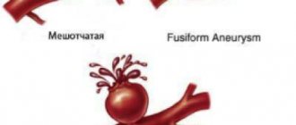
Angiospasms can occur in different vascular regions, mainly in diseases characterized by damage to the arteries/nervous apparatus ( aneurysms , atherosclerosis , vasculitis , etc.) or disorders of the neurohumoral regulation of vascular tone ( neuroses , hypertension , pathological menopause , hypothalamic syndrome , etc. .). Angiospasm often shapes the dynamics of clinical symptoms in organic vascular diseases, such as thrombosis , atherosclerosis , embolism, etc.), significantly complicating their course. Prolonged chronic or acute arterial insufficiency in the heart (coronary vasospasm), brain (cerebral vasospasm), and in the retina (retinal vasospasm) can cause the development of acute vascular insufficiency. As a rule, acute arterial obstruction is the cause of such serious conditions as myocardial infarction , stroke , and visual impairment.
Vascular stenosis develops in most cases with atherosclerotic damage to the arteries, which is caused by the deposition of cholesterol plaques on the walls. Chronic arterial insufficiency of the lower extremities is also widespread. What are the types of vascular diseases in the legs? In the lower extremities, so-called “occlusive vascular diseases” are quite common, provoked by narrowing/clogging of the iliac arteries/abdominal aorta as a result of the deposition of atherosclerotic plaques on their walls, which significantly impairs the blood flow of the lower extremities. These are obliterating diseases of the peripheral arteries of the legs ( atherosclerosis obliterans , thromboangiitis obliterans , endarteritis obliterans , diabetic angiopathy ), characterized by a steadily progressive course with the transition of increasing intermittent claudication to a constant pain syndrome. Critical leg ischemia leading to gangrene is an extremely severe outcome of long-term arterial insufficiency.
Pathogenesis
The pathogenesis of vasospasm has not yet been fully studied, which is largely due to its non-identity for arteries of different vascular regions. It is generally accepted that a common factor in the mechanism of development of vasospasm includes functional disorders of the membranes of smooth muscle cells of the vessel, the essence of which is a violation of the exchange of potassium, calcium, and sodium ions through the membranes, which disrupts the alternation of muscle contraction/relaxation phases. Enhanced/accelerated depolarization of membranes, as well as an increased influx of free calcium ions into cells, contribute to intense muscle contraction, and a delay in membrane repolarization (preceding muscle relaxation) contributes to prolonged contraction of the arterial wall.
Disturbances of this kind may be caused by disturbances in the innervation of vascular walls, an imbalance in the production/destruction of humoral regulators of tone, increased sensitivity of vascular wall receptors to the effects of vasoconstrictors, and a disorder of humoral regulators of vascular tone. Often, stenosis can develop against the background of local changes in the vessel wall in areas of thrombus , vascular inflammation, scars (for example, near the area of coronary artery thrombosis/location of an aneurysm or atherosclerotic plaque).
Main signs of seizure syndrome
During cramps, certain muscle groups contract or twitch. The duration of the convulsive syndrome varies from a few seconds to ten minutes. A patient who has suffered a stroke most often suffers from short-term cramps throughout the body. They can occur in the legs, arms, neck or face. After an attack of convulsions, the patient loses consciousness or falls asleep as a result of the brain being in a state of shock. Seizures may manifest differently. Some patients experience mild tremors, while others suffer from severe seizures.
Convulsions during and after a stroke can occur:
- short-term contractions of the facial muscles (in this case, one half of the face may become distorted and a mask-like face may appear);
- numbness of the muscles of the limbs, complete loss of control over them, the appearance of a feeling of a wooden arm or leg;
- contraction of the facial muscles against the background of numbness of the limbs.
Causes
The causes of vascular spasms have not been sufficiently studied, however, a number of diseases can be identified that are accompanied by vasospasm in different vascular regions:
- Atherosclerosis at the stage of pronounced atherosclerotic changes in the vascular bed.
- Osteochondrosis of the spine in the cervical region.
- Hormonal disorders in diseases of the endocrine system ( diabetes mellitus ).
- Diseases of the cardiovascular system ( hypertension , atrial fibrillation ).
- Dysfunction of the autonomic nervous system.
- Traumatic brain injuries.
- Frostbite.
- Stress and overwork.
- Brain hemorrhages.
- Vasculitis is inflammation of the artery (vascular walls).
- Brain tumors.
- Chronic intoxication (poisoning with lead/carbon disulfide compounds).
The most common/significant risk factors for the development of vasospasm are: arterial hypertension , smoking, alcohol abuse, male gender, old age, dyslipidemia , physical inactivity and excess body weight. Among them, the greatest importance is given to disorders of fat metabolism and arterial hypertension.
Obviously, the causes of vasospasm in different vascular regions differ, that is, the causes of spasms of cerebral vessels and the causes of vasospasm of the retina or lower extremities are different.
Treatment of seizures after stroke
What should you do if a patient experiences seizures after a stroke? You need to call an ambulance immediately. Before doctors arrive, first aid is provided by people who are close to the patient at the time of the attack. It is necessary to provide the patient with oxygen access. If this happened indoors, then the windows should be opened; otherwise, tight clothing should be removed from the patient. The patient needs to place a pillow or cushion under his head.
If there are dentures in the mouth, they need to be removed and the oral cavity cleaned of food debris. If the patient begins to breathe hoarsely, he should be placed on his side and checked to ensure that nothing is obstructing his breathing. If pain develops in the muscles, you need to get a massage. It is advisable to lubricate the skin with olive oil before the massage.
Seizures are treated in the intensive care unit. Relief of seizures begins with intravenous administration of anticonvulsants. If ineffective, they switch to a combination of anticonvulsants - parenterally and through a tube:
- diazepam 0.15 - 0.4 mg/kg intravenously at an administration rate of 2-2.5 mg/min, if necessary, repeat the administration of 0.1-0.2 mg per 1 kg of body weight per hour;
- valproic acid intravenously 20-25 mg per 1 kg of weight for the first 5-10 minutes, then carry out a continuous infusion of the drug at a rate of 1-2 mg per 1 kg per hour or bolus administration 4 times a day at a daily dose of 25-30 mg per 1 kg of weight per day;
- in case of refractory status epilepticus and in case of ineffectiveness of diazepam, sodium thiopental is used under the control of external respiration function.
The patient's condition is alleviated with finlepsin or carbamazepine. Finlepsin has the following side effects:
- reduces intellectual activity when using the drug for a long period of time;
- causes powerlessness, apathy;
- provokes the development of osteoporosis or increased bone fragility (this problem is eliminated with the help of calcium supplements);
- increases the risk of bleeding when combined with anticoagulants.
Finlepsin treatment is carried out for persons no older than 65 years. A maximum of 400 mg of the drug can be consumed per day. If a loved one who has suffered a stroke develops seizures, call an ambulance and call the Yusupov Hospital. The clinic’s doctors will provide professional specialized medical care aimed at relieving convulsive syndrome and prescribe further treatment for the patient.
Symptoms
Symptoms of vascular spasm are determined by its localization and are manifested by manifestations of dysfunction of ischemic tissue of various organs. Let's look at just a few of them:
Symptoms of vascular disease in the legs (using the example of obliterating atherosclerosis ). Depending on the severity of insufficiency of arterial blood supply to the limb, several stages of clinical manifestations of the disease are distinguished:
- Stage of functional compensation . At this stage, cramps, chilliness and paresthesia of the lower extremities are characteristic, and less often - burning/tingling in the fingertips, leg fatigue, and increased fatigue. As the skin cools, it becomes pale and cold to the touch. As a rule, when walking on level ground after 1000 meters or more, intermittent claudication appears due to insufficient blood supply to the muscles, accumulation of under-oxidized metabolic products in tissues and disruption of the process of oxygen utilization.
- Subcompensation stage . There is an increase in the intensity of intermittent claudication, which already occurs after traveling a distance of about 200 m. The skin of the legs/feet becomes dry, loses elasticity and flakes, and hyperkeratosis . Characterized by a slowdown in hair growth (patches of baldness appear) and nails, which become dull, brittle, thickened and acquire a matte color. Initial signs of atrophy of the foot muscles and subcutaneous fat are noted.
- Stage of decompensation . Against the background of ongoing spasm of the vessels of the legs of the affected limb, pain appears at rest, and walking is possible at a distance not exceeding 25 meters. Progressive atrophy of the leg/foot muscles is noted, the color of the skin of the legs changes, and the skin turns pale when the limb is raised and redness appears when it is lowered. The skin becomes easily vulnerable and various types of minor injuries (abrasions/ bruises ) lead to the formation of non-healing chronic wounds , cracks , and painful superficial ulcers . The patient has limited ability to work. With severe pain syndrome, disturbances in night sleep are observed, patients take a forced position - the “doll” pose.
- Stage of destructive changes . At this stage, vasospasm of the lower extremities causes the development of necrobiotic processes. And the rate of their increase is determined by the level of the gap between the amount of blood flow to the tissues and their need for oxygen. The pain in the fingers and feet is extremely intense. The resulting ulcers are located mainly in the distal parts of the legs (usually on the fingers). The bottom/edges of the ulcers are covered with a gray-dirty coating without granulation, and there is inflammatory infiltration around them. Swelling of the foot/leg develops. Characteristic is the development of wet gangrene of the fingers and feet. The ability to work at this stage is completely lost.
Symptoms of cerebral vascular spasms
Angiospasm of cerebral vessels, contributing to the development of chronic circulatory failure of brain structures (dyscirculatory encephalopathy) can develop with damage to the main arteries (stenosis of the vertebral/carotid arteries), narrowing/thickening of intracerebral arteries against the background of arterial hypertension and due to narrowing of the lumen due to thickening of the walls of small arteries.
There are several stages of development of dyscirculatory encephalopathy. The initial symptoms of the disease may be latent for some time. At this stage, the disease manifests itself with complaints of noise in the head, headaches , dizziness , and decreased non-professional memory and performance. Patients may be tearful, distracted, irritable, and depressed. They experience difficulty transitioning from one type of activity to another.
At the next stage, non-professional/professional memory impairments progress, a narrowing of the range of interests, a decrease in intelligence, and fixation on a specific problem are noted. The patients are quarrelsome, and personality changes are increasing. Such patients sleep poorly at night and are drowsy during the day. Neurological symptoms increase, spasms in the head are noted, movements slow down and their coordination is impaired, staggering when walking/mild speech impairment is characteristic, and performance is significantly reduced.
Subsequently, against the background of ischemia, gross changes in the tissue of certain brain structures occur, which intensifies the manifestations of neurological symptoms, and mental disorders develop. Patients stop recognizing loved ones, can get lost while walking, perform inappropriate actions and almost completely lose their ability to work.
Angiospasm of the retina
Bilateral retinal vasospasm is typical, less often a unilateral process is noted. Patients complain of the appearance of “fog” before the eyes and the flickering of “floaters”. With a short-term spasm, blurred vision is possible, but it is transient. Distortion of visual perception in the form of meta/photomorphopsia is possible. In some cases, a feeling of discomfort appears in the orbital area; patients may feel pulsation in the temples, headache and dizziness . After the attack is over, the patient’s condition returns to normal and visual function is restored. In severe cases (acute obstruction of the central retinal artery) can lead to a pronounced/irreversible decrease in visual acuity.
Treatment
Neurologists at the Yusupov Hospital provide complex treatment for epilepsy. It is aimed at reducing the frequency of epileptic seizures and stopping medications during remission. According to statistics, in 70% of cases, adequately selected treatment helps to almost completely relieve paroxysmal activity in patients.
To achieve optimal results in treatment, patients are prescribed drugs for epilepsy of the following properties:
- anticonvulsants – help relax muscles, they are prescribed to both adults and children;
- tranquilizers - allow you to remove or reduce the excitability of nerve fibers; the drugs have shown a high degree of effectiveness in the fight against minor attacks;
- sedatives – help relieve nervous tension and prevent the development of severe depressive disorders;
- injections - used for twilight states and affective disorders.
Idiopathic focal epilepsy is benign. It requires symptomatic treatment. In focal forms with seizures that appear in series 2-3 times a month and are accompanied by an increase in mental disorders, neurosurgeons perform surgery.
Is epilepsy curable?
Before starting treatment, a diagnosis should be determined, because loss of consciousness and various convulsions can occur due to a sharp drop in blood sugar, anemia, poisoning, high fever, cerebrovascular accident, calcium deficiency and other conditions. Antiepileptic drugs cannot be prescribed to such patients immediately. The diagnosis of epilepsy is made only when epileptic seizures recur.
With adequate treatment, according to generalized statistics, a good therapeutic effect is achieved in 80-85 percent of patients, and in 60-70 percent of patients seizures can be eliminated completely. And this is a very good reason to stop antiepileptic drugs.
The percentage of patients with epileptic seizures could be significantly lower if patients took medications regularly and did not stop treatment on their own. The decision to discontinue medications can only be made by a doctor, and not earlier than after 5 years of treatment, if during this time the patient has not had epileptic seizures. At the Yusupov Hospital, an epileptologist always monitors how the patient’s illness develops.
Tests and diagnostics
The diagnosis of “angiospasm” is based on the presence of characteristic symptoms of vasospasm and their dynamics, characteristic of the development of transient ischemia in the area of the spasmodic artery. To establish a diagnosis, the following is carried out:
- Ultrasound of leg vessels (Doppler/duplex scanning).
- CT angiography (with contrast agent) of cerebral vessels, which allows you to clearly determine the diameter of the lumen of the vessels and visualize the places of narrowing.
- Dopplerography of cerebral vessels (to assess the speed of blood flow in the intracranial arteries in various areas).
- MRI of the brain/cervical spine.
- Doppler ultrasound of the brachiocephalic arteries.
- Fundus ophthalmoscopy.
- Functional tests (Goldflam/thermometric, etc.).
Laboratory methods include biochemical blood tests, determination of blood cholesterol levels, and coagulogram. In the presence of cognitive impairment - neuropsychological testing.
Diagnostic methods
Neurologist
It is not enough to interview and examine the patient to identify narrowing of the blood vessels of the spine; in any case, additional examinations will be prescribed. These include:
- Duplex scanning
- despite the fact that this is a rather dangerous diagnostic method in this situation, it is informative and accessible, it allows you to determine the degree of narrowing and its nature. - Angiography - the process uses a contrast agent to examine the vessels.
- and MRI
- allow you to obtain layer-by-layer 3D images, with which you can make a diagnosis.
All this will allow the doctor to determine not only the patient’s health status, but also the reasons that led to the occurrence of stenosis.
Prevention
Prevention of vascular diseases, including vasospasm, consists of maintaining a healthy lifestyle, including:
- Balanced diet.
- Active lifestyle.
- Stop smoking.
- Body weight control.
- Monitoring blood pressure , cholesterol /sugar levels.
- Periodic medical examinations.
The basis for the prevention of thrombosis and vascular diseases is the strengthening of blood vessels, which is achieved by regular practice of procedures such as contrast showers/douches, contrast foot baths, bath procedures, lymphatic drainage massage, pine/turpentine baths.
Consequences and complications
The consequences of vasospasm are determined by the duration/severity of ischemia in the area of the spasmodic artery, the sensitivity of tissues/organs to oxygen deficiency, as well as the development of collateral circulation in the affected organ. The brain and heart muscle, kidneys, and spleen are especially sensitive to hypoxia. Accordingly, ischemia of these organs is accompanied by a high risk of developing ischemic stroke and myocardial infarction . Prolonged vasospasm of the arteries of the lower extremities can cause the development of obliterating diseases ( atherosclerosis obliterans , thromboangiitis obliterans , endarteritis obliterans , diabetic angiopathy ), retinal vasospasm - visual disorders, and so on.
First aid for seizures
Proper care for an epileptic seizure reduces the risk of complications and injuries. The person who happens to be next to the patient should catch him and prevent him from falling. You also need to do the following:
- Place a blanket, pillow or cushion of clothing under your head;
- Free your neck and chest from constricting objects and clothing (tie, shirt, scarf);
- Carefully turn the patient's head to the side to minimize the risk of inhalation or passive reflux of vomit or own saliva;
- Open your mouth and put a cloth or handkerchief in it to prevent the patient from biting his tongue;
- Do not forcefully open your mouth;
- If breathing stops for a long time, perform artificial ventilation from the mouth to the nose or mouth.
During an attack, the patient may experience involuntary urination or bowel movements. This manifestation of epilepsy should not cause fear in others. After an attack, patients usually experience drowsiness and severe weakness.
List of sources
- Krylov V.V., Gusev S.A., Titova G.P., Gusev A.C. Vascular spasm in subarachnoid hemorrhage. M.: Medicine; 2000.
- Avksentyeva M.V., Krysanov I.S., Chupin A.V. Pharmacoeconomic aspects of the treatment of obliterating diseases of the peripheral arteries of the lower extremities // Angiology and Vascular Surgery. 2012. T. 18, No. 4. P. 16-21.
- Drozhzhin E.V., Darwin V.V. The role of rheological disorders in the pathogenesis of the obliterating process in the arterial system // Collection of scientific papers of Surgut State University. Vol. 12. Natural sciences. Surgut: Surgut State University Publishing House, 2003. pp. 67-69.
- Zudin A.M., Zasorina M.A., Orlova M.A. Epidemiological aspects of chronic critical ischemia of the lower extremities // Surgery. 2014. No. 10. P. 78-82.
- Savelyev V.S., Koshkin V.M. Critical ischemia of the lower extremities. M.: Medicine, 1997. - 160 p.