This term unites a group of chronic neuromuscular diseases accompanied by primary muscle damage. Progressive muscular dystrophies are manifested by various disorders in the structure and metabolism of muscle tissue, leading to a decrease in strength and the appearance of progressive weakness in the muscles. Pathologies are characterized by a steadily progressive course.
Etiology and pathogenesis
Primary forms of diseases are caused by genetically determined disorders in the functioning of mitochondria, ion channels of myofibrils, disruptions in the synthesis of muscle proteins and enzymes that regulate metabolism in muscle tissue. The defective gene that causes progressive muscular dystrophy can be inherited dominantly, recessively, or in an X-linked form of inheritance.
Acquired forms of diseases can develop against the background of endocrine pathologies, chronic intoxications, vitamin deficiency, malabsorption, severe chronic diseases of internal organs, and tumor processes.
Symptoms of muscular dystrophy
- Decreased tone of the affected muscles.
- Disorders of gait and posture.
- Muscle atrophy with preserved skin sensitivity.
- Fast fatiguability.
- An increase in the volume of the limbs due to the growth of connective tissue in place of the muscles.
- Cardiomyopathy.
- Breathing problems.
- Mental retardation.
- Impaired joint mobility.
- When facial muscles are damaged, facial expressions become poorer and speech becomes less intelligible.
Muscular dystrophy in children is manifested by the loss of physical skills acquired before the disease: the child stops walking, holding his head up, and sitting.
Common types of disease
- Duchenne muscular dystrophy.
- Becker muscular dystrophy.
The disease develops in children aged 3–5 years. It begins with damage to the lower extremities and gradually rises higher. This is a rapidly progressive form that leads to muscle atrophy after 5 years. It is characterized by heart damage and the development of mental disorders.
The process also begins from the lower extremities. Weakness in the legs and fatigue first appear at the age of 15–20 years. The transition of the disease to the upper limbs due to muscular dystrophy is observed in adults after 50 years.
Classification of myopathies
Myopathies are divided into two large subgroups - primary and acquired.
Primary myopathies are caused by genetic disorders. They are characterized by a gradual progressive course. Known hereditary myopathies are juvenile Erb myopathy, Duchenne myopathy, Landouzy-Dejerine myopathy, Pompe disease (for which pathogenetic therapy is available)1.
Primary forms include distal forms (distal muscular dystrophy). This is a group of rare, progressive genetic disorders that cause wasting (atrophy) and weakness of muscles away from the center of the body - the arms and legs. These include distal myopathy Miyoshi, distal muscular dystrophy with vacuoles “in the frame” (Nonaka), Welander, Lane, tibial muscular dystrophy Udd, others2.
Primary forms are associated with one or more genetic mutations that are responsible for defects in proteins that provide muscle tone and contraction, as well as some enzymes. For example, one of the most common hereditary forms, Nemalin myopathy, is caused by mutations in 10 genes. All of them are related to the production of proteins3. Another rare disease, Pompe disease is caused by a congenital deficiency of the enzyme and the associated accumulation of glycogen in muscle tissue.
The defective gene can be inherited recessively or dominantly, and sometimes it is inherited linked to the X chromosome.
Acquired myopathies are divided into inflammatory (dermatomyositis, polymyositis), infectious, metabolic, endocrine, medicinal, and toxic. They develop against the background of concomitant diseases and pathological conditions, for example:
- Taking medications (glucocorticosteroids);
- Drinking alcohol;
- Ingestion of toxins into the body (toxic form);
- Endocrine disorders (dysfunction of the thyroid gland, etc.);
- Avitaminosis;
- Heart failure;
- Liver disorders;
- Malignant diseases, etc.
Diagnostics
A neurologist treats and examines patients with muscular dystrophy. The patient's symptoms and complaints allow one to suspect the disease. To clarify the diagnosis, use:
- determination of the level of the enzyme CPK (creatine phosphokinase) in the blood. In case of illness, this indicator increases several times;
- electromyography. The disease is indicated by a decrease in the electrical activity of the muscles;
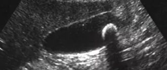
- biopsy. The method involves taking material and subsequent analysis under a microscope. The study helps exclude other diseases;
- genetic analysis.
Genetic testing is the end point of diagnosis. The LAMA2 gene is being sequenced. To confirm the diagnosis, it is necessary to identify homozygous or two compound heterozygous mutations. If only one point mutation is detected in a heterozygous state in the presence of a characteristic clinical picture or a significant increase in the level of CPK in the blood and a hypersignal from the white matter on T2 according to MRI of the brain, it is advisable to conduct a chromosomal microarray analysis in order to search for large deletions and duplications that occur with high frequency - according to various sources, from 18% to 30–40% of mutations in the LAMA2 gene. Genotype-phenotype correlations Prognosis of disease severity depends on the age of onset of symptoms, the presence or absence of laminin-α2 expression on immunohistochemical testing of muscle biopsies, the type of mutation and its effect on protein function. For MD-MD, acquisition of independent walking is a critical clinical measure of disease severity. The type 1A AMD phenotype is associated with mutations that lead to the cessation of protein synthesis. Basically, these are nonsense variants in which a stop codon is formed. Such patients are characterized by a severe course of the disease with early onset, respiratory disorders, often with the need for respiratory support, and lack of independent walking. According to the biopsy, a complete absence of laminin-α2 expression in the muscles and skin is determined.
Forms of the disease with onset in childhood or adulthood, a milder course, and the achievement of independent walking are caused by homozygous or compound-heterozygous missense substitutions, as well as large deletions with restoration of the reading frame. These mutations result in decreased or nearly normal expression of laminin-α2 and affect merosin polymerization. Most studies confirm a direct correlation between genotype and phenotype. Despite this, F. Geranmayeh et al. reported on a family whose members had homozygous missense mutations in the LAMA2 gene, and not all acquired the ability to walk independently.
The differential diagnosis of Merosin-negative AMD should be differentiated from diseases accompanied by muscle weakness and hypotension from the first months of life. First of all, these are other forms of congenital myopathies (Ullrich AMD, rigid spine syndrome, central core disease, nemaline, centronuclear myopathies, dystroglycanopathies, Fukuyama AMD), congenital myasthenic syndromes, spinal muscular atrophy. Distinctive features of type 1A AMD are the highest CK levels among all congenital myopathies, 2–17 times higher than normal, and changes on MRI of the brain (hyperintense signal from the white matter in T2 mode, structural abnormalities). In Fukuyama AMD and dystroglycanopathies, partial merosin deficiency may be observed, detected by muscle tissue biopsy; Antibodies to the long fragment of laminin α2 chain help distinguish primary from secondary merosin deficiency. Structural changes in the brain that occur in AMD with glycosylation defects (muscular-eye-brain syndrome, Fukuyama AMD, Walker-Warburg syndrome) are usually more significant and are accompanied by severe mental retardation and a high incidence of epilepsy. The combination of developmental delay and changes on MRI often makes it necessary to differentiate AMD type 1A from leukodystrophies. High levels of CPK, slow progression with stabilization after the 1st year of life in merosin-negative AMD and, on the contrary, a rapid, steadily progressive course characteristic of leukodystrophies, help distinguish these two conditions. Differential diagnosis of later forms of LAMA2-related MD is more difficult. I. Nelson et al. reported on 4 patients, two of whom were observed with a diagnosis of Bethlem myopathy, two with Emery-Dreyfus myodystrophy. Patients diagnosed with Bethlem myopathy had a typical phenotype of collagenopathy (skin changes, contractures of large joints). Patients with Emery-Dreyfus muscular dystrophy had contractures in the elbow joints, dilated cardiomyopathy and severe heart rhythm disturbances such as ventricular tachycardia and fibrillation. The MRI picture of the muscles and the increase in the level of CPK in the blood also corresponded to the expected diagnoses. Two patients suffered from drug-resistant epilepsy. An MRI of the brain revealed pathological changes in 3 people. All patients underwent genetic testing, which revealed homozygous (2 cases) and compound heterozygous (2 cases) mutations in the LAMA2 gene. Based on this observation, the authors proposed that all patients with myopathy, including adults with contractures in large joints, changes on MRI of the brain, and even isolated cardiomyopathy, include the LAMA2 gene in the diagnostic search. Also, mild forms sometimes have to be differentiated from neuropathies, in which nerve damage comes to the fore in the clinical picture, and mild proximal muscle weakness is of little concern to patients and is detected only during neurological testing.
Patient management tactics
In 2010, the International Consensus on Standards of Care for Patients with AMD was published, detailing the scope and manner of care they need. Children should be observed by a multidisciplinary team of specialists, including a pediatrician, neurologist, orthopedist, pulmonologist, nutritionist, cardiologist, and physical therapist. Patients under 1 year of age and older with severe or progressive forms (refractory epileptic seizures, severe hypotension, nutritional problems) should be examined by specialists every 3–4 months. Children over 1 year of age in stable condition require routine examination every 4–6 months. Evaluation includes assessment of nutritional status, cardiac function and orthopedic complications, detection of gastroesophageal reflux, pulmonary function tests, overnight pulse oximetry or polysomnography in frail children with recurrent respiratory infections. Because the vast majority of patients with AMD do not gain the ability to walk independently and have a progressive decrease in muscle mass, these children are often low weight. Consultations with a gastroenterologist and nutritionist are recommended 2 times a year. The goal is not to normalize body weight to standard values; positive dynamics in annual weight gain is sufficient. Adequate weight gain in such children can be achieved by additional intake of hypercaloric mixtures. Patients with swallowing disorders, gastroesophageal reflux, or aspiration pneumonia may require a nasogastric tube or percutaneous gastrostomy tube. A visit to a pulmonologist to evaluate respiratory function should be done at least annually. Respiratory tests include spirometry and cough assessment and are performed from 4–6 years of age. Indicators of forced vital capacity (FVC) <60% of the predicted value are associated with breathing disorders during sleep, FVC <40% of the age norm is associated with a high risk of nocturnal hypoventilation. For such patients, it is necessary to measure the level of saturation and hypercapnia during night sleep; in case of pathological indicators, non-invasive ventilation is required. For patients with severe spinal deformities and recurrent respiratory infections, a chest CT scan is recommended to assess the presence of chronic atelectasis and compression of the airways by vertebral bodies. Classes with a physical therapist are aimed at reducing muscle hypotonia, increasing the patient’s motor capabilities, and preventing contractures and respiratory dysfunction. These include daily vertical positioning in orthopedic devices, stretching exercises and swimming in the pool. An orthopedic examination and x-ray of the spine should be performed at least once a year. More frequent assessment is warranted during periods of rapid growth and progression of spinal deformities. Orthoses and splints are used to prevent the progression of joint deformities; the use of a corset is mandatory to maintain posture.
Cardiological examination in the absence of complaints is carried out at 5 and 10 years, then every 2 years and includes electrocardiography and echocardiography. Patients with severe respiratory failure who are on mechanical ventilation support are required to undergo annual echocardiography. Patients with complaints of rapid heartbeat and increased fatigue additionally undergo Holter heart rate monitoring.
Treatment Strategies
Several options are currently being explored to restore basement membrane structure in MD-MD: Transgenic expression of cDNA encoding laminin-α1 using the chicken β-actin (CAG) promoter. Conducted in the dy3K/dy3K mouse model, which showed significant muscle and peripheral nerve recovery with increased strength. These studies proved that laminin-α1 can completely replace laminin-α2. However, this approach cannot be applied as gene therapy to be delivered by viral vectors because the laminin protein is too large for DNA. Parenteral administration of recombinant laminin-111. Difficulties for use in humans also lie in the large size of the protein. Use of linker proteins. Laminin-411 expression is increased in MD-MD patients, but this protein has low affinity for α-dystroglycan and α7β1-integrin. A smaller version of the agrin protein (mini-agrin, or mag) has been developed that significantly improves binding to α-dystroglycan. Initiation of laminin polymerization. To restore the polymerization of laminin, a linker protein was created, consisting of fragments of laminin and nidogen αLNNd). There was a restoration of strength in dy2J/dy2J mice and a reduction in fibrosis according to histology. This treatment option is only suitable for a small group of patients whose laminin-α2 expression is only slightly reduced.
Use of CRISPR/Cas9 technologies. There was a restoration of the synthesis of full-length laminin-α2 in the dy2J/dy2J mouse model, an increase in muscle strength, and a decrease in fibrosis. Given the promising results, funding and further development of LAMA2 CRISPR technologies is a high priority for research. In addition to treatment aimed at restoring the structure of the basement membrane, therapy is being developed to prevent the consequences of muscle damage. Phase I of an open-label clinical trial of omigapril for patients with LAMA2- and COL6-related muscular dystrophies (CALLISTO) has been completed. Omigapril is an inhibitor of apoptosis by blocking glyceraldehyde-3-phosphate dehydrogenase. It reduces weight loss, muscle fiber degeneration, especially in the respiratory muscles, which helps improve respiratory function and prevent complications. Another drug, losartan, is an angiotensin II type I receptor blocker. It affects the activity of transforming growth factor, reducing fibrosis and improving clinical manifestations. Other drugs (bortezomide, prednisolone) did not show their effectiveness or had significant side effects in type 1A AMD. Although therapies aimed at restoring the structural defect show a greater degree of clinical improvement in patients with MD-MD than pathogenic therapies, it cannot be certain that one treatment method will be sufficient for patients. It is likely that protocols combining these two treatment groups may be most successful.
Conclusion
MD-AMD is the most common form of AMD. It is extremely heterogeneous genetically and clinically, manifesting itself, in the vast majority of cases, as severe non-ambulatory phenotypes with a complete absence of merosin expression in tissues, but mild KP forms with a late onset of the disease associated with a reduced content of laminin-α2 in muscles and peripheral nerves are also found . This heterogeneity leads to diagnostic difficulties, especially for mild forms of the disease. Knowledge of the features of the clinical course and pathogenesis of LAMA2-related muscular dystrophies is especially important in the context of actively developing specific methods for their therapy.
Treatment
To date, scientists have not invented any means to get rid of this disease.
All existing methods and drugs can only reduce the severity of symptoms and alleviate the condition:
- corticosteroids (prednisolone). Increase muscle strength and delay the development of certain forms of the disease;
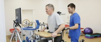
- physical exercise. Allows you to maintain joint mobility and normal posture longer;
- mobile devices. Used to support weakened muscles and facilitate movement.