Features of the course of Landouzy-Dejerine muscular dystrophy
The neuromuscular disease is characterized by progressive damage to the facial muscles, shoulders and shoulder blades. Muscles in other parts of the body are also affected, but much more slowly and less severely. A feature of the flow is the asymmetry of the destructive process. Often the muscles of the shoulder girdle are the first to atrophy, gradually spreading to the facial muscles, leading to facial expression, weakness and the inability to raise the arms.
Unlike other muscular dystrophies, with Landouzy-Dejerine myopathy, sports and physical activity do not slow down the pathological process, but rather accelerate the destruction of muscle tissue. In the future, cardiac dysfunction is observed, vision loss and hearing loss may occur.
This myopathy was first described by the French doctors Landouzy and Dejerine in 1884.
It is known that the diagnosis is genetically determined, with an autosomal dominant mode of inheritance. Most patients have gene mutations, but some patients do not have genetic disorders.
Landouzy-Dejerine glenohumeral-facial myopathy is a rare pathology, with an incidence rate of 1-1.5 per 100,000 population. Due to hereditary predisposition, it is often detected in blood relatives. The first symptoms appear in childhood. The peak incidence and severity of clinical manifestations are adolescence, pregnancy and menopause.
The pathology continues to be studied, but the cause of the defect and the development of atrophy is not known. There is currently no pathogenetic treatment, which is why the disease continues to be classified as a serious disease. Despite the fact that patients remain able to work for a long time and can care for themselves, in most cases the diagnosis leads to their disability.
Psychotherapy
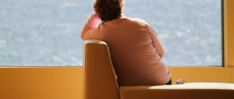
The onset of clinical manifestations and subsequent deterioration are usually provoked by mental stress. Moreover, severe muscle disease is, in itself, a serious mental stress. Against the background of the disease, secondary depression often develops with apathy and reluctance to take care of one’s health. And this leads to progression of the disease and possible death of the patient.
If necessary, we provide our patients with psychotherapeutic treatment. Our patients become more resistant to mental irritants, become more active, have a positive attitude towards treatment, and this immediately affects muscle function.
In addition, we teach patients special psychotechniques for working with their own muscles.
If there are signs of depression, it is possible to prescribe modern antidepressants (selective serotonin reuptake inhibitors, such as Cipralex, Fluoxetine, etc.). These antidepressants do not reduce, but increase the patient’s activity and mood, and do not cause muscle weakness.
Reasons for development
The primary causes of the biochemical defects leading to myopathy are unknown. But, analyzing statistical data, scientists identify several factors in the development of myopathy:
- hereditary factor - often members of the same family are affected;
- genetic predisposition - many have a defect in chromosome 4;
- viral diseases in the mother during pregnancy;
- pathology of pregnancy in the mother.
In some patients, myopathy occurs against the background of normal health for no apparent reason. In this case, the diagnosis is classified as idiopathic myopathy.
Due to the lack of information about the initial processes of pathology development, doctors are practically unable to stop them and restore lost body functions.
Symptoms
The first signs of the disease appear at 10-20 years of age, when excessive muscle weakness, fatigue and slow development of the muscles of the shoulder girdle are noted. Patients find it difficult to raise their arms and perform physical exercises. Due to atrophy of muscle tissue, their shoulder blades protrude excessively, acquiring a wing-shaped shape, the gap between them is widened, and the chest is flattened. Muscular weakness contributes to the development of scoliosis.
Dystrophy of the muscles of the shoulder, chest and trapezius muscles leads to the development of the symptom of a drooping shoulder. A common complication in this case is dislocation of the shoulder joint. Patients experience difficulties in everyday life and at home. It is difficult for them to comb their hair, wash their face, and brush their teeth. Due to previously habitual loads, they get tired too quickly, to the point of being unable to perform them.
As myopathy progresses, it involves the facial muscles in the process. The face becomes amicable. Due to damage to the orbital muscles of the mouth and eyes, it is difficult or impossible to close your eyes and purse your lips. This leads to frequent eye inflammation and corneal injury.
A common complaint is speech impairment - unintelligible, slow. This significantly impairs the quality of life, especially social life and work activity. Difficulties also arise during meals. Patients have difficulty eating solid food, chewing it and swallowing it.
Characteristic signs of the disease are: inverted lips “tapir lips”, a transverse smile “Gioconda’s face”, a polished forehead.
In the future, progressive muscular dystrophy in other parts of the body is possible. When the tissues of the buttocks and thighs are affected, muscle weakness contributes to excessive fatigue from walking, which can lead to lameness. Damage to the calf muscles is accompanied by the symptom of foot drop, severe weakness when walking and the inability to run.
Depending on which symptoms are more pronounced and what is the order of their occurrence, the following forms of Landouzy-Dejerine myopathy are distinguished:
- facioscapulohumeral;
- facioscapular-humeral-peroneal;
- facioscapulohumeral-gluteofemoral;
- facioscapulohumeral-gluteal-femoral-peroneal;
- facioscapulohumeral-peroneal-gluteofemoral;
- infantile form.
The most severe is the infantile form. It occurs in children under 5 years of age. It is characterized by symmetrical paresis of the facial muscles, damage to the shoulders, chest, and absence of tendon reflexes. Due to rapidly increasing weakness, children experience respiratory failure up to the need for artificial ventilation.
Important! Often, parents of children with excessive muscle weakness try to restore the body with the help of sports sections and physical activity. As a result, the disease progresses even faster.
Journal "Child's Health" 1 (28) 2011
Facioscapulohumeral myodystrophy of Landouzi-Dejerine was described by Landouzi and Dejerine in 1884. Most domestic pediatric neurologists first became acquainted with this pathology in the manual of L.O. Badalyan [1], which until now serves as a reference book for many specialists. At the same time, it should be noted the significant contribution of a team of scientists who have been and are studying the diagnosis, development mechanisms, clinical manifestations and treatment of neuromuscular diseases for many years [2, 5].
Landouzi-Dejerine muscular dystrophy is inherited in an autosomal dominant manner with high penetrance and occurs with a frequency of 0.9–2 per 100,000 population. The genetic heterogeneity of facioscapulohumeral myodystrophy has been established: in 90–95% of families linkage is found with the 4q35 locus (type 1A of the disease), and in the remaining 5–10% with the 10q26 locus (type IB). The primary biochemical defects are currently unknown [4, 8, 11].
In the classic version, the first signs of the disease appear mainly at the age of 10–20 years. Atrophy and muscle weakness are localized in the area of facial muscles, shoulder blades and shoulders. First, atrophy is observed in the shoulder girdle, followed by spread to the face. Typically, the initial manifestations are difficulty raising the arms above the head, protruding “wing-shaped” shoulder blades and scoliosis. As the process progresses, the orbicularis muscles of the mouth and eyes suffer severely - it is not possible to close the eyes tightly and purse the lips. Due to atrophy, the face becomes hypomimic. As a rule, patients themselves note a change in their facial expressions; their speech becomes unintelligible. It should also be noted that there are characteristic symptoms in the form of a transverse smile (“Gioconda’s smile”), everted lips (“tapir lips”), and a “polished” forehead. Atrophy of the biceps and triceps muscles of the shoulder, pectoralis major, serratus anterior, and trapezius muscles can cause symptoms of loose shoulder girdles, “wing-shaped” shoulder blades, the appearance of a wide interscapular space, flattening of the chest and scoliosis. In some cases, atrophy spreads to the leg muscles. In this case, weakness is most noticeable in the peroneal muscle group along the foot drop, but can also be in the proximal parts of the legs [5, 6, 10]. Depending on the sequence and nature of the spread of the process, the facial-scapular-humeral form itself, the facioscapular-humeral-peroneal, the facioscapular-humeral-gluteofemoral, the facioscapular-humeral-gluteofemoral-peroneal and the facioscapular-humeral-peroneal-gluteofemoral forms are distinguished. In the early stages of the disease, muscle tone is reduced in the proximal muscle groups, deep reflexes are reduced mainly in the biceps and triceps brachii muscles [1].
It is necessary to note the asymmetry of atrophy , which is a characteristic clinical feature of this pathology. In some cases, muscle pseudohypertrophy occurs. Contractures and retractions are moderately expressed. Cardiomyopathy occurs in rare cases. Angioretinography may reveal abnormalities of the retinal vessels. In many cases, severe ocular manifestations include telangiectasia, edema, and retinal detachment. Coagulation of telangiectasia prevents the development of blindness. Hearing loss may also occur. The above symptoms are considered as part of the phenotypic manifestations of this pathology. The course of the disease is in most cases relatively favorable. We especially want to focus on the fact that physical overload, intense sports activities and irrationally conducted physical therapy can contribute to a more severe course of the disease [9].
Despite certain advances in molecular genetics and other achievements of medical science, the diagnosis of this disease at the present stage is based primarily on clinical features (mainly the facial-brachial localization of the myodystrophic process) and on genealogical analysis (autosomal dominant type of inheritance).
Differential diagnosis should be carried out with other progressive muscular dystrophies (Erb-Roth, Becker, etc.) and with secondary myopathic syndromes that occur against the background of inflammatory, vascular, toxic and metabolic processes.
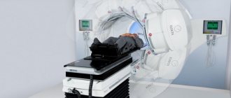
To clarify the diagnosis, a biochemical study with the determination of creatine phosphokinase (CPK), electroneuromyography (ENMG) and muscle biopsy are used. The level of CPK can increase 5 times, but in some cases the enzyme content is normal. ENMG records both myopathic and denervation potentials. When conducting histological studies, minimal changes are revealed in many muscles of the extremities; the largest number of pathological signs are observed in the suprascapular muscles, where the phenomena of progressive degeneration and slight marginal denervation are found [5, 7, 9].
We would like to draw your attention to the fact that monographs by foreign authors in recent years have also described the infantile form of facioscapulohumeral myodystrophy, which rapidly progresses and leads to severe disability. Symptoms appear in infancy, but no later than 5 years of life in the form of bilateral paresis of the facial muscles, which can simulate congenital damage to the facial nerves. Subsequently, rhinolalia and sometimes ptosis develop. Progressive proximal muscle weakness occurs 1–2 years after the onset and primarily affects the shoulders and then the pelvic muscles. Pseudohypertrophy of the legs may also be observed. Tendon reflexes are reduced and disappear over time. There is a rapid increase in weakness, which leads to death due to respiratory failure before the patient reaches 20 years of age. Very rarely, weakness does not progress over a long period of time, and severe disability does not occur until adulthood. Half of the patients' relatives have retinal telangiectasia and a high incidence of hearing loss. The diagnosis of this pathology should be suspected in every child with progressive facial diplegia, but in each case it is necessary to exclude myasthenia gravis and brainstem glioma [8, 10].
We present a table that presents data on the localization of genes responsible for hereditary myopathies, indicating the primary biochemical defects and a brief functional characteristics of these proteins (Table 1) [12]. As noted above, the primary biochemical defects in the case of Landouzy-Dejerine facioscapulohumeral myodystrophy are currently unknown.
Currently, therapeutic options for Landouzi-Dejerine myodystrophy are very limited. Symptomatic treatment is aimed primarily at preventing the development of contractures, maintaining existing muscle strength and reducing the rate of atrophy. The main task is to maximize the period during which the patient is able to move independently, since contractures, scoliosis, and respiratory disorders quickly increase in a supine position. The therapeutic complex should include therapeutic exercises, massage, orthopedic measures, drug therapy, and a balanced diet. In the drug treatment of progressive muscular dystrophies, it should be noted the prescription of courses of metabolic drugs aimed at improving and maintaining the metabolic and energy processes of intact myocytes, cardiomyocytes (cardonate, elkar, vitamin E, methionine, cytoflavin, ATP-long, mildronate, cocarboxylase). Correcting the patient's diet is of particular importance. A diet with a high protein content and low fat content, with a reduced calorie content and an optimal content of vitamins and microelements is recommended [5, 6, 8]. Psychological support for the patient, continued education, and proper professional guidance also play an important role.
In the clinic of pediatric psychoneurology of the State Institution "IPAG AMS of Ukraine", where, along with common severe and difficult to diagnose neurological diseases in children, rare neurological nosologies are also concentrated, over the past five years, 7 patients with progressive Landouzy-Dejerine muscular dystrophy have been observed. We provide a description of the medical history of a patient who underwent examination and treatment in our department. We believe that this clinical case is of significant interest, given the family history of this disease (11 cases of the disease in the family).
Patient M., 16 years old, was admitted to the department with complaints of weakness in the upper extremities - the inability to raise his arms up. The patient has been noticing weakness in her arms for a year, while a mother suffering from a similar disease noticed a decrease in strength in her son’s upper limbs three years ago. Weakness gradually increases. A noticeable deterioration was noted after physical activity - sports activities on the horizontal bar and lifting weights.
From the life history: a boy from the 2nd normal pregnancy, 2 births with weakness of labor, was born with a weight of 4000 g, length 58 cm. He developed according to his age, studies in the 9th grade, satisfactorily.
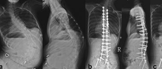
Neurological status: intelligence preserved. The function of the oculomotor nerves is not impaired. Facial hypomia and hypotrophy of the muscles of the shoulder girdle and “wing-shaped” shoulder blades are pronounced (Fig. 1–3). Muscle strength in the proximal parts of the upper extremities was reduced to 3 points, more on the left, in the distal parts - 5 points, in the lower extremities - 3 points. Muscle tone is reduced. Tendon reflexes on the upper and lower extremities are lively and uniform; abdominal reflexes are alive. There are no pathological foot signs. The gait is not changed. Coordinator tests are performed accurately.
Examination: general clinical tests without pathological changes; serum enzyme activity - ALT - 23 U/L (N - 8-40 U/L), AST - 34 U/L (N - 10-41 U/L), CPK - 148 (N < 270 U/L) ; ENMG - signs of primary muscle damage.
Consulted with a geneticist: the diagnosis of progressive Landouzi-Dejerine muscular dystrophy was confirmed.
Family history: the mother of the proband, 41 years old, began to have a disease at the age of 14, manifested by weakness of the muscles of the shoulder girdle; weakness of facial muscles appeared at the age of 35–36 years. A similar disease is observed in her father, in 5 of 10 siblings and in 4 nephews (Fig. 4). At her place of residence, a woman receives social benefits due to progressive Erb-Roth muscular dystrophy.
Analyzing the family history, we draw attention to the fact that the disease is hereditary in nature with an autosomal dominant type of inheritance. Juvenile Erb-Roth limb-girdle myodystrophy is inherited in an autosomal recessive manner. The difference between the Erb-Roth muscular dystrophy clinic is the predominantly ascending (starting from the lower extremities) and not the descending nature of the lesion, as is observed in all sick members of the described family. Therefore, we believe that the diagnosis of the boy’s mother is not Erb-Roth muscular dystrophy, but Landouzy-Dejerine muscular dystrophy.
The case we present demonstrates a rare form of progressive muscular dystrophy with an autosomal dominant mode of inheritance—Landouzy-Dejerine facioscapulohumeral myodystrophy. A special feature of the case is a burdened family history of this disease (11 patients in the family), which, however, did not cause alertness among medical workers when the first symptoms appeared in the patient. It is important to note that with timely diagnosis and the necessary limitation of physical activity, it would be possible to avoid the rapid progression of the disease.
Thus, muscular dystrophies are characterized by progressive muscle weakness, muscle atrophy and movement disorders. However, various forms of myopathies are distinguished by different types of inheritance, variability in the age of onset of the disease, predominant localization of muscle damage and other signs that it is necessary to be able to diagnose and differentiate in a timely manner. It is obvious that future success in the treatment of progressive muscular dystrophies will depend on the latest developments in the field of molecular genetics and gene therapy. Therefore, timely treatment will prevent rapid progression of the disease, improve the patient’s quality of life and increase his life expectancy.
In conclusion, we would like to note that the publication in a widely read pediatric journal of an article devoted to purely neurological pathology is due to the fact that patients primarily turn not to a neurologist, but to pediatric doctors, and the future fate of the patients in many cases depends on their tactics.
We consider it necessary to recall that back in the 60s of the last century, complex issues of various diseases of the nervous system, in the origin of which hereditary predisposition plays an important role, were dealt with by the world-famous scientist S.N. Davidenkov, who described the scapular-peroneal form of myopathies. In his works, for the first time in the Soviet Union, he raised the problem of possible prognosis of offspring for certain hereditary diseases. Since then, the diagnosis of many hereditary diseases has risen to the required level thanks to the latest molecular genetic technologies. However, the methods for determining the type of inheritance of pathology remain the same, and it is important for every doctor to remember that in the case of the dominant type “... the risk of transmitting the disease to offspring is so great that sick family members should refrain from childbearing. On the contrary, family members who remain healthy can freely have children without any danger of passing on a family disease to them” [4].
Diagnostic methods
Despite the frequent detection of genetic mutations during genetic testing, in some patients with severe clinical symptoms they are absent. Because of this, a genetic blood test is not the main criterion for the disease. In most cases, Landouzy-Dejerine myopathy is diagnosed on the basis of characteristic clinical manifestations, primarily damage to muscles typical of the disease.
The following types of examination are also additionally carried out:
- biochemical blood test to determine the level of creatine phosphokinase;
- muscle biopsy;
- electroneuromyography.
A significant increase in the level of creatine phosphokinase indicates progressive atrophy of muscle tissue. Patients have weak myocyte activity. For histological examination, tissue samples are taken from different parts of the body. The examination reveals characteristic signs of atrophy. Additionally, patients need an ophthalmological examination and consultation with a neurologist.
Important! The course of some diseases is similar to this myopathy. Doctors must carry out differential diagnosis with neurological diseases, autoimmune and connective tissue pathologies.
Accompanying illnesses
Muscle diseases can progress very quickly if they are complicated by other diseases that reduce mobility. Therefore, we make sure to treat concomitant diseases.
Major diseases that can make you feel worse:
- Diseases of the joints and spine (arthrosis, herniated disc, osteochondrosis, etc.) block the mobility of the back and limbs. They can develop due to a shift in the body’s centers of gravity due to muscle weakness. Here we successfully use gentle manual therapy, massage, and Karipazim electrophoresis.
- Chronic tonsillitis, pharyngitis, sinusitis, adenoiditis. The constant presence of pathogenic microbes in the respiratory tract causes constant intoxication, and toxins negatively affect the muscles, especially the heart. Therefore, if necessary, we carry out a course of treatment for these diseases.
- Other chronic infections (kidney, liver, lower respiratory tract).
- Overweight. It is dangerous due to excessive stress on muscles, joints, and spine. Often causes immobility. We identify the causes of obesity and select a treatment plan for it.
- Disorders of the thyroid gland. Metabolic disorders with decreased or increased thyroid function can cause decreased muscle strength. If necessary, we monitor the level of thyroid hormones in the blood and involve an endocrinologist in treatment.
- Vascular diseases (arterial hypertension, cerebrovascular accidents) contribute to heart overload and the rapid development of myopathy of the heart muscle, and hence heart failure. If necessary, we perform cardiac stress tests and select treatment.
- Bronchial asthma leads to oxygen deficiency in muscle tissue, which contributes to muscle weakness and the progression of myopathy. If necessary, we recommend a course of treatment for bronchial asthma.
Treatment
The disease is an incurable diagnosis. There is no pathogenetic treatment. It is also quite difficult to stop rapidly progressive atrophy. Treatment of Landouzy-Dejerine myopathy is aimed primarily at slowing down dystrophic changes that lead to the inability to move and care for oneself, restoring lost functions and generally improving the patient’s condition. Drug therapy is based on the use of medications that support myocytes, improve metabolic processes in them, and have a beneficial effect on the heart. The most commonly prescribed drugs are: ATP-long, Mildronate, Methion, vitamin E, Cytoflavin, cocarboxylase.
The following factors are also important for improving the condition:
- nutrition – it is recommended to increase the amount of proteins and reduce fats;
- physiotherapy – patients are prescribed massage, electrophoresis and other procedures;
- physical therapy – moderate physical activity and a special complex is necessary to prevent the development of scoliosis and contractures.
Considering that myopathy often occurs in childhood, special attention must be paid to the mental state of patients. Often the pathology leads to depressive disorders, nervous breakdowns and emotional disorders. To prevent psychological disorders and improve the emotional state of patients, they need the help of a psychologist or psychotherapist.
For socialization, it is necessary to help patients choose a profession in order to maintain their ability to work.
Watch the video on the topic of the article!
Possible complications
Despite careful study, Landouzy-Dejerine myopathy continues to be a severe pathology. In many ways, the prognosis depends on the characteristics of the course and speed of development of the pathological process. Hypomia and amymia not only limit physical capabilities, but also cause psychological discomfort and difficulties with socialization.
Progression and involvement of other parts of the body in the pathological process leads to significant restrictions, including the inability to move.
In some cases, with damage to the orbicularis oculi muscles and frequent inflammatory processes, patients' vision deteriorates to the point of blindness.
Often, following the doctor’s recommendations helps preserve life expectancy, but in extremely severe forms of the disease, death is possible due to severe respiratory failure.
Massage for myopathy and amyotrophy
Standard massage techniques are not entirely suitable for the treatment of myopathy and amyotrophy. In addition, the effect of massage directly depends on the correctness of its implementation. Unfortunately, we regularly see patients after a course of massage with problematic muscles that are completely untreated during massage.
Proper massage begins with identifying inelastic, tight, weakened muscle areas. It is on these areas that the main efforts should be directed during massage. Moreover, a combination of a tonic massage on weakened areas with a relaxing, stretching and absorbing massage on tight areas of muscles is required .
If there is weakness of the respiratory muscles, chest massage is performed to facilitate breathing movements.
We perform massage in the clinic and teach patients’ relatives how to perform massage independently at home. If the patient is undergoing treatment without an accompanying person, we teach self-massage.
What do you need to remember?
- Landouzy-Dejerine myopathy is characterized by progressive atrophy of the muscles of the face, shoulders and shoulder blades.
- The main cause of the disease is genetic predisposition.
- Signs of the pathology are hypomimicness and amymicity, muscle weakness and loss of muscle tissue.
- Treatment is aimed at inhibiting the development of the disease and preventing its progression.
- Muscular dystrophy leads to disability of the patient due to severe limitations in his capabilities.
- The prognosis for recovery is unfavorable, since the disease is incurable.
Literature
- Asanov A. Yu. Fundamentals of genetics and hereditary developmental disorders in children: Textbook. manual for university students. - M.: Academy, 2003. - P. 216.
- Badalyan L. A. Child neurology: textbook. allowance. 4th ed. - M.: MEDpress inform, 2021. - 608 p.
- Vlodavets D.V., Sukhorukov V.S., Kharlamov D.A., Belousova E.D. A method for treating congenital structural myopathies and congenital muscular dystrophies by correcting secondary mitochondrial changes: Abstract of thesis. dis. ... Ph.D. - M., 2009. - 28 p.
- Grinio L.P. Atlas of neuromuscular diseases. - M.: ANS, 2004. -167 p.
- Erokhina V. A. Rehabilitation of children with hereditary myopathies // Bulletin of the Moscow University of the Ministry of Internal Affairs of Russia. - 2015. - No. 12. - P. 304-308.
- Arvanitidis A., Henriksen K., Karsdal MA, Nedergaard A. Neo-epitope Peptides as Biomarkers of Disease Progression for Muscular Dystrophies and Other Myopathies // J Neuromuscul Dis. - 2016. - No. 30. - R. 333-346.
- Carroll MB, Newkirk MR, Sumner NS Necrotizing Autoimmune Myopathy: A Unique Subset of Idiopathic Inflammatory Myopathy // J Clin Rheumatol. - 2021. - No. 22. - R. 376-80.
- Inoue M., Nishino I. Diagnosis of Idiopathic Inflammatory Myopathy: A Muscle Pathology Perspective // Brain Nerve. - 2021. - No. 68. - R. 1431-1441.
Diagnosis: myopathy (amyotrophy). What to do?
Today there is no panacea for hereditary muscular dystrophies. However, proper treatment can significantly slow down muscle atrophy and increase regeneration and growth of new muscle tissue, and even restore some lost capabilities . Treatment of myopathy and amyotrophy requires the daily implementation of a number of medical procedures, so we not only provide medical care, but also train the patients’ relatives and/or the patients themselves to independently perform the necessary procedures.
Treatment at our clinic includes the following:
- Taking medications according to a schedule prescribed for a period of 3-12 months;
- Medical nutrition;
- Physiotherapy;
- Massage;
- Gymnastics;
- Psychotherapy and spiritual practices;
- Neuropsychological development (for children lagging behind in intellectual development).
Detailed training is provided for each of the above points. We insist on training to independently perform procedures, because... We strive to make regular and sufficient treatment also accessible and cheap.