Vegetative-vascular dystonia is one of the most frequently diagnosed diagnoses in neurological practice, especially at the outpatient stage of treatment. However, I personally am increasingly asked the question of what it is if there is information on the Internet that “there is no such diagnosis in the West.” That is why I decided to touch upon the topic of vegetative dystonia on this site and consider this issue within the framework of the symptom of dizziness. And also at the same time tell why there is no point in using information on the Internet without having the proper medical data for its full processing and understanding.
What is unsteadiness of movements?
Unstable movements – permanent or temporary loss of coordination. If they occur systematically, this indicates the presence of abnormal disorders in the body. Extra-systemic indicate that inflammatory processes are occurring in the body.
Most often, unsteadiness of walking manifests itself in combination with other symptoms:
- headache;
- unexplained sudden weakness;
- tingling in the legs;
- dizziness.
To better understand, it is necessary to know why a person may stagger when walking and why this is so important.
Movement is achieved through a healthy bone structure, developed muscles and healthy joints. The cerebellum is the part of the brain responsible for coordination; it is also controlled by vision and the vestibular apparatus.
Impulses pass through the spinal cord, sending a signal to the lower parts of the body that a movement needs to be performed. Only after this the muscles begin to work.
When these impulses are disrupted, the entire system is disrupted. This is where the gait disorder comes from. A transmission failure causes the legs to become unresponsive.
Failures in the musculoskeletal system lead to exactly the same result. The legs cannot correctly carry out the necessary “commands”.
If the functioning of the cerebellum is disrupted, the impulse itself does not reach, therefore, the command does not arrive at all. Consequently, a person cannot move, his legs do not obey.
Features of the ataxic gait
Due to imbalances, patients with pathological processes in the cerebellum try to increase the area of support, spread their legs widely, stagger and sway randomly when walking.
Movements of the upper and lower extremities lose synchrony. Instability persists in a standing position, regardless of the presence or absence of visual control. With unilateral damage to the cerebellum, one half of the body suffers, and falls on the affected side are possible. When cortical structures are involved, the clinical picture resembles that of cerebellar ataxia. Instability increases when turning, the patient often “falls” in the direction of the lesion, unsteadiness of gait correlates with the severity of the damage to the cortex. In people with damage to the ventrolateral thalamus, unsteadiness and instability occur on the opposite side, and there is a tendency to fall backward or to the healthy side.
Old age
Often, older people (over 60 years old) experience swaying, as well as “floaters” before the eyes. It is also common to experience tinnitus and dizziness when walking. Headaches appear sharply, and sometimes even fainting occurs. As a rule, all these symptoms are ignored and blamed on age.
In some cases, this may indicate problems with the body, so you should consult a doctor. This way you can ensure your usual full life.
One of the reasons is a malfunction of the cardiovascular system of the brain. Atherosclerosis appears (as mentioned above, a disease of the second category).
With age, the functioning of the vestibular apparatus is impaired.
Gait disturbance may also be due to heart disease. For example, ischemia can reduce blood circulation in the area of the ear where the organ responsible for balance is located. Also, insufficient blood circulation leads to uncertain movements. Other diseases also have a detrimental effect (arrhythmia, hypertension).
It has a great influence on gait and blood viscosity, because it supplies oxygen to the organs. How quickly it reaches the desired place, including the cerebellum, depends on its speed.
Injuries and diseases of the spine also make it difficult to transmit signals. Therefore, movement disorders are possible. Elderly people are often susceptible to sleep disorders, which leads to diseases of the third category.
The most common occurrence of maladaptation is observed. The person feels extremely insecure, there is a feeling that no one needs him, inexplicable fears and anxiety. As a result, he may skid when walking, become dizzy, have dark vision, and his condition worsens every day. For treatment, you must definitely contact professionals and it is better not to delay, because the consequences can be disastrous.
Based on all of the above, we can conclude that many factors can lead to an unsteady gait and they should not be ignored.
If the cause is eliminated, the consequences will also be eliminated. Life will be familiar, fulfilling and healthy. Therefore, it does not matter how old a person is, treatment is necessary regardless of age.
Treatment
Conservative therapy
Treatment tactics are determined taking into account the underlying disease. In case of infectious-inflammatory genesis of ataxic gait, antibiotic therapy or antiviral therapy is indicated. For patients with vascular diseases, depending on the nature of the pathology, anticoagulants, antiplatelet agents, thrombolytics, angioprotectors, and vasodilators may be recommended. Patients with ataxia and unsteady gait due to intoxication require detoxification therapy.
Pathogenetic therapy for hereditary ataxia has not been developed. The same symptomatic measures are taken as for ataxias of other etiologies. Prescribe B vitamins, anticholinesterase drugs, ATP, piracetam, meldonium. Exercise therapy classes are conducted to improve the general condition and strength of muscles, and reduce the severity of incoordination. Patients are referred for massage.
Surgery
Surgical interventions are required for large processes and circulatory disorders. Taking into account the etiology of the ataxic gait, the following techniques are used:
- Neoplasms
: removal of tumors of the cerebellum, cortex, brain stem. - Abscesses
: drainage, removal along with the capsule. - Hematomas
: transcranial and endoscopic removal, stereotactic aspiration. - Occlusive hydrocephalus
: ventriculoatrial and ventriculoperitoneal shunting, external ventricular drainage.
When should you sound the alarm?
Impaired coordination in some cases is a consequence of serious illnesses. It is recommended to consult a specialist if:
- stumbling has become more frequent, often occurring even out of the blue;
- falls due to weakness in the legs;
- impaired control of movements;
- unnatural gait;
- after a long walk there is a sudden stop, then there is a feeling that it is impossible to move your leg;
- feeling of “cotton” legs;
- difficulty walking on stairs;
- when moving, most of the weight increasingly moves to the heel area;
- when trying to get up, a person falls;
- difficulty raising a leg or starting to move after a long rest;
- the appearance of dizziness, pounding in the temples, darkening in the eyes.
Often a person begins to be afraid and panic.
It is absolutely impossible to ignore such symptoms, since they indicate the presence of diseases.
Chronic cerebral circulatory failure (syn.: dyscirculatory encephalopathy - DEP, chronic cerebral ischemia - CIM) remains one of the most common and socially significant neurological problems. According to statistics, at least 15% of cognitive impairment (CI) in the Russian population is associated with cerebrovascular diseases (CVD), while the overall prevalence of CI in outpatient neurological appointments is more than 80% among people over 60 years of age [1, 2]. Among elderly outpatients, patients with chronic CVD make up at least 12%.
The most common and significant manifestation of DEP for the patient’s quality of life is K.N. They are characterized by such features as a decrease in the rate of cognitive activity, lack of concentration, impairment of executive functions (planning and control), visuospatial dysgnosis and dyspraxia [3, 4]. In our opinion, it is specific changes in higher brain functions that should serve as the main guideline in the clinical diagnosis of DEP syndrome.
However, it would be absolutely wrong to reduce the clinical picture of DEP to changes in higher mental functions only. On the contrary, with rare exceptions, other neurological disorders are observed in DEP. The presence of non-cognitive neurological symptoms is the most important differential diagnostic feature of DEP, allowing one to distinguish between this syndrome and primary degenerative diseases of the brain (such as Alzheimer's disease, Pick's disease, etc.) [5, 6].
In the past, much attention in diagnosing DEP syndrome was paid to the presence of so-called focal microsymptoms: the appearance of reflexes of oral automatism, asymmetry of nasolabial folds, increased tendon reflexes, tremor when performing coordination tests, etc. However, clinical experience indicates the nonspecificity of these changes, many of which are variant of the age norm or are associated with other diseases. In particular, as an example, we can point to intention tremor when performing a finger-nose test, which, in the absence of other discoordination disorders, may reflect the presence of a disease such as essential tremor.
Non-cognitive neurological symptoms more specific to DEP syndrome are pseudobulbar syndrome, movement disorders and dysfunction of the pelvic organs. At the same time, movement disorders affecting the area of maintaining balance and walking develop earlier and most often. Disturbances in gait and postural stability are observed in more than 70% of patients with DEP [7–9]. This type of neurological disorder is most typical for advanced and late stages of DEP. Thus, the presence of gait and postural stability disorders is mentioned as one of the specific diagnostic signs of Binswanger’s disease [10]. This term refers to the advanced (dementia) stages of vascular brain damage caused by microangiopathy (arteriolosclerosis) [10, 11].
Here are the diagnostic criteria for Binswanger's disease [10]: dementia; the presence of less than two of the following: underlying vascular disease or risk factor (arterial hypertension, diabetes mellitus, previous myocardial infarction, cardiac arrhythmias, heart failure); previous stroke or corresponding focal neurological symptoms (hemiparesis, hemihypetesia, etc.); symptoms of subcortical damage: gait disturbances, increased muscle tone, the phenomenon of “countercontinence”, pelvic disorders; bilateral leukoaraiosis according to CT or MRI (focal changes more than 2´2 sq. mm); the diagnosis is unlikely in the presence of multiple or bilateral cortical lesions, severe dementia (MMSE<10).
Identification of disorders of postural stability and gait of vascular origin is important not only for differential diagnosis, but also for providing effective care to patients with DEP. There is no doubt about the impact of these disorders on quality of life. Common consequences of balance and gait disorders are falls, injuries, hip fractures, intracranial hemorrhages and other serious complications.
Syndromes of postural stability and gait disorders in DEP.
Disturbances in gait and postural stability in DEP are described in the literature within the framework of such neurological syndromes as frontal ataxia, gait apraxia, vascular parkinsonism of the lower body and frontal dysbasia.
Frontal ataxia ( syn
.: cortical ataxia, Bruhn's ataxia, frontal astasia-abasia) was first described in patients with bilateral lesions of the anterior frontal lobes of the brain by an intracranial tumor. It was noted that in some cases the gait of patients with local lesions of the frontal lobes resembles the gait of patients with cerebellar pathology. It is characterized by a wide base of walking, staggering and frequent falls, and in some cases, the inability to stand and walk (astasia-abasia) in the absence of paresis and sensory disorders. Subsequently, similar gait and balance disorders were described in DEP and normal pressure hydrocephalus in the elderly [12, 13].
The term “frontal ataxia” reflects the idea that vascular disturbances in postural stability and gait are based on impaired coordination of movements, which is similar in pathophysiological mechanisms and clinical manifestations to cerebellar, sensory, and vestibular ataxia. It is assumed that the basis of frontal ataxia is a disruption of the connection between the frontal lobes and the cerebellum, caused by ischemic or other vascular damage to the fronto-ponto-cerebellar pathways. As a result of such a lesion, secondary cerebellar dysfunction develops, which causes symptoms [13].
With frontal ataxia, there are no other discoordination disorders, for example, in the arms or legs when performing the heel-knee test in the supine position, which does not fit well with the idea of secondary cerebellar dysfunction. To explain this contradiction, the concept of “gait apraxia” was proposed, according to which disturbances in postural stability and gait in chronic cerebrovascular disease DEP only superficially resemble ataxia. At the same time they are apraxia ,
associated with the loss of the walking skill acquired in early childhood [14]. The argument in favor of this concept is the specific clinical characteristics of gait in DEP syndrome, indicating the insufficiency of the central mechanisms of programming and regulation of movements. Such features include difficulties initiating walking, stopping the execution of a motor program, perseveration, difficulties dividing attention (for example, the inability to talk while walking), etc.
The same objections can be raised against the idea of gait apraxia as against frontal ataxia. In the vast majority of patients with severe disturbances in gait and postural stability of vascular etiology, as well as with frontal dysfunction of another cause (local damage to the anterior parts of the brain, normal pressure hydrocephalus), the neurological status does not show signs of any other (except for gait) dyspractical disorders. There is no apraxia in the arms and, as a rule, in the legs. The absence of the latter is evidenced by the preserved ability to depict certain learned actions in a lying position (for example, to show how to hit a ball while playing football or how to pedal a bicycle) [14].
Although the structure of gait and postural stability disorders undoubtedly contains signs of insufficiency of the central link of movement regulation, true apraxia, from our point of view, can only be spoken of in a limited number of cases, mainly in the late stages of DEP in patients with severe vascular dementia. A significant risk factor for loss of walking skills in dementia patients is prolonged abstinence from walking (for example, as a result of injury or illness, the patient is on bed rest).
The term “vascular parkinsonism” has been proposed to describe movement disorders in patients with suspected atherosclerotic dementia. Vascular parkinsonism is characterized predominantly by axial hypokinesia (in the face, trunk muscles), a bilateral symmetrical increase in muscle tone of the plastic type without the cogwheel symptom, a shuffling gait and the absence of tremor. Gait disturbances and hypokinesia in the legs are much more pronounced than extrapyramidal disorders in the arms. The latter circumstance gave rise to the term “parkinsonism of the lower body.” It is believed that the main cause of lower body parkinsonism is chronic cerebrovascular insufficiency [15, 16]. The following are the diagnostic criteria for vascular parkinsonism [17]:
- presence of parkinsonism;
- at least 2 points on the following scale:
- morphological or angiographic signs of diffuse vascular disease - 2;
— clinical manifestation of parkinsonism within 1 month after stroke — 1;
- 2 or more strokes in history - 1;
- 2 or more risk factors for cerebrovascular accidents - 1;
- neuroimaging signs of CVD in 2 or more vascular territories - 1
Clinical-neuroimaging and clinical-morphological comparisons indicate the presence of a connection between vascular parkinsonism syndrome and changes in white matter (vascular leukoencephalopathy) and expansion of perivascular spaces in the area of the striatum. As a result of these changes, the striatum during morphological examination becomes similar to cheese with holes ( fr.
.:
etait crible
or Swiss cheese syndrome). Vascular extrapyramidal disorders are caused by direct damage to the striatum or disruption of their connections with other parts of the brain [15, 16].
Axial hypokinesia in combination with gait disturbances does not always have a vascular etiology (pseudovascular parkinsonism). At the same time, with severe vascular and non-vascular pathology of the brain, behavioral disorders are often formed that can imitate parkinsonism, but in their pathogenesis do not belong to extrapyramidal disorders (vascular pseudoparkinsonism, pseudovascular pseudoparkinsonism) [16].
Differential diagnosis of vascular parkinsonism
Pseudovascular parkinsonism
: atypical Parkinson's disease, progressive supranuclear palsy, dementia with Lewy bodies;
normotensive hydrocephalus; local damage to the frontal lobes or subcortical basal ganglia. Vascular pseudoparkinsonism:
vascular damage to the brain with the development of apathy syndrome, severe depression with psychomotor retardation or severe dementia.
Pseudovascular pseudoparkinsonism:
apathy syndrome, severe depression with psychomotor retardation, severe dementia of non-vascular etiology, akinetic mutism.
From our point of view, disturbances in gait and postural stability in DEP have a complex pathogenesis, including dysfunction of the extrapyramidal and coordinating motor systems, as well as insufficiency of the central mechanisms of movement regulation, elements of loss of the skill of walking. The formation of both primary motor and central dysregulatory and dyspractical disorders is mediated by dysfunction of the anterior parts of the brain. From our point of view, the most suitable term to denote disturbances in gait and postural stability in DEP syndrome is frontal dysbasia-dystasia.
Pathomorphology and pathophysiology of gait and postural stability disorders in DEP.
The development of disturbances in postural stability and gait in DEP is primarily associated with diffuse damage to the white matter (vascular leukoencephalopathy). Thus, in the works of N.N. Yakhno et al. [18], I.V. Damulina et al. [9], using the method of clinical neuroimaging comparison, showed the correspondence between the severity of balance and walking disorders and the severity of leukoaraiosis. The connection between damage to the white matter of the brain and the risk of falls in old age is also indicated by the results of the European long-term prospective study LADIS [19]. According to this study, changes in the subcortical nucleus region do not increase the risk of falls [19]. Thus, the most significant factor for the development of vascular disorders of postural stability and gait is damage to the white matter of the brain.
It is assumed that vascular leukoencephalopathy leads to disruption of communication between the frontal lobes of the brain and the subcortical basal ganglia on the one hand, and between the frontal lobes and the cerebellar hemispheres (fronto-ponto-cerebellar pathways) on the other. Normally, the frontal lobes and striatum are integrated into closed functional systems—frontostriatal circles [20]. The circulation of excitation along them plays an important role in programming cognitive activity and motor acts. Violation of the integrity of the frontostriatal systems in vascular leukoencephalopathy leads to secondary dysfunction of the frontal cortex, which underlies classic subcortical vascular CI (decreased rate of cognitive activity, lack of concentration, etc.) [3, 4, 21].
A qualitative analysis of the characteristics of disturbances in postural stability and gait in DEP indicates that vascular movement disorders may be based on the same pathophysiological mechanisms that are responsible for the development of vascular CIs. The results of clinical and stabilographic studies indicate that disturbances in postural stability and gait in advanced stages of DEP are associated with insufficiency of central integration and coordination of the components of the static comotor system (sensory, pyramidal, extrapyramidal, cerebellar, peripheral) [7-9, 18].
The following mechanisms that control voluntary motor activity are most affected [7–9, 14, 22, 23]: the ability to initiate a motor program; generation of walking rhythm; the ability to quickly take into account information coming from sensory systems about changes in external and internal conditions; the ability to quickly make adjustments to the motor program depending on changing conditions; the ability to stop performing a motor program or change it to another.
Important for understanding the pathophysiological mechanisms of vascular disorders of postural stability and gait is a reliable connection between their severity and the degree of CI [7–9]. The parallelism of the increase in the severity of cognitive and motor disorders can be considered as an indirect confirmation of the commonality of their anatomical and physiological substrate. It is assumed that for the occurrence of vascular CIs, disruption of connections between the basal ganglia and the dorsolateral parts of the frontal cortex is critical [20], and for the occurrence of vascular disorders of postural stability and gait, connections with the supplementary motor cortex [14]. Disorders of gait and postural stability and vascular CIs are not only united by a common pathogenesis, but also directly affect the severity of each other. Thus, attention disorders in the structure of vascular CIs aggravate the risk of falls: the patient does not notice obstacles on the way, stumbles, etc.
Clinical picture
The clinical picture of disturbances in postural stability and gait in DEP is characterized by the following features [7-9, 12-16, 18, 22, 23]: difficulty in starting to walk - more time passes from the internal command to the first step than normal; sometimes there is marking time before the first step; decreased walking speed; expansion of the walking base, ataxic gait; shortening the length of the step, shuffling, mincing gait; decrease in step height, “magnetic gait” - the patient’s feet “stick” to the floor, he can tear them off the surface with great difficulty; “skier’s gait” - the patient slides his feet along the surface without lifting them, as if skiing; a frozen straight posture (“as if he had swallowed a stick”) or deviation of the body back (Henner’s symptom) while walking; inactivity of the trunk muscles (axial hypokinesia); violation of postural reflexes that maintain balance and, as a result, frequent falls with a minimal shift in the center of gravity; pro-, retro- and lateropulsion during push tests; turns while walking, which are carried out as a single block, with frequent imbalances and falls; freezing while walking, problems due to small obstacles, for example, if it is necessary to step over a threshold.
Depending on the stage of the disease and the characteristics of its course, different features of postural stability and gait in different patients can be expressed to a greater or lesser extent and are observed in approximately 67% of patients with DEP. These disorders are absent only in the earliest stages of DEP. Initial walking disorders take the form of a so-called cautious gait, when the step becomes somewhat shorter than normal, but the normal walking pace is maintained. A cautious gait was observed in 33% of patients with DEP, usually in stages I or II of this syndrome. The risk of falls is small, but increases in the presence of certain external factors (poor lighting, slippery walking surface, etc.) [7–9].
Advanced stages of DEP are characterized by the development of a shuffling or “magnetic” gait, when there is not only a shortening of the step, but also a decrease in pace, difficulty in starting to walk, and sudden freezing. This type of gait was observed in 17% of patients with stages II or III DEP. The risk of falls in these patients is significantly higher, and external provoking factors no longer played such an important role. Later, expansion of the walking base, disturbance of its rhythm and loss of postural reflexes occurred. The risk of unprovoked falls in this group of patients was greatest. There was a positive relationship between the severity of gait and postural stability disorders and the degree of CI, which indicates the pathogenetic commonality of the motor and cognitive manifestations of DEP (see figure) [7-9].
The most characteristic changes in gait at different stages of DEP syndrome.
Differential diagnosis of postural stability and gait disorders in DEP
As follows from the above description, gait disturbances in DEP partly resemble the gait of patients with parkinsonism (vascular parkinsonism of the lower half of the body). However, along with the similarities, there are also a number of important differences. Disturbances in gait and postural stability in DEP are characterized by the preservation of friendly arm movements when walking (the absence of acheirokinesis characteristic of Parkinson's disease). Another difference is the ineffectiveness or significantly less effectiveness of external psychosensory programming of the act of walking. External or internal commands, rhythmic counting, visual marking of the route, which significantly facilitate walking for patients with Parkinson's disease, have little effect on the gait of patients with DEP. Finally, in DEP there is no positive therapeutic response to dopaminergic therapy [15, 16, 24–26].
A wide base of walking, staggering and frequent falls, characteristic of vascular gait disorders, cause a natural association among neurologists with ataxia (frontal ataxia). However, with primary cerebellar disorders, the step length of the same patient varies significantly during the act of walking, while with frontal dysbasia, the patient's steps are shortened evenly (shuffling, mincing gait). The slowing of gait characteristic of DEP syndrome is also not observed in primary ataxic disorders [13–15].
Very often, patients with impaired postural stability and gait of vascular etiology complain of dizziness, which makes the differential diagnosis with vestibular disorders relevant. Typically, patients with DEP and problems with balance and walking refer to dizziness as a subjective feeling of unsteadiness. They often describe this feeling in the following expressions: “leading”, “staggering”, “storming”, “throwing from side to side”, etc. Such dizziness is non-systemic, in contrast to systemic (vestibular) dizziness, which is not felt in sitting or lying position, is not accompanied by the illusion of rotation or flight. In this case, there is no nausea, vomiting, vegetative disorders, and there is no nystagmus in the neurological status. Non-systemic dizziness due to disturbances in postural stability and gait of vascular etiology is constantly felt when the patient moves [27-29].
Contrary to popular belief, DEP is not accompanied by true vestibular disorders. In the vast majority of cases, vestibular disorders in elderly patients with arterial hypertension or other vascular diseases are a comorbid pathology and are associated with degenerative or inflammatory changes in the inner ear (benign positional paroxysmal vertigo, vestibular neuronitis, etc.) or much less often associated with acute cerebrovascular accidents [ 27-29].
Treatment of patients with impaired postural stability and gait due to DEP
The traditional recommendation for the management of patients with DEP is to achieve as much control as possible over the underlying vascular disease. If there are appropriate indications, antihypertensive, antiplatelet or anticoagulant and lipid-lowering therapy should be carried out. In addition to drug therapy, it is necessary to optimize the lifestyle of patients (giving up bad habits, increasing mental and physical activity, reducing excess body weight). Adequate treatment of concomitant cardiac, endocrine and other somatic diseases is of great importance.
There is no doubt about the effectiveness of these measures in relation to primary and secondary prevention of acute cerebrovascular accidents. Their influence on the risk of occurrence and progression of clinical manifestations of chronic CVD has been less studied. According to some data, the administration of antihypertensive drugs that do not increase daily blood pressure variability helps prevent the increase in CI [30, 31]. However, this effect was not demonstrated in all studies conducted [32–35]. To date, there is no convincing evidence of the preventive effect of antiplatelet (anticoagulant) therapy or statins against vascular CI [37, 38].
The effect of therapy for underlying vascular disease on postural stability and gait disorders has not been studied to date. Theoretically, controlling vascular disease can stop or slow the progression of vascular movement disorders, but is unlikely to lead to regression of already formed symptoms. To reduce the severity of clinical manifestations of DEP, it is advisable to influence cerebral reparative processes. As is known, there is no strict correspondence between the severity of morphological changes in the brain and clinical symptoms. This is due to the individual premorbid characteristics of the patient, his initial “motor talent” (the so-called cerebral reserve) and the activity of neuroreparative processes. The latter are based on functional restructuring of the brain, the formation of new connections between neurons and the formation of new neuronal systems in order to compensate for the emerging functional defect. The activity of neuroreparative processes depends on the severity of structural brain damage, the patient’s age, the presence of concomitant diseases, and the adequacy of neurorehabilitation measures.
For the purpose of drug support of reparative processes in patients with DEP syndrome, it is advisable to carry out neurometabolic therapy. Since neuroreparation is based on the formation of new synapses, a positive effect is expected from drugs with membranotropic action. In everyday clinical practice, Ceraxon (citicoline) has proven itself well in the treatment of patients with chronic cerebrovascular insufficiency. The drug has a dual mechanism of action: firstly, Ceraxon replenishes the deficiency of acetyline, which is present in both neurodegenerative and vascular diseases of old age. Secondly, ceraxon, being a precursor of phosphatidylcholine, protects against damage and activates the synthesis of neuronal membranes. The latter is of key importance for synaptogenesis and anatomical and functional restructuring of neurons. These processes underlie neuroreparative processes after brain injury. Ceraxon also reduces the synaptic concentration of glutamate during acute ischemia and, conversely, increases its concentration in the recovery period, respectively, providing neuroprotective effects in the acute period of ischemia, and activating neuroreparative effects in the recovery period. The drug has an antioxidant effect and is able to limit the increase in cytotoxic cerebral edema [39–44].
Numerous studies are devoted to the effectiveness of Ceraxon in elderly patients with CI of vascular and neurodegenerative nature. Against the background of its use, reliable and pronounced improvements in memory, concentration, rate of cognitive activity and executive functions are observed. At the same time, positive dynamics were noted in the emotional and behavioral sphere. Considering the pathogenetic commonality of neuropsychiatric and movement disorders, one can expect a beneficial effect of Ceraxon on disorders of postural stability and gait in DEP [45–48].
The work of D. Petrova et al. was devoted to the study of the effectiveness of Ceraxon in cases of postural stability disorders and dizziness. We observed 40 patients with balance disorders due to DEP (average age 55.5±4.2 years), who received Ceraxon at a dose of 1000 mg (20 patients) or 2000 mg (20 patients) intravenously for 5 days, and then orally dose of 500 ml/day for another month. In both groups, a significant improvement in stability and a decrease in the severity of balance disorders were noted, both according to the results of clinical tests and according to stabilography. At the same time, a decrease in the severity of systemic and non-systemic dizziness and positive dynamics of electronystagmography were observed. The effect was dose-dependent: in the group of patients receiving a higher dose of the drug, a significantly greater regression in the severity of postural stability disorders and dizziness was recorded [49].
Thus, currently available data allow us to recommend Ceraxon for the correction of disorders of postural stability and gait in patients with DEP syndrome. In conclusion, it should be emphasized that the correction of neurological complications in DEP should begin as early as possible, with relative preservation of the cerebral reserve. In these cases, the combined use of basic, neuroprotective and neuroreparative therapy, as well as the use of non-drug approaches, will allow patients and their relatives to maintain independence and high quality of life for as long as possible.
Doctor's help
It is impossible to make a diagnosis yourself when it sways from one side to the other. This is done exclusively by specialists. To begin with, doctors monitor the patient’s movements using the following methods:
- observation of movements directed forward with the face and forward with the back;
- doctors watch how the patient moves in a straight line with his right and left sides;
- alternate change of step rhythm;
- comparison of walking with eyes closed and with eyes open;
- watching a person climb stairs;
- movement around an object (for example, a chair);
- making turns while driving;
- It is also suggested to walk on your toes and heels.
Based on the results, the doctor prescribes further examination and treatment:
- undergoing magnetic resonance imaging;
- X-ray;
- CT scan;
- blood donation (general analysis and biochemical);
- A biopsy of muscle tissue is performed.
In addition, the patient should be examined by other doctors:
- ENT;
- ophthalmologist;
- endocrinologist
After going through all the stages, the doctor can diagnose the patient and prescribe the necessary treatment.
You should turn to traditional medicine only after examination and consultation with a doctor.
Any medications are prescribed only by a certified specialist! All drugs are prescribed based on the individual characteristics of each organism. Medicines that help one person can cause irreparable harm to another.
Diagnostics
The diagnosis of vegetative-vascular dystonia is still rather a diagnosis of exclusion. If the neurological status does not have any focal neurological symptoms, pathological reflexes, and there is vegetative stigmatization (hyperhidrosis, marbling of the skin color, increased dermographism, etc.), and there is not the slightest evidence for the presence of other diseases that could would cause this condition, we can talk about VSD. Relatively reliable studies that confirm the presence of autonomic dysfunction are REG, EEG, as well as stress autonomic tests (orthoclinoplasty and others).
Most often, “real” vegetative-vascular dystonia occurs at the age of 14-30 years, as well as in the age period of 45-55 years in women.
Who should I contact?
There are often situations when the patient himself understands that he needs the help of a specialist. However, he has no idea which doctor to see. To do this, you need to carefully monitor the symptoms and act in accordance with them:
- Cardiologist. It is worth contacting him if, along with impaired gait, there is also increased blood pressure or in the presence of diseases of the cardiovascular system.
- Neuropathologist. A neurologist is consulted if a person often experiences stress, nervous or other mental disorders.
- Traumatologist/orthopedist. You have been injured, there is pain in the muscles or joints.
- Surgeon. You should see a surgeon if you receive serious injuries.
If it is impossible to identify the symptoms on your own, you should consult a general practitioner.
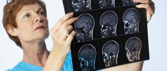
Often the cause of symptoms is sought using magnetic resonance imaging. This diagnostic method allows you to identify the cause of the development of pathology, as well as make an accurate diagnosis.
Choosing a clinic is easy to do using the service. A single registration center for MRI or CT allows you to select a medical center based on specified parameters: type of examination, metro station, and also sort the search results by price, rating, work schedule.
Patients have access to online registration through the website or telephone registration with a discount on the study in the amount of up to 1,000 rubles.
Symptoms
The symptoms of vegetative dystonia can be practically loved, without exaggeration. These may include changes in blood pressure, increased sweating, rapid heartbeat, sleep disturbances, bowel movements (vomiting, constipation, etc.), headaches, general weakness, and mood swings. There may be muscle pain, transient, numbness and crawling that do not have a clear localization. The person may experience discomfort in the chest and a lump in the throat. The symptoms of autonomic dysfunction are diverse and multifaceted. However, as part of the site about dizziness, I wanted to dwell on this symptom in more detail.
Dizziness with VSD may occur. However, nevertheless, it is rarely (almost never) systemic in nature, but fits more into the framework of non-systemic dizziness in the form of a fainting state or general lightheadedness, and may have signs of psychogenic dizziness. It does not have a fixed time frame and often depends on stress factors and weather conditions. Most often, it is not accompanied by tinnitus or nausea leading to vomiting, although nausea may be present.


