The expansion of the external cerebrospinal fluid spaces of the brain in adults can develop in the same way as in small children. In childhood, it is provoked by a violation of the formation of central nervous system structures during the prenatal period of fetal development, trauma during childbirth, and intrauterine infection. In adults, hydrocephalus is usually a consequence of head injury or infectious diseases of the central nervous system.
What it is
Externally, the brain is reliably protected from external influences by the bones of the skull. Cerebrospinal fluid is responsible for its internal protection: it prevents injury to the organ on the inner surface of the skull, ensures the maintenance of optimal intracranial pressure and regulates the internal state of the water-electrolyte environment.
In addition, cerebrospinal fluid is responsible for metabolic processes between blood and brain matter and removes the products of its metabolism.
Thus, cerebrospinal fluid plays a secondary, but no less significant role in the functioning of all brain structures. Therefore, any disturbances in its composition or deviations in concentration inside the skull immediately affect a person’s well-being.
The production of cerebrospinal fluid occurs in the ventricles of the brain through the epithelium lining them inside. From there, the fluid is delivered through the cerebrospinal fluid pathways to the outer surface of the organ, where it is collected in tanks, washes it and is absorbed through the venous sinuses and granulations of the arachnoid membrane.
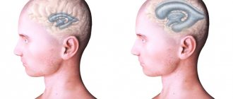
The expansion of the external cerebrospinal fluid spaces of the brain in adults is characterized by the accumulation of cerebrospinal fluid on the surface of the organ. This disease is called external hydrocephalus. It manifests itself in the expansion of the external brain cavities and deformation of brain tissue due to their compression.
The following external liquor spaces of the brain are prone to such changes:
- Tanks (located in the subarachnoid space in the areas of divergence of the arachnoid and pia mater);
- Subarachnoid fissures (located under the arachnoid membrane).
The accumulation of cerebrospinal fluid in them leads to an increase in intracranial pressure and the appearance of signs of hypertensive-hydrocephalic syndrome. It manifests itself in neurological abnormalities: convulsions, cephalgia, deterioration of hearing and vision, and in especially severe cases – paresis, paralysis, and decreased mental abilities. These manifestations are caused by increased pressure on neurons and their dysfunction.
Structural features of the brain
To understand the essence of this pathology, it is important to know what membranes cover the brain. There are three of them:
- arachnoid;
- hard;
- soft.
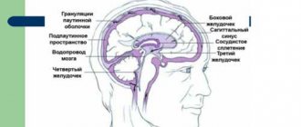
The subarachnoid space is located between the arachnoid and soft membranes. The first covers the entire surface of the brain, which in turn is enveloped by the endometrium. To communicate with other tissues, plexuses under the arachnoid membrane are used - membranes. The system of ventricles of the spinal cord and brain consists of the subarachnoid vascular plexuses. It consists of 4 reservoirs in which cerebrospinal fluid constantly circulates.
Subarachnoid spaces are small cavities in the brain filled with a special fluid (CSF). Their job is to nourish and protect the brain. The cerebrospinal fluid contains nutrients that are used to maintain the vital activity of nerve cells and the ventricles of the brain. Tissue waste products are also removed through the cerebrospinal fluid.
Liquor continuously circulates in the cavities of the brain. This is ensured by heart contractions, breathing, and body position. Normally, the volume of liquid filling the cerebrospinal fluid spaces should not exceed 140 ml.
Causes and symptoms
The expansion of the cerebrospinal fluid spaces of the brain is caused by impaired circulation of cerebrospinal fluid along the cerebrospinal fluid pathways.
This may happen for the following reasons:
- Increased cerebrospinal fluid production;
- Delays its absorption into the circulatory system;
- Due to the blocking of the main pathways for fluid removal by various formations: tumor, blood clots, cyst.
In young children, expansion of the functional cavities of the liquor system can be provoked by:
- Intrauterine infection;
- Head injuries;
- Inflammation of the meninges;
- Exposure to toxic chemicals;
- Congenital anomalies of the structure of the central nervous system structures;
- Presence of intracerebral tumors, cysts;
- Ischemic damage to brain structures due to prolonged hypoxia.
With the expansion of the subarachnoid convexital spaces (in the area of the central sulcus), children experience more serious neurological disorders, including heart rhythm disturbances. In adults, expansion of external liquor spaces most often becomes a consequence of head injury, infectious diseases of the central nervous system, and tumors of the brain.
Pathology can be suspected by the following manifestations:
- Morning headache;
- Nausea and vomiting that does not depend on food intake;
- Dizziness;
- Drowsiness, apathy;
- Hearing and vision impairment;
- Decreased mental abilities;
- Memory problems;
- Movement disorders: apraxia, limb tremor;
- Convulsions, paresis, paralysis.
In especially severe cases, increased pressure on brain structures leads to a displacement of important functional centers of the nervous system. This condition is life-threatening for the patient: for example, when the trunk is displaced, serious problems with breathing and cardiac activity are observed.
Symptoms of expansion of subarachnoid spaces
In children and adults, the pathology manifests itself differently. So, in children of the first year of life, the following signs help to suspect something is wrong:
- Negative reaction to noise, medium-level sounds, light;
- Frequent regurgitation;
- Poor sleep, anxiety;
- Strabismus, different pupil diameters;
- Head growth that does not correspond to age;
- Bulging and slower overgrowth of the fontanel;
- Weather sensitivity, anxiety when weather changes;
- Trembling, tremors of the chin, arms or legs.
As you can see, the symptoms are quite non-specific. They may more likely indicate hydrocephalus with intracranial hypertension secondary to expansion of the subarachnoid space. These signs cannot make an accurate diagnosis, but they should prompt an immediate visit to a neurologist or pediatrician.
In adults, one of the main symptoms of expansion of the subarachnoid space is headache, which can be quite intense and prolonged. Patients often complain of severe dizziness and nausea, inability to perform work duties, weakness, restlessness, and anxiety.
Cranialgia is especially pronounced early in the morning and may be accompanied by vomiting, dizziness, hypersensitivity to light and sounds. During the day, patients are often drowsy, at night they cannot sleep, or their sleep is restless and intermittent. An increase in headache, its pulsating nature, and the appearance of vomiting at the height of pain are characteristic symptoms of increasing intracranial pressure.
Diffuse expansion of the subarachnoid space and an increase in the volume of the lateral ventricles will sooner or later cause atrophic and dystrophic changes in the cerebral cortex, which will manifest itself as symptoms of encephalopathy - decreased memory and attention, visual impairment, and intelligence. Autonomic symptoms are often associated in the form of tachycardia, pressure surges, unmotivated anxiety and even fainting.
Movement disorders are more typical for cerebellar localization of pathology. In this case, symptoms may include unsteadiness and uncertainty in gait, dizziness, loss of coordination and fine motor skills.
Constant fatigue, repeated attacks of severe headaches, and the inability to get a good night's sleep disrupt the emotional state of patients, who can become irritable, anxious, and prone to depression. Secondary changes in the brain impair the ability to work and limit active life.
compression of the brain due to subarachnoid hemorrhage
Uneven expansion of the subarachnoid space is possible after trauma, brain surgery, or with tumors that locally compress the membranes of the brain and the cerebrospinal fluid pathways. In this case, focal neurological symptoms are observed in the form of disturbances in sensitivity, speech, hearing, etc. A severe course of the pathology is indicated by convulsive syndrome or epilepsy.
Depending on the width of the intershell space, there are light (1-2 mm expansion), moderate (up to 4 mm) and severe (over 4 mm) degrees of expansion. To a certain extent, such a division is arbitrary, especially with uneven or local changes in the liquor spaces.
The described symptoms usually occur with severe variants of the pathology, severe hydrocephalus and intracranial hypertension. Most often, mild and even moderate degrees of impairment are manifested only by periodic headaches, fatigue and autonomic dysfunction.
Treatment
Treatment tactics for neurological disorders caused by expansion of external liquor spaces depend on the form and stage of the disease:
- Acute hydrocephalus of the brain threatens the patient’s life and therefore requires radical measures: neurosurgical surgery. During this procedure, craniotomy is performed, followed by the installation of an external drainage system, which will ensure continuous outflow of excess cerebrospinal fluid. One of the advantages of this method of treatment is the possibility of administering drugs directly to the brain structures, for example, blood thinners and anti-inflammatory drugs. At the moment, this treatment option is considered outdated and is rarely used in practice due to the inconvenience of the drainage system design and the high risk of patient infection.
- Treatment of chronic expansion of external liquor spaces involves performing liquor shunt operations. This type of surgery involves the installation of a complex system of valves and catheters through which excess cerebrospinal fluid is discharged into the natural cavities of the body - the abdominal, pelvic or atrium.
If the patient has been diagnosed with the initial stage of expansion of the external liquor spaces, then conservative treatment is used.
It consists of taking the following medications:
- To reduce intracranial pressure, diuretics (Diacarb, Mannitol) are prescribed - drugs that can remove excess fluid from the cerebrospinal fluid spaces through urine. They are used only when the natural outflow of cerebrospinal fluid is preserved.
- Maintaining the required potassium concentration in the body (Panangin).
- To restore brain activity and improve metabolic processes in brain tissue, nootropic drugs (Cebrolysin, Cavinton, Actovegin) are prescribed.
These measures are aimed at reducing intracranial pressure, normalizing the main functions of the central nervous system, as well as providing brain tissue with the necessary nutrients. If conservative therapy is ineffective and the patient’s condition continues to deteriorate, then surgical treatment is indicated.
Principles for diagnosing convexital dilatation
Let's consider the main diagnostic methods:
- Neurosonography is an ultrasound examination of the brain structure through open slits of the fontanelles. It is carried out for each child after birth;
- Computed tomography (CT) measures the size of the cerebral ventricles, subarachnoid space;
- Comprehensive MRI of the head – monitors soft tissue changes. Layer-by-layer mapping of brain areas followed by three-dimensional remodeling shows the spatial structure of objects;
- Cisternography – determines the direction of movement of the cerebrospinal fluid, the type of dropsy (hydrocephalus).
Additional diagnostic methods are contrast angiography, PET/CT. Positron emission tomography is a radiation examination, therefore it is performed according to strict indications to limit the level of radiation exposure to tissues.
Disseminated encephalomyelitis
Disseminated encephalomyelitis (DEM) is an acute pathology of the central nervous system. In this disease, the myelin sheath of nerve fibers is destroyed. The perivascular Virchow-Robin spaces are dilated due to damage to the white and gray matter. The MRI image shows foci of demyelination.
This pathology is of autoimmune origin. The clinical picture of the disease resembles signs of multiple sclerosis. Patients experience gait and movement disturbances, speech disorders, dizziness, and inflammation of the optic nerve.
Unlike many other demyelinating diseases, REM is treatable. Patients are prescribed corticosteroids to suppress the autoimmune reaction:
- "Prednisolone";
- "Dexamethasone";
- "Metypred."
After a course of therapy, 70% of patients experience complete recovery. In advanced cases, patients may still experience the consequences of the disease: impaired sensitivity of the limbs, gait disturbances, and visual disturbances.
Prevention
How to prevent the above pathologies? It can be concluded that older patients are more susceptible to such diseases. Therefore, all people over 60 years of age need to regularly visit a neurologist and undergo an MRI examination of the brain.
It is also important to constantly monitor blood cholesterol levels and blood pressure. After all, diseases accompanied by pathological changes in the white matter most often develop against the background of atherosclerosis and hypertension.