Retinal angiopathy develops against the background of organic and functional disorders of the fundus vessels. They lead to circulatory failure and subsequent degeneration of retinal tissue. Most often, angiopathy is a secondary pathology that develops against the background of other ophthalmological or general systemic diseases.
Causes of angiopathy
The following diseases can lead to disruption of the vascular system of the eye:
- hypertension or hypotension;
- atherosclerosis;
- diabetes mellitus and other endocrine disorders;
- eye injuries;
- autoimmune diseases;
- genetic predisposition.
In most cases, disorders of the vascular system affect the entire body, and angiopathy is combined with other diseases. Only angiopathy of traumatic origin can be local in nature.
The developing pathology causes serious functional and trophic disorders of the tissues and structures of the eye:
- dystonia of the vascular wall;
- vascular spasms;
- formation of atherosclerotic plaques;
- thrombosis of veins and capillaries.
Angiopathy always carries the risk of ischemic changes in the retina. Dysfunction of the circulatory system leads to slow metabolism, accumulation of metabolic products in tissues and hypoxia. Over time, this causes degeneration and functional failure of the light-sensitive cells of the eye and, as a consequence, loss of visual acuity.
Development of angiopathy during pregnancy
This is a common phenomenon that occurs in both new and experienced mothers during the second or third semester. In most cases, the disease appears towards the end of pregnancy.
The appearance of retinal angiodystonia is associated with high blood pressure, atherosclerosis or diabetes mellitus. As the embryo grows, blood volume increases, pressure jumps (especially in a stressful situation), which causes the vascular walls to stretch. As a result, the risk of thrombosis, hemorrhage, retinal detachment, and blindness increases.
It is worth highlighting the characteristic differences of hypertensive angiopathy during pregnancy:
- Periodic narrowing of the arteries. The cause is toxicosis.
- Sclerosis of blood vessels.
- Impaired blood circulation.
- The visual system recovers quickly after the birth of a child.
If such a pathological condition develops during pregnancy, the doctor may prescribe a planned cesarean section. Natural childbirth can cause retinal vessels to rupture, and a woman can completely lose her vision.
Types of retinal angiopathy
The classification of angiopathy is based on etiology. There are 5 types of angiopathy:
Hypertensive. General vascular disorders in hypertension affect the functioning of the veins and capillaries of the fundus. The retina that feeds from them does not receive the necessary substances in full and accumulates metabolic products. This causes tissue degeneration, functional failure of light-sensitive cells, and then irreversible organic changes. Angiopathy caused by vascular hypertonicity is further complicated by atherosclerotic changes in the lumen of blood vessels.
Hypotonic. Constantly low pressure in the vascular system is also not beneficial, since low tone causes slower blood flow. The consequence of this is sluggish metabolism, nutritional deficiency and general functional weakness of both the eye muscles and light-sensitive elements.
Diabetic. Elevated blood glucose levels have a toxic effect on the walls of blood vessels. Their elasticity and permeability are impaired. Diabetes is often accompanied by atherosclerosis, which causes the formation of plaques that narrow the lumen of blood vessels, which also impairs trophism.
Traumatic. Impaired blood supply to the fundus can be caused by mechanical damage. Especially often, angiopathy develops after traumatic brain injury and as a result of a violation of the integrity of the upper parts of the spine.
Youthful. Angiopathy that occurs before the age of 30 is considered “youthful.”
The reasons for such early development of pathology, as a rule, are chronic, sluggish infections (toxoplasmosis, tuberculosis and others). Over time, the disease leads to vasculitis - inflammatory changes in the vascular system. The consequence of this is insufficient blood supply and retinal dystrophy.
Stages of development of diabetic angiopathy disease
There are several types of diabetic angiopathy. She may be:
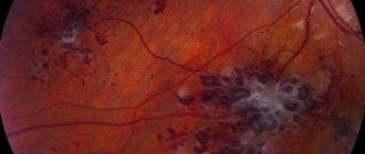
Non-proliferative. The vessels in the fundus are gradually affected, microscopic aneurysms form, and small hemorrhages develop. At this stage, the retina swells and the iris becomes redder.
Preproliferative. The retinal veins are damaged. Rupture of the vessel causes hemorrhage and the formation of venous infiltrate, which causes significant visual impairment.
Proliferative. It is the most difficult stage. New capillaries are formed, which are fragile. As a result, multiple hemorrhages appear, which cause retinal detachment.
Traumatic. If the head, neck or eyes are injured, blood vessels are compressed and intracranial pressure increases. As a result, leukocyte emboli are formed.
Youthful. It is a rare and dangerous form of the disease. The etiology is not fully understood. The development of pathology is observed before the age of 25 years. The manifestation of juvenile angiodystonia is the inflammatory process, hemorrhage, and proliferation of connective tissues. As a result, various complications arise.
Congenital. In most cases, premature babies suffer from this form of the disease due to the fact that their vascular system is underdeveloped.
To make the most accurate diagnosis and begin appropriate treatment, it is necessary to determine the type of retinal angiopathy. Treatment measures should begin when the initial stage is present. Without adequate treatment, the patient may go blind.
Symptoms of angiopathy
Developing angiopathy may not manifest itself for a long time. This complicates diagnosis in the early stages. Most often, the disease is detected by complications that have already arisen, which give clear symptoms. Only with further research does it become clear that the disorders are secondary, and the underlying cause was the existing angiopathy.
Characteristic symptoms in the presence of angiopathy may be:
- episodic blurring of the visual field;
- headache;
- unpleasant and painful sensations in the eye area;
- loss of visual acuity;
- narrowing of the field of view, loss of peripheral vision areas.
These symptoms in any combination are a serious reason for immediately contacting an ophthalmologist. Even if the cause is not angiopathy, such disorders indicate existing organic disorders or developing pathological processes.
What is the result?
Your nutrition of the eye muscles and tissues will improve. Vascular tone is normalized. The vessels and capillaries will strengthen and restore normal blood supply to the retina and other organs. The retina of the eyes will be strengthened. The unpleasant symptoms of “cloudness” and decreased visual acuity will disappear. The risk of serious complications will be significantly reduced.
Powerful and effective treatment with the Neurodoctor device will quickly eliminate the clinical manifestations of the disease, localize the foci of pathology, and reduce the dose of medications. This will allow you to live a full life and forget about your illness forever.
You will strengthen the retina of the eyes, normalize blood circulation in the vessels and get rid of discomfort. In addition, the device will prevent the development of dangerous complications that can lead to partial loss of vision or complete blindness.
Diagnostics
Even completely healthy people are recommended to have an annual preventative visit to an ophthalmologist. If you have risk factors, you should visit your doctor twice a year. This is especially important for identifying angiopathy and other eye diseases that do not have obvious symptoms in the early stages. Many of them have a favorable prognosis only with timely treatment.
The basis for diagnosing angiopathy is examination of the fundus. At the same time, even minor disorders are quickly and painlessly identified, threatening serious complications in case of untimely treatment.
Our ophthalmological center carries out a full range of diagnostic measures aimed at early diagnosis of angiopathy. After examining the fundus, experienced doctors, if necessary, conduct a more thorough examination using modern equipment. To clarify the diagnosis, the following may be prescribed:
- duplex study of brachiocephalic vessels;
- Ultrasound of the eyeballs;
- other hardware and instrumental methods for studying the vascular system of the eye.
Therapy
Diabetic angiopathy
The leading role is played by therapy of the underlying disease. It is necessary to follow a strict diet, quit smoking and alcohol, and control body weight.
Since most diabetic angiopathy is associated with damage to the vessels of the lower extremities, special attention should be paid to the condition of the legs (feet). If the doctor suspects a patient has a “diabetic foot” (deformation, infection of soft tissues, the appearance of trophic ulcers), hospitalization and inpatient treatment in the clinic are carried out. Physiotherapy is used during the rehabilitation period. In case of severe pathologies, surgical intervention is indicated.
Retinal angiopathy
This type of angiopathy responds better to conservative treatment than others (taking into account the underlying disease). The use of medications, laser therapy, physiotherapy, and massage of the collar area can be effective. In advanced cases, surgical intervention is necessary.
Hypertonic, striatal and CAA
Hypertensive angiopathy is treated by stabilizing blood pressure and normalizing blood circulation.
For striatal angiopathy, drugs to improve cerebral metabolism and hemodynamics are prescribed. If there is no effect, minimal surgical intervention is required.
For cerebral amyloid angiopathy, symptomatic treatment is indicated; it is not possible to achieve positive dynamics in the treatment of the underlying disease.
Treatment of angiopathy
The first task in the treatment of angiopathy is to identify and eliminate the negative factors that served as the background for the development of the disease. As a rule, these are chronic diseases. It is necessary to monitor blood pressure and prevent hypertensive crises. Diabetics must maintain optimal blood sugar levels. These measures are ensured by appropriate treatment regimens prescribed by specialized specialists. The ophthalmologist monitors how the treatment affects the blood vessels of the eyes, and also prescribes treatment aimed at eliminating hypoxia, normalizing the blood supply to the optic nerve head, and strengthening the walls of the blood vessels of the eye. Vitamins, antioxidants, neuroprotectors and mineral complexes can also provide good support to tissues experiencing dystrophy.
Specialists of the Moscow Ophthalmological Center will offer an individual treatment regimen, which may include drugs in the form of:
- tablets;
- injections;
- parabalbar injections into the eye area;
- intravenous drips.
Additionally, hardware treatment programs, physiotherapeutic procedures and massage are prescribed.
Surgical care can be aimed mainly at eliminating complications that have already developed. Most often, laser surgery is used to treat and prevent retinal detachment caused by angiopathy.
Do you need effective treatment, but still have doubts?
“Neurodoctor” is today considered one of the most effective methods of treating diseases of very different etiologies. It combines perfectly with traditional treatment methods and allows the body to recover faster. Today the Neurodoctor device is:
List of used literature
- Babaeva A.R., Tarasov A.A., Davydov S.I., Emelyanova A.L. // Bulletin of VolSMU. - 2006. - T. 19, No. 3. - P. 18-23.
- Voskanyants A. N., Nagornev V. A. // Cytokines and inflammation. - 2004. - T. 3, No. 4. - P. 10-13.
- Gurevich V.S. // Diseases of the heart and blood vessels. - 2006. - No. 4. - P. 4-8.
- Demyanov A.V., Kotov A.Yu., Simbirtsev A.S. // Cytokines and inflammation. - 2003. - T. 2, No. 3. - P. 20-35.
- Dedov I.I., Aleksandrov A.A. // Consilium med. — 2004. —T. 6, No. 9. - P. 620-624.
- Dedov I. I., Antsiferov M. B., Galstyan G. R., Tokmakova A. Yu. Diabetic foot syndrome. — M.: Federal Diabetology Center of the Ministry of Health of the Russian Federation, 1998
Medicines
If the problem of treating the underlying disease is solved, drugs such as pentoxifylline (Pentyline), Trental, Vazonit (help to improve blood flow) are used in treatment.
Xanthinol nicotinate and ginkgo biloba preparations (Bilobat, Tanakan) improve the condition of the vascular wall. To normalize microcirculation, take vitamins C, A, E, B.
ATP-long, actovegin, cocarboxylase drugs have a metabolic effect: they activate blood circulation and improve metabolism.
Soft antiplatelet agents: Magnicor, dipiradamole prevent the formation of blood clots.
Since all such pathologies seriously affect the quality of life, and in some cases are threatening, drug therapy should be agreed with the attending physician.


