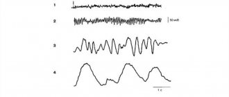
About the treatment of multiple sclerosis. Interferons
Interferons (IFNs) are a group of biologically active proteins (glycoproteins) produced by various cells of the body in response to a viral infection or exposure to certain chemical and biological substances. The binding of interferons to cellular receptors leads to a significant positive effect.
A number of intracellular proteins are produced, which:
- have an antiviral effect ;
- stabilize the immune system ( immunomodulation);
- controls cell division ( antiproliferation); .
There are alpha, beta, and gamma interferons.
Interferon beta is recognized as the most effective in the treatment of MS . Their use reduces the number of exacerbations and slows down the development of the disease.
interferon beta drugs is to suppress the formation in the human body of substances that damage the myelin sheath of nervous tissue. At the same time, interferons promote the activation of other cells that reduce the effect of antibodies to myelin , thereby reducing the activity of the inflammatory process.
In addition, beta interferons protect myelin layers from further destruction, and also reduce the likelihood of damage to the blood-brain barrier (BBB) , preventing the penetration of immune complexes into brain cells.
There are two types of beta interferons:
- Interferon beta-1b;
- Interferon beta-1a.
The main clinical effects of drugs from both groups include:
- reduction in the average annual frequency of exacerbations;
- slowdown in disability (increase in EDSS);
- improving the quality of life of patients[xvii].
interferon preparations are administered by the patient independently subcutaneously or intramuscularly, after prescription and recommendations from a doctor. Injections of the drug are quite low-traumatic and do not cause difficulties to the patient. As a rule, courses of interferon are long. Since unjustified cessation of interferon beta treatment leads to a return of symptoms of multiple sclerosis, its cessation is justified only if the drug is not sufficiently effective or serious side effects develop as a result of treatment.
Due to the fact that interferon are involved in the activation of the body's defenses , after administration, reactions reminiscent of a “cold” (flu-like syndrome) may develop, as well as local reactions in the injection area in the form of itching, redness and swelling.
contraindications for treatment with interferon beta :
- pregnancy;
- hypersensitivity to the drug;
- therapy with anticonvulsants.
Interferon beta therapy is currently used throughout the world to treat relapsing and secondary progressive forms of multiple sclerosis[xix].
Studies have shown that long-term use of interferon beta is quite safe and effective. The use of the drug in adults with relapsing-remitting and progressive forms of multiple sclerosis reduced the frequency of exacerbations, the severity of symptoms and extended the duration of remissions. The formation of new lesions also decreased significantly decreased .
Material and methods
Study No. BCD-054−2 is conducted in accordance with the regulatory framework of the participating countries. All CT documents were reviewed and approved by the Ethics Board and local ethics committees of all study centers.
A total of 399 patients with RRMS were examined and randomized into four groups: 114 patients each into three active treatment groups and 57 into a placebo group.
Study design:
multicenter, double-blind, comparative, randomized, placebo-controlled, prospective, parallel group study followed by an open-label phase with SamPEG-IFN-β1a.
Patients were recruited in the Russian Federation (27 centers) and the Republic of Belarus (3 centers). Before any screening procedures began, patients signed an informed consent (IC). The information for the patient contained all the information about the present CI necessary to make an informed and independent decision. The last patient was included in the study on July 2, 2021.
Inclusion criteria:
age from 18 to 60 years, a definite diagnosis of MS (according to the 2010 MacDonald criteria), relapsing-remitting, in whom no new or worsening of previously existing neurological symptoms were observed within 30 days before signing the informed consent (IC), or During this period, complete stabilization of the condition was observed after the last exacerbation, with a score on the EDSS scale of 0−5.5, having not previously taken IFN-β drugs that alter the course of MS.
Sample calculation
The required number of patients to test the hypothesis of non-inferiority of the study drug to the comparator drug for the end point “time to first exacerbation” was estimated using data on the time of onset of the first exacerbation in patients during 48 weeks of therapy based on the results of previous studies with similar populations patients [4, 5]. According to additional calculations, this number of patients was also sufficient to test the hypothesis of superior efficacy of the study drug over placebo according to the CUA (combined unique active lesions) effectiveness indicator, which reflects the cumulative assessment of new and enlarged lesions on MRI after 20 weeks of therapy. Patients were stratified depending on the number of exacerbations over the previous year and the number of lesions accumulating T1 contrast.
Investigational therapy
Patients enrolled in the SamPEG-IFN-β1a treatment group receive study drug at doses of 180 mcg and 240 mcg for 52 weeks, alternating between placebo injections once every 2 weeks to blind therapy. In the comparison drug group, participants received low-dose interferon beta-1a (NIB, Avonex, Biogen Idec Limited, UK) 30 mcg once a week. Patients in group 4 (placebo group) were administered placebo once a week during the first 20 weeks. All injections were performed intramuscularly. During the first weeks, all patients underwent titration of the drug dose until its full dose was reached by the 4th week of therapy, then the drugs were administered at full dosage until the 52nd week of the study.
After the end of the main period (52 weeks) of follow-up treatment/observation, patients enrolled in the BCD-054 groups continued to receive SamPEG-IFN-β1a therapy for 48 weeks. Patients receiving NIB were transferred to the follow-up period (from weeks 53 to 56) after completion of 52 weeks of therapy. For patients receiving placebo, the observation period began after the last placebo dose and continued from weeks 21 to 24 of the study.
Primary endpoint for assessing effectiveness
SamPEG-IFN-β1a compared with placebo after 20 weeks of therapy was an indicator of the cumulative number of new contrast-enhancing lesions on T1-weighted MRI and new lesions in T2-weighted mode or cases of enlarged lesions in T2-weighted mode without double addition - CUA. Parameters reflecting the dynamics of brain MRI parameters, as well as clinical indicators associated with exacerbation and level of disability after 20 weeks of therapy, were selected as additional endpoints.
Endpoints for Security Assessment
after 20 weeks of treatment, we included patients who had registered the development of AEs and SAEs, including AEs of grade 3-4 severity, determining the frequency of cases of early termination of participation in the study due to the development of AEs or SAEs, as well as the dynamics of indicators on the scale Beck's depression. The severity of AEs and laboratory abnormalities was assessed in accordance with the CTCAE v.4.03 classification [6].
Statistical data processing
For statistical analysis, the SAS 9.4 software environment and the programming language for statistical data processing R were used. The choice of descriptive statistics and statistical comparison method was determined by the type of data and type of distribution. Means and standard deviations were used to describe normally distributed quantitative variables; For statistical processing, two-sample Student's test and analysis of variance were used. For non-normally distributed quantitative data, mean values were described using medians and interquartile ranges; For statistical processing, the Mann-Whitney test with Benjamini-Yekutili, Wilcoxon, Kruskal-Wallis, and Friedman corrections was used. Percentages or proportions were used to describe categorical data. Statistical comparisons of categorical data were performed using Fisher's exact test or Pearson's χ2 test, corrected for multiple comparisons by Benyamini-Yekutili. Statistical significance of differences was determined at p
<0,05.
Testing the statistical hypothesis of the study based on the results of 20 weeks of therapy was carried out by comparing the 95% classical confidence interval (CI) for the difference in the arithmetic mean values of CUA between the SamPEG-IFN-β1a and placebo groups and the interval –0.3407–0.3407, where –0. 3407 is the superiority margin calculated using the 95% CI for the difference in the means between the active treatment and placebo groups from previous studies, ensuring that the superiority margin metrics are consistent with the calculated CI.
Efficacy and safety analyzes after 20 weeks were conducted in the mITT population, which included 397 patients who received at least one administration of BCD-054/NIB/placebo: group 1 - 113 patients, group 2 - 113, group 3 - 114, 4th group - 57.
Due to the fact that this study involves blinded therapy, during the first 52 weeks, when analyzing a series of data regarding the effectiveness and safety of repeated use of BCD-054, information about patients' belonging to a particular treatment group is not provided; groups are coded by numbers in any order in order to eliminate the hypothetical risk of unblinding of treatment groups. The placebo group is unblinded.
References
- Lifshits, V.M., Sidelnikova, V.I. Medical laboratory tests. - M.: Triad X, 2007. - 312 p.
- Nesterova, I.V. Congenital and acquired interferonopathies: differentiated approaches to interferon-corrective therapy, Federal State Autonomous Educational Institution of Higher Education "Russian Peoples' Friendship University" of the Ministry of Education and Science of the Russian Federation, Moscow, 2021. - V. 16(2). — P. 50-53.
- Savenkova, M.S., Karashtina, O.V., Shabat, M.B. and others. Interferon status and choice of interferon inducers in frequently ill children. - Children's infections, 2021. - No. 2. - P. 45-51.
results
A total of 399 patients were randomized into the study: 114 in the active treatment group and 57 in the placebo group. Before the first administration of the drug, 2 patients from groups 1 and 2 were excluded from the study; they were excluded from the mITT (modified intent-to-treat) population. During the 20-week CT after the first administration of the drug, another 28 patients dropped out: 20 due to IS withdrawal, 2 due to adverse events (AEs)/serious AEs (SAEs), 3 due to non-compliance, 1 due to protocol violations (violation of the sequence of using syringes when administering the drug), 2 - due to pregnancy. Thus, 369 patients completed 20 weeks of therapy (Fig. 1).
Rice. 1. Distribution of patients into study groups, the number of participants who dropped out of the study, indicating the reason for dropping out.
Analysis of effectiveness after 20 weeks of therapy
The medians of demographic indicators, general and biochemical blood tests, as well as physical and other data in all patients were comparable in all groups at screening. Analysis of the characteristics of the underlying disease at screening did not reveal differences between the groups in the duration of the disease, the number of exacerbations recorded during the entire period of the disease and during the 12 months preceding the signing of I.S. In all groups, the absolute majority were patients who had no experience taking drugs that modify the course of MS (DMT): 71.68, 72.57, 75.44 and 63.16% in groups 1, 2, 3 and the placebo group, accordingly ( p
=0.4104, Pearson χ2 test). Among patients with experience of therapy, the most frequently used drugs were glatiramer acetate and laquinimod; no statistically significant differences were found between the groups in the range of drugs used. The median total EDSS score at baseline in patients of all groups was 2 points, which met the inclusion criteria.
In each group, the majority of patients had at least 1 Gd+ lesion in T1 mode or 1 new lesion in T2 mode detected according to brain MRI within 12 months before signing the IP: 64.60, 62.28, 61.06 and 66.67% in groups 1, 2, 3 and placebo group, respectively ( p
=0,8841).
As part of screening to assess brain damage before starting medication, all patients underwent MRI of the brain with contrast; an independent specialist assessed the number of lesions and lesion volume in T2-WI and T2-FLAIR modes, as well as the number of active Gd+ lesions and their volume in T1 mode. In pairwise comparisons, all groups were comparable according to MRI findings.
Because CUA is analyzed over time, CUA was assessed starting with the second MRI in the study, starting at “Visit 4/Week 12” and then at “Visit 5/Week 16” and “Visit 6/Week 16”. 20". Dynamic observation of the complex CUA indicator during the analyzed period showed a tendency towards its decrease over 20 weeks of therapy in groups of patients receiving therapy with IFN-β drugs. With a statistically significant difference between the scores in group 2 and the placebo group ( p
=0.0021) and in group 3 and placebo group (
p
=0.0115) (Fig. 2).
Rice.
2. Dynamics of average CUA values over 20 weeks of therapy in the study groups (mITT population). Here and in Fig. 3 * - p<0.05 versus placebo In addition to assessing the CUA indicator, a per scan
, which consists of estimating the average value of the indicator based on all MRI assessments performed in each patient.
According to the analysis, the median CUA per scan
after 20 weeks of therapy in group 1 was 0.333 [0.0;
1.0], in the 2nd - 0.0 [0.0; 0.667], in the 3rd - 0.333 [0.0; 0.667], in the placebo group - 1.0 [0.0; 2.0]. The average CUA per scan
after 20 weeks in group 1 were 0.986±2.046, in group 2 - 0.619±1.055, in group 3 - 0.665±1.165, in the placebo group - 1.673±2.376.
When pairwise comparison of median CUA per scan
after 20 weeks of the study, there was a statistically significant difference between CUA in groups 2, 3 and the placebo group (
p
= 0.0067).
At the same time, the medians of the CUA per scan
between the 2nd and 3rd groups, as well as the 1st group and the placebo group did not have statistically significant differences (
p>
0.05).
When compared by primary endpoint, CI for mean difference CUA per scan
between the 1st group (BCD-054 180 mcg) and the placebo group was [–1.584, –0.5232], between the 2nd group (BCD-054 240 mcg) and placebo [–1.5511, –0. 4643].
The results obtained show that the upper limit of the CI in both groups does not exceed the predefined superiority limit of –0.3407. When comparing arithmetic means of CUA per scan
between the BCD-054 180 μg and placebo groups, as well as BCD-054 240 μg and placebo, a statistically significant difference is observed (
p
= 0.0001 for comparison of the BCD-054 180 μg group and placebo;
p
= 0.0003 for comparison of BCD-054 240 mcg and placebo groups).
Due to the fact that the calculated upper limit of the CI for both groups does not exceed the predefined superiority limit (=δ–0.3407), the hypothesis of the superiority of BCD-054 in two dosages (180 and 240 mcg) over placebo according to the primary endpoint CUA per scan
after 20 weeks of blinded use.
When assessing the number of foci accumulating contrast in T1 mode, when performing MRI of the brain during the 20-week treatment period, positive dynamics were revealed in the form of a decrease in the number of foci, which was statistically significantly expressed in the active treatment groups. Thus, in group 1, the average value (±standard deviation) of T1 Gd+ lesions at screening was 1.460±2.806, at visit 6/week 20 - 0.515±1.092, in group 2 - 1.522±3.836 and 0.358±1.532, in group 3 - 1.307±3.382 and 0.269±0.791, placebo group - 1.158±2.789 and 1.077±1.998, respectively. It should be noted that there was no statistically significant difference in screening between all groups ( p
=0.9076).
After 20 weeks, statistically significant differences were found between the median number of lesions accumulating contrast in T1 mode in groups 2 and placebo, in groups 3 and placebo, as well as in groups 1 and 3 ( p
< 0.05) (Table 1).
Table ١.
Dynamics of MRI parameters within the analyzed ٢٠-week period (n=397) The proportion of patients without lesions accumulating contrast in T1 mode was statistically significantly lower in group 2 compared to the placebo group, in group 3 compared to group 1, as well as placebo group ( p
<0.05). The proportion of patients without lesions accumulating contrast in T1 mode, according to the results of MRI performed at visit 6/week 20, in group 1 was 62.8% (71 out of 113 patients), in group 2 - 77.9 % (88 out of 113), in the 3rd group - 78.9% (90 out of 114) and placebo group - 54.4% (31 out of 57).
Analysis of the number of enlarged lesions in T2WI mode demonstrated the absence of statistically significant differences between groups and in the dynamics in groups during the first 20 weeks of blinded therapy ( p>
0.05). It should be noted that the average values of the number of enlarged lesions in T2WI mode decreased by week 20, with the most pronounced dynamics in the active treatment groups. Thus, the average number of enlarged lesions in T2WI mode in group 1 at visit 4/week 12 was 0.376±1.341, and at visit 6/week 20 - 0.139±0.584, in group 2 - 0.240±0.898 and 0.123± 0.492, in the 3rd group - 0.317±1.117 and 0.106±0.392, in the placebo group - 0.340±0.960 and 0.288±0.848, respectively.
During the period of equilibrium concentrations after repeated administration of BCD-054, there was an increase in the level of MxA protein by 2-3 times compared with the initial one, determined before the first injection in the study, regardless of the dose of the drug. At the same time, in the NIB comparison group, a similar increase was less than 1.7 times. In the placebo group, there was no increase in MxA protein levels during 20 weeks of CI. When assessing the area under the effect-time curve of MxA protein in groups taking into account the frequency of injections, it was noted that repeated intramuscular administration of the study drug BCD-054 at a dose of 180 or 240 mcg provided approximately 2 times greater exposure of MxA protein to compared with non-PEGylated IFN-β1a (Fig. 3).
Rice. Fig. 3. Median values of AUEC0−168.SS and AUEC0−336.SS of MxA protein in 4 groups of patients with RRMS 168 and 336 hours after repeated administration of drugs in the 16th week during the period of equilibrium concentrations (nmol/l) h.
Safety analysis after 20 weeks of therapy
The range of AEs observed during the first 20 weeks of the study was comparable both quantitatively and qualitatively. In most cases, clinical and laboratory abnormalities recorded as AEs were of the same type and were expected according to the known data on the safety of the use of IFN-β1a drugs in patients with MS1, 2 (Table 2).
Table 2. The most common adverse reactions Note: 1 at p<0.05 between all groups (*) pairwise comparison of groups was carried out: 1.2 – nos. 1 and 2, 1.3 – nos. 1 and 3, 1, pl . –№№ 1 and placebo, 2,3 –№№ 2 and 3, 2, pl.. – №№ 2 and placebo, 3, pl. – No. 3 and placebo.
The most common AE during the first 20 weeks of the main study period was influenza-like syndrome: 75.22, 66.37, 78.07% of patients in groups 1, 2 and 3, respectively ( p
>0.05).
In the group of patients receiving placebo, 19.3% of patients also had manifestations of influenza-like syndrome of 1st and 2nd severity. When comparing the proportion of patients with influenza-like syndrome (all severity and severity 2) between the placebo group and the other active treatment groups, as expected, there was a statistically significant difference ( p
< 0.0001). About 70% of cases of influenza-like syndrome developed during the first 12 weeks of therapy. Most cases of influenza-like syndrome were grade 1 and 2, with the exception of 4 cases of grade 3. In 90% of cases, to reduce and/or regress the clinical manifestations of influenza-like syndrome, patients were prescribed oral administration of non-steroidal anti-inflammatory drugs or other drugs with antipyretic and analgesic effects. About 95% of all cases of influenza-like syndrome had resolved without any negative consequences at the time of writing, about 5% were ongoing. The duration of manifestations of influenza-like syndrome less than 7 days was observed in more than 85% of cases.
Injection site reactions, reported in less than 10% of patients in groups 2 and 3, were also expected given the study therapy. In 40% of cases, oral antihistamines and external ointments/gels with anti-inflammatory activity were used to eliminate reactions at the injection site. More than 85% of all cases had resolved without any consequences at the time of writing, about 15% were ongoing.
As is known, IFN therapy can have multidirectional effects on thyroid function. An increase in the level of thyroid-stimulating hormone (TSH) was detected in all groups with a frequency of no more than 3% and reached grade 1-2. In no case was hormonal therapy required. More than 55% of cases of TSH level deviations were transient in nature and resolved without negative consequences.
A number of laboratory abnormalities were also recorded in general and biochemical blood tests. The frequency of these abnormalities in patients was comparable in the groups. The most common abnormalities were increased activity of transaminases and γ-glutamyl transpeptidase (GGT), decreased numbers of leukocytes, neutrophils and lymphocytes, and increased TSH levels. Laboratory abnormalities were mostly grade 1-2 and were transient and asymptomatic.
It should be emphasized that in patients in whom a decrease in the levels of leukocytes, neutrophils, and lymphocytes was determined, the development of any manifestations of infectious processes was not noted.
Psychoemotional disturbances were observed in no more than ≈3% of patients in each active treatment group. Only 1 patient discontinued participation in the study early. According to the study physicians, no SAEs related to the therapy were recorded.