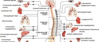
Phakomatosis - main symptoms:
- Skin rashes
- Convulsions
- Skin spots
- Impaired movement coordination
- Insomnia
- Epileptic seizures
- Hearing loss
- Visual impairment
- The appearance of a tumor formation
- Baldness
- Mental retardation
- Mental development disorder
- Facial skin pigmentation
- Daytime sleepiness
- Dementia
- White eyelash color
- Antisocial behavior
- White hair color
- Premature aging
- White eyebrow color
What is phakomatosis
Phakomatosis is a multicomponent concept that unites a number of congenital pathologies that affect the eyes, nervous system and human skin. The type of inheritance of the disease is autosomal dominant. Phakomatoses provoke the formation and progression of various tumors and hematomas - benign neoplasms that develop in organs.
Phakomatosis is a very serious congenital disease that is diagnosed in children at an early age. The symptoms are multifaceted, however, rash and tumor-like formations are its main characteristic. If the tumor is localized in the brain, then with its development the patient may experience psychological abnormalities.
Phakomatoses are also distinguished by the presence of hemianopsia - a person partially sees the space around him. Scientists can accurately diagnose the problem only by the age of 7-10 years, despite the fact that the general clinical picture of phakomatosis in children is reflected in the first years of life. However, cases have been recorded where the pathology did not manifest itself for decades.
In the International Classification of Diseases (ICD-10), phakomatosis is coded Q85.
↑ NF1
Diagnostic criteria
To establish the diagnosis of NF1, the following criteria were defined. At least two of them must be present to make a diagnosis.
- Five or more café au lait pigment spots more than 5 mm in diameter in a prepubertal child and six or more café au lait pigment spots with a diameter of more than 15 mm in a postpubertal patient.
- Two or more neurofibromas of any type or one plexiform neurofibroma.
- Axillary or groin spots.
- Optic nerve glioma.
- Two or more Lisch nodules.
- Characteristic bone defects (pseudoarthrosis of the tibia or dysplasia of the sphenoidal wing) (Fig. 24.1).
- Close relatives suffering from NF1.
Prevalence
The prevalence of neurofibromatosis has been found to be 1:3000-1:5000, making this disease one of the most common autosomal dominant disorders. Penetrance is almost complete; as a result of the high level of spontaneous mutations, new mutations are detected in 50% of patients with neurofibromatosis. The affected gene is localized on the proximal long arm of chromosome 17 (17qll.2).
General manifestations
- Pigment spots on the body the color of coffee with milk. However, these changes are not a pathognomonic sign of neurofibromatosis and can occur in healthy people.
- Small spots are localized mainly in the folds of the skin - in the armpits, in the groin area and under the breasts in women.
Peripheral neurofibromas
Almost all patients with NF1 by the age of 16 develop peripheral neurofibromas of the skin and, in more rare cases, neurofibromas of the subcutaneous tissue, palpated along the peripheral nerves.
Plexiform neurofibromas
Peculiar new formations of soft consistency. Pathognomonic symptom for NF1. Characteristic signs are hypertrophy of surrounding tissues, local tissue proliferation and hypertrichosis in the affected area. When the process is localized in the orbit, a significant decrease in vision is possible due to direct compression of the optic nerve or amblyopia developing due to ptosis and/or strabismus caused by the tumor.
Decreased learning ability
Although mental retardation is rare in NF1, there may be a slight decrease in visual perception.
Ophthalmic manifestations
An examination by an ophthalmologist of a patient with suspected neurofibromatosis is important not only to confirm the diagnosis, but also to identify complications in the visual organ and, if possible, prescribe treatment as early as possible.
Pathological changes in the organ of vision can be localized in the orbit and include:
- optic nerve glioma;
- meningioma of the optic nerve;
- orbital neurofibroma;
- orbital bone defects.
Exophthalmos may be associated with a change in the position of the eyeball, which is subsequently followed by strabismus and amblyopia.
Orbital tumors often cause changes in the optic nerve, manifested by congestive nipples, atrophy, and, somewhat less commonly, optic nerve hypoplasia. Additional changes include:
- optociliary shunts (especially for metingioma of the optic nerve);
- folding of the choroid;
- amaurosis, determined by the direction of gaze. Neurological studies are indicated to differentiate the various causes of exophthalmos in NF1.
Eyelids
- Most often, the eyelids are affected by plexiform neurofibroma with a characteristic 5-shaped deformation of the edge of the upper eyelid. Strabismus and/or ptosis resulting from these changes can lead to amblyopia.
- Congenital ptosis occurs even in the absence of an orbital tumor.
Iris Lisch
nodules In NF2 they are rare. Their prevalence in NF1 increases with age. In early childhood, Lisch nodules are not often observed, but by the age of 20 they are found in almost 100% of patients. Optic nerve
Involvement of the optic nerve in the pathological process is manifested by optic nerve gliomas (astrogynomas) (Fig. 24.2). 70% of all optic nerve gliomas occur in patients with NF1. The true frequency of their prevalence in NF1 is difficult to determine due to the lack of symptoms and therefore the subclinical course. In approximately 15% of patients with NF1 and normal visual acuity, optic nerve gliomas are detected radiographically. These neoplasms are divided into two categories.
Anterior (orbital gliomas)
These gliomas are represented by exophthalmos, loss of vision and, occasionally, changes in the position of the eyeball. Involvement of the optic nerve in the process is expressed in its atrophy, dysplasia, direct damage by the tumor and congestive nipple. Sometimes optociliary vascular shunts are formed on the side of the tumor. As a result of the changes, strabismus often occurs.
Posterior (chiasmal fibroids)
Symptoms of these gliomas include hydrocephalus, endocrine pathology, and decreased vision combined with nystagmus. Nystagmus can be vertical, rotatory or asymmetrical (occasionally imitates nutane spasms). Dissociated vertical nystagmus often occurs.
Conjunctiva
Neurofibromas of the conjunctiva are rare and are usually located in the limbal zone.
Cornea
In NF1, thickening of the corneal nerves occurs, but this symptom is not pathognomonic. Much more often this disorder occurs in multiple endocrine neoplasia syndrome.
Uveal tract
Pigmented hamartomas of the choroid occur in 35% of patients. Diffuse neurofibromas cause thickening of the entire uveal tract, leading to glaucoma.
Retina
In NF1, the retina is rarely involved in the pathological process. There are isolated reports of cases of astrocytic hamartomas of the retina and its pigment epithelium.
Research
- Computed tomography (CT) or magnetic resonance imaging (MRI) of the brain and orbit can detect bone pathology, meningiomas, and optic nerve gliomas.
- Visual evoked potentials (VEPs) help in assessing the state of the chiasm, as well as in monitoring the dynamics of chiasmal gliomas.
Management tactics
The management tactics of patients with NF1 remain controversial to this day. Treatment is usually complex. An integral component in the overall set of prescriptions is genetic consultation of all members of the patient’s family.
Treatment of complications of neurofibromatosis from the organ of vision is complex and includes:
Plexiform neuroma
Chemotherapy and radiation therapy are ineffective. Surgical removal is technically difficult and has a high complication rate. If amblyopia is suspected, occlusion is recommended.
Gliomas of the optic nerve and chiasm
Treatment issues remain relevant to this day. Conservative treatment is indicated for small tumors and intact visual functions. In older patients, radiation therapy is sometimes recommended (in younger children, radiation therapy may be complicated by mental retardation).
Surgical intervention is advisable in the blind eye with severe exophthalmos. Surgical removal of chiasmal gliomas is technically impossible. In some cases, only accompanying cysts are removed. Chemotherapy is becoming increasingly popular in the treatment of chiasmal gliomas when combined with hypothalamic dysfunction. Shunt surgery is indicated for some patients with chiasmal gliomas with increased intracranial pressure. It is advisable to conduct an endocrinological examination of all patients with chiasmal gliomas.
Glaucoma
Usually requires surgery and has a poor prognosis.
NF2 (two-way acoustic) (Fig. 24.2)
This form of the disease is less common than NF1. The gene responsible for the disorder is localized near the center of the long arm of chromosome 22.
General manifestations
- Cafe-au-lait spots on the body occur in approximately 60% of patients.
- Skin neurofibromas are observed in approximately 30% of cases. Plexiform fibromas are rare.
Manifestations from the central nervous system
A hallmark of neurofibromatosis are bilateral acoustic neuromas. Other cranial nerves may also become involved as the tumor grows, especially the V, VI, and VII nerves. Gliomas, meningiomas, and schweinomas are common.
Manifestations from the organ of vision
- Lith's nodules, if they occur, are rare.
- Posterior subcapsular cataracts occur frequently but have little effect on visual acuity.
- Combined hamartomas of the pigment epithelium and retina.
- Epiretinal membranes with slight decrease in vision.
In most cases of ocular manifestations of NF2, no treatment is required. The need for treatment arises when bilateral meningioma of the VIII pair of cranial nerves appears, especially if the tumor is small.
Causes of phakomatosis
The main cause of phakomatoses is considered to be a mutation in the TSC1 and TSC2 genes. Most often, phakomatoses occur spontaneously: the development of the proteins tuberin and hamartin, which are responsible for proper cell growth and division, is disrupted. Many tumors and neoplasms begin to develop in the body in those people who have insufficient quantities of such proteins.
Phakomatoses are inherited, however, due to the autosomal dominant type of pathology, the probability of its occurrence in the next generation ranges from 0% to 50%.
Why do children's enamel deteriorate?
The following factors contribute to the destruction of tooth enamel:
- addiction to carbonated drinks;
- frequent consumption of fruit juices;
- insufficient salivation and dry mouth;
- diseases of the gastrointestinal tract;
- use of aspirin, antihistamines;
- hereditary predisposition;
- consuming large amounts of starch and sugar-containing foods.
It is important to know. The beginning of the process of enamel destruction is indicated by the following signs: increased tooth sensitivity, the occurrence of cracks and chips, and changes in the color of the enamel.
Classification
Currently, the classification includes about thirty different forms of phakomatosis.
The most common and well-studied forms are the following:
- tuberous sclerosis;
- neurofibromatosis;
- phakomatosis fifth;
- encephalotrigeminal phakomatosis;
- Ito hypomelanosis;
- Louis-Bar syndrome;
- Klippel syndrome;
- Bourneville disease;
- Sturge-Weber syndrome;
- albinism;
- neurofibromatosis type 2.
In tuberous sclerosis, unexpected gene mutations in chromosomes can be observed.
This form has three obvious signs:
- epilepsy attacks from birth, convulsions;
- abnormalities of mental and mental development;
- the appearance of colorless spots on the skin from birth.
Changes are recorded in the brain from birth, since some convolutions can be distinguished from healthy ones.
Defects in the internal organs of a sick child are also observed: a benign neoplasm in the heart muscle, which leads to death, as well as tumors of other organs.
Ataxia-telangiectasia (Louis-Bar syndrome)
Ataxia-telangiectasia (Louis-Bar syndrome) is a phakomatosis characterized by severe immune disorders, cerebellar pathologies, and vascular dilations located throughout the body. The disease also creates favorable conditions for the subsequent development of oncology (about 15% of patients suffer from leukemia and other oncologies).
Symptoms : Damage to the cerebellum, which becomes obvious when the child learns to walk. Spider veins in a child appear before the eyes, and then spread to the face, neck and beyond. Skin changes usually appear between 3 and 5 years of age. The child is constantly tormented by infectious diseases that are caused by immune disorders.
Neurofibromatosis
Neurofibromatosis is characterized by the appearance of neurofibromas under or on the skin - tumors that can reach a meter in diameter.
The formations affect nerve tissue and cause dangerous consequences, such as:
- breathing problems;
- swallowing disorder;
- blurred vision;
- disruption of the functioning of vital organs.
Mental disabilities are also present.
Symptoms of fifth phakomatosis are most often observed in children aged about ten years. Small tumors appear, which over time develop into malignant neoplasms accompanied by ulcers.
Encephalotrigeminal angiomatosis is characterized by the presence of vascular tumors on the back of the head and face of a person. On the face, tumors appear as burgundy-colored spots located on one side of the face. From birth, convulsions on the opposite side of the face, as well as epileptic seizures, are noted.
Ito's hypomelanosis is often accompanied by dementia, baldness, and light spots on the skin that resemble marble stains.
Symptoms of Ito hypomelanosis.
Severe immune disorders, vasodilation and cerebellar pathology are characteristic of Louis-Bar syndrome. About 15% of people who have a history of such phakomatoses often suffer from various forms of cancer.
Klippel syndrome can be diagnosed by the following signs:
- the presence of flat tumors on the leg;
- the presence of a flaming nevus on the lower limb;
- aneurysms and vascular dilations appear at the site of the lesion;
- the limb becomes enlarged and lengthened.
Bourneville disease can also be called tuberous sclerosis. Characterized by pronounced signs of dementia, it is the most severe form of phacomatous disease. The presence of adenoma of the sebaceous glands is noted.
Sturge-Weber syndrome is manifested by a delay in human intellectual development and the appearance of characteristic pigment spots in the face.
Albinism as a form of phakomatosis is caused by a defective gene, due to which melanin is not observed in the body. It is characterized by the presence of pale white skin, white eyelashes, hair and eyebrows.
Symptoms of albinism.
Neurofibromatosis type 2 is an extremely rare type of phakomatosis. It is characterized by an asymptomatic course, accompanied by the appearance of slowly growing tumors.
Gum diseases
Gingivitis
This is the most common form of gum inflammation. Gingivitis itself is not dangerous, but if left untreated it quickly progresses to periodontitis. And this is a more serious disease that requires surgical intervention. Gingivitis is characterized by point inflammation of the mucous membrane, but the dental tissues are not affected and the teeth are not destroyed.
Periodontitis
The disease is characterized by destruction of the supporting apparatus of the tooth. The gum gradually moves away from the tooth and a pocket appears in which food debris accumulates. Bleeding, swelling, and excessive sensitivity occur during chewing. Teeth become loose and fall out over time.
Periodontal disease
Pathology occurs due to impaired blood supply to dental tissues, which causes their degeneration. The necks of the teeth become exposed, and the gaps between them increase. The gums swell and begin to slowly deteriorate. Often the inflammation affects both jaws at once. Local treatment will not bring the desired result; complex therapy will be required.
Symptoms of the disease
Phakomatoses of any type have fairly similar clinical symptoms, which are expressed in the presence of a characteristic rash and defects in the central nervous system and internal organs of the patient.
The clinical picture is as follows:
- convulsive epileptic seizures, starting in childhood;
- antisocial behavior;
- visual impairment;
- deterioration of hearing function;
- impaired coordination of movements;
- insomnia at night, drowsiness during the day.
Premature aging is periodically observed in people diagnosed with phakomatosis.
Diagnostics
The following specialists can diagnose phakomatoses:
- neurologist;
- pediatrician;
- ophthalmologist;
- gastroenterologist;
- nephrologist;
- cardiologist;
- geneticist;
- endocrinologist
The clinician must:
- study the clinical picture of the patient;
- conduct a thorough examination for symptoms of phakomatosis;
- study the patient's complaints.
In almost every case, the doctor prescribes the following tests:
- electroencephalography;
- magnetic resonance therapy;
- CT scan;
- angiography;
- echocardiography;
- blood chemistry;
- Analysis of urine;
- genetic testing;
- ophthalmoscopy;
- ultrasonography;
- electrocardiogram.
Reverse ophthalmoscopy
Phakomatoses have different symptoms at different ages, which significantly complicates the diagnosis of the disease, however, after passing all the necessary examinations, the doctor will be able to confirm or deny the presence of pathology in the patient.
Treatment of phakomatosis
Treatment of phakomatosis is divided into:
- medicinal;
- surgical;
- psychocorrective.
Drug treatment is aimed at removing visible symptoms.
Various drugs are used:
- anticonvulsants (Levetiracetam, Carbamazepine);
- dehydrating (Acetazolamide);
- neurometabolic (group of vitamins B).
In the presence of epileptic seizures, taking neurometabolics that have a stimulating effect is not allowed.
Surgical treatment of this pathology is used to excise the malignant neoplasm. If the tumor is in the brain, then the operation is performed by neurosurgeons.
Psychocorrection occupies not the last position in the list of treatment methods for getting rid of phakomatoses. It helps the child adapt socially, as well as develop intellectual and mental abilities.
Phakomatoses have an unfavorable prognosis. The result of therapy depends on the form of the disease, its severity and the age of the patient. Injury or infectious disease can contribute to the development of complications.
If you think you have Phakomatosis
and symptoms characteristic of this disease, then doctors can help you: neurologist, pediatrician, ophthalmologist.
Source
Did you like the article? Share with friends on social networks:
Prognosis and prevention
As a rule, phakomatoses in children have an extremely unfavorable prognosis. However, the final outcome largely depends on the type of disease developing, the age of the patient at which the disease began to manifest itself, as well as the severity of the pathology. Infectious pathologies or injuries can worsen the course of the disease.
The consequences of phakomatosis include such possible unfavorable factors as:
- Severe symptomatic epilepsy;
- Profound mental retardation;
- The occurrence of malignant tumors.
The patient's death may be caused by:
- Brain edema;
- Status epilepticus, when seizures occur one after another for 30 minutes or more;
- Cancer cachexia;
- Sepsis.
Prevention of phakomatosis primarily involves eliminating the possibility of giving birth to a sick baby. For this purpose, consultations are carried out on the genetic characteristics of the father and mother planning a pregnancy. If the results indicate a probable chance of having a child with phakomatosis, then parents may be advised not to have the baby.
Patients with existing phakomatous disease are observed by doctors of various specialties throughout their lives. That is why high information content on this pathology is very important. The choice of an effective method of therapeutic support for patients is possible only with the coordination of the actions of all medical personnel.
Author of the article: neurologist Lidiya Rashidovna Magdoteva