16 September 2020
67711
0
4 out of 5
Radicular syndrome is not a separate disease, but is a complex set of symptoms that occur when spinal nerves are compressed at the points where they branch from the spinal cord. This can be observed in many diseases, but in any case, the appearance of radicular syndrome requires immediate diagnosis of the causes of development and the beginning of treatment adequate to the situation. Indeed, in addition to the fact that it is accompanied by severe pain, prolonged compression or compression of the nerve roots can lead to irreversible changes and ultimately necrosis of the nerve fibers, resulting in loss of sensitivity and motor disorders in those parts of the body for the innervation of which they were responsible.
General information
In official medicine, damage to the spinal roots is also called radiculopathy. It is always based on compression of the nerve. This may be caused by an increase in size or displacement of certain anatomical formations bordering the spinal cord, the emergence of neoplasms or a reflex reaction of the body to irritants, manifested by muscle spasms, etc. There are many, and quite diverse, reasons for the development of radicular syndrome, but more on them later .
Radiculopathies have always been and remain common. But if previously they occurred mainly in people aged 40-60 years, today they are often diagnosed at a younger age, including teenagers. Damage to the spinal nerves and bundles can be observed in any part of the spine, on the basis of which radiculopathy is distinguished:
- cervical;
- chest;
- lumbosacral spine.
In all cases there is pain of varying severity and nature. But the place of its concentration and the area to which it will irradiate (give off) directly depends on which specific spinal root is infringed. This also has a direct impact on the nature of other symptoms that will accompany radicular syndrome. After all, the nerve roots extend in pairs at the level of each of the 33 vertebrae, each of which is responsible for transmitting sensory and motor impulses to the corresponding part of the body.
The pain associated with radiculopathy can be very similar to that characteristic of heart disease, including myocardial infarction, or other life-threatening conditions, such as peritonitis. This significantly complicates diagnosis and often leads to the prescription of treatment that is completely inappropriate for the patient.
Pain in radicular syndrome can reach such intensity that it deprives the patient of rest and completely immobilizes them. This can be combined with spastic muscle tension, forced posture, the appearance of sensory disturbances, for example, a feeling of numbness in the corresponding part of the body, impaired mobility up to paralysis, and other symptoms. Without intervention, this can lead to irreversible changes in the structure of the nerve and disability, so it is important to diagnose radicular syndrome as early as possible, determine the cause of its development and eliminate it conservatively or surgically, depending on the complexity of the situation.
Diagnostic measures
If signs of radicular osteochondrosis appear, you should urgently contact a neurologist. During diagnosis, the specialist determines the stage of the pathology and tries to distinguish radicular osteochondrosis from other pathologies (oncology, diseases of the gastrointestinal tract, kidneys, heart).
The study begins with a patient interview and visual examination. During the survey, the doctor tries to determine the nature of the pain syndrome and provoking factors. Then he identifies pain sensitivity, checks tendon reflexes, and evaluates muscle tone. During a visual examination, the specialist pays attention to the patient’s posture, the configuration of the spine, and his gait.
In some cases, you may need to consult with specialized specialists: gastroenterologist, cardiologist, oncologist, therapist, gynecologist, urologist.
To clarify the diagnosis, radiography is performed. This study allows you to identify symptoms of osteochondrosis, curvature of the spine, displacement of the vertebrae, deposition of calcium salts, and signs of a hernia.
If necessary, an MRI or CT scan is prescribed. These are highly informative studies that help determine the location of protrusions or hernias, swelling of nerve bundles.
Reference. Magnetic resonance or computed tomography is required before surgery.
If necessary, the doctor may prescribe ultrasound, myelography, and laboratory tests to clarify the diagnosis.
Causes
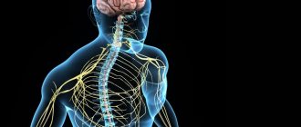
The spinal cord is an organ of the central nervous system. It is located in the spinal canal, on one side formed by the vertebral bodies together with the cartilaginous layers separating them (intervertebral discs), and on the other by the bony processes of the vertebrae. Thus, nature took care of protecting such a delicate and important organ for human life as the spinal cord.
The vertebral processes connect at several points, thereby forming natural openings called foraminal openings, through which the nerve roots extending from the spinal cord exit. They are large nerve fibers. As they move away from the spinal cord, they branch more and more into smaller fibers and reach all organs and parts of the human body, ensuring the transmission of appropriate signals to them that regulate their functioning.
Each spinal root is formed by sensory and motor nerve fibers. The first ones depart from the spinal cord from the side of the vertebral processes, i.e., closer to the back of the body and are therefore called posterior. The motor roots are located almost adjacent to the vertebral bodies and intervertebral discs and are called anterior. Thus, from their names it is clear that the anterior roots are responsible for motor capabilities and are responsible for muscle contraction, while the posterior ones transmit signals to receptors that must respond to the action of various kinds of irritating factors: touch, rough blows, etc.
As already mentioned, the spinal roots exit on both sides of the spine through natural openings formed by the arches and processes of the vertebrae. Therefore, any changes in the condition of the spine and the surrounding soft tissues can affect the condition of such important nerves and lead to the development of radicular syndrome.
The bulk of cases of radiculopathy occur in diseases accompanied by degenerative changes or inflammatory processes in the spine and surrounding tissues. In the latter case, it is provoked by a reflex muscle spasm that occurs in response to pain, or swelling of the soft tissues.
The most common causes of radicular syndrome are:
- osteochondrosis;
- protrusion and intervertebral hernia;
- spondylosis;
- spondyloarthrosis;
- spondylolisthesis;
- traumatic injury to the spine;
- infectious diseases of the spine;
- neoplasms.
The significant “rejuvenation” of radicular syndrome is largely due to changes in the lifestyle of modern people for the worse. It is excess weight, a sedentary lifestyle, bad habits, stress and lack of habit of maintaining posture that become the main reasons for the development of the above diseases, which in turn cause the occurrence of radiculopathy. All of them contribute to poor circulation, which becomes a “trigger” for the occurrence of degenerative changes in the cartilage tissue of the intervertebral discs and the development of corresponding pathologies.
Osteochondrosis
Osteochondrosis is a chronic disease in which there is a gradual destruction of the intervertebral discs against the background of degenerative-dystrophic changes occurring in them, i.e., the cartilage located between the vertebral bodies, acting as shock absorbers and ensuring the flexibility of the spine. They gradually become thinner and lose their natural elasticity. This leads to the fact that the bodies of neighboring vertebrae come closer together, and the diameter of the foraminal openings decreases, which can result in pinching of the spinal roots.
Protrusions and intervertebral hernias
Protrusions and hernias are one of the consequences of osteochondrosis, accompanied by deformation of the shape of the intervertebral disc and its protrusion. Initially, a protrusion is formed. This means that the disc is already deformed, but its outer shell (annulus fibrosus) still retains its integrity. If untreated, it ruptures in the most vulnerable place, which leads to the loss of internal contents (nucleus pulposus) into the spinal canal or the area from the front of the spine, i.e. the formation of a full-fledged intervertebral hernia.
Both protrusion and hernia can compress the spinal roots, either just one of them or both at once. This directly determines on which side neurological disorders will be observed. Large hernias and sequestered ones, that is, separated from the intervertebral disc, can also compress the spinal cord itself, which is fraught with serious complications. If the sequester begins to move along the spinal canal, this will be accompanied by the appearance of signs of damage to those nerve roots to the level of which it descends. If it reaches the lumbosacral region and the large bundle of nerves located there, called the cauda equina, it can lead to paresis and paralysis of the lower extremities.
Spondylosis
Spondylosis is another complication of osteochondrosis, in which the intervertebral discs wear out and become so thin that the vertebrae come together to a critically close distance. Against this background, their deformation occurs, which is accompanied by the formation of bone outgrowths called osteophytes on their edges. Over time, adjacent vertebral bodies can firmly fuse together, which leads to severe pinching of the spinal roots.
Spondyloarthrosis
This disease means the destruction of the facet joints, i.e., the place where the processes of the vertebrae meet. It involves damage to all elements of the joint, including its capsule, ligaments, surfaces and even periarticular muscles. This may also be accompanied by the formation of osteophytes and pinched nerve roots.
Spondylolisthesis
Spondylolisthesis is a pathology of the spine in which the body of one or more vertebrae is displaced relative to the underlying one by a certain distance. Since this disrupts the spinal axis and displaces all the anatomical structures in this area, this leads to compression of the spinal cord roots or even the spinal cord itself.
Traumatic injuries
Spinal injuries are far from uncommon. Strong blows, falls from heights, road traffic accidents and other factors can lead to vertebral subluxations, fragmentation and compression fractures, which is almost always accompanied by the development of radicular syndrome.
Infectious diseases
From the point of view of the occurrence of radiculopathy, the greatest danger is posed by syphilitic and tuberculosis infections, as well as osteomyelitis and nonspecific bacterial meningitis. In such cases, damage to the nerves by pathogenic viruses and bacteria may occur, which will lead to their inflammation.
Neoplasms
The spine and spinal cord, just like other organs, can be subject to the formation of benign and malignant tumors. In addition, with oncological diseases of other organs, metastases can separate from the maternal tumor, which, through the bloodstream, enter the tissues surrounding the spine and can also provoke pinching of the nerve roots and the occurrence of corresponding symptoms.
One of the very common benign tumors of the spine is hemangioma. Many people are already born with such a vascular neoplasm located in the vertebral body, or acquire it with age. It is not dangerous, but it significantly increases the risk of spinal compression fracture.
Physiotherapy
Physiotherapeutic methods are widely used for the treatment of thoracic osteochondrosis after the completion of the acute stage of the inflammatory process. They can further reduce the severity of back discomfort, and also have a number of other positive effects on the body.
Most often, for thoracic osteochondrosis, the following are prescribed:
- electrophoresis with the introduction of drugs - this method allows for deeper penetration of the drug components into the tissues and enhances their therapeutic effect through the use of a weak electric current;
- magnetotherapy is a method of physiotherapeutic treatment, which is based on the beneficial effects of a magnetic field on the body, which helps stimulate blood circulation in the area of influence, which leads to the activation of metabolic processes, a decrease in pain and swelling;
- laser therapy is a method that allows you to achieve a pronounced anti-inflammatory and vasodilator effect, which will also lead to an improvement in the condition of the intervertebral discs and a reduction in pain;
- Ultrasound therapy is a physiotherapeutic procedure that provides an anti-inflammatory and analgesic effect;
- diadynamic currents are an effective method of physical influence, thanks to the use of which there is a decrease in the severity of pain, an increase in metabolic rate and an improvement in the condition of muscle tissue.
As a rule, physiotherapeutic procedures are prescribed in courses of 10-15 sessions. But each of them has its own contraindications, which must be taken into account when choosing a specific type of exposure.
Electrophoresis
Symptoms
The nature of the manifestations of radicular syndrome depends on the degree of compression of the nerve, the characteristics of its location, shape, thickness and a number of other individual characteristics. But radicular syndrome is always characterized by the presence of 3 components: pain, sensory and motor disorders.
The first signs of radiculopathy appear suddenly. As a rule, this occurs after making a sudden movement or heavy physical work. Initially, the pain may be mild or moderate, but it tends to intensify after any movement, laughing, coughing, sneezing and subside when taking a certain body position. In this case, the pain syndrome will be combined with:
- motor disorders with the development of cramping syndrome, i.e. short-term involuntary painful muscle contractions like cramps;
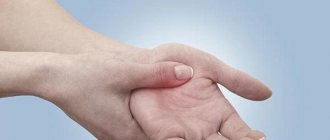
- sensory (sensitive) disorders in the form of paresthesia, i.e. the appearance of a tingling sensation, crawling, limited local sensation of cold or heat.
Pain with radicular syndrome is different from others. They are burning, baking, shooting. Their characteristic feature is spread from the spine to the extremities, irradiation into neighboring areas of the body, for example, legs, thigh or heart area, arms, etc., which is determined by the level of damage.
Pain syndrome causes reflex tension in the muscles and ligaments in the affected area, which leads to an even greater increase in pain. To alleviate his condition, a person tries to take a forced position of the body and avoids movements that will involve the affected segment of the spine.
In this case, the muscles will be more tense on the side of the compressed root, which can be determined by palpation, i.e. palpation, of the back. This can cause the body to tilt in one direction or another. If the spinal roots are pinched at the level of the cervical vertebrae, over time this can lead to the development of torticollis, which will cause curvature of the spine.
If left untreated, the patient's condition will progressively worsen. Over time, compression of the spinal root will lead to:
- paresis (weakness) of the muscles innervated by the affected root;
- reduction of corresponding tendon reflexes;
- reduction or even complete loss of sensitivity of the corresponding part of the body (dermatitis);
- disruption of nutrition of the tissues of the innervated area, which will result in a decrease in muscle size, and then their atrophy, as well as thinning of the skin and an increase in its susceptibility to damage.
The spinal roots pass at the level of each vertebra. In which spinal motion segment one or both of them will be infringed completely determines the nature of the resulting disorders and their localization (on one side or both). If several roots are compressed at the same time, the symptoms will be mixed and combine the symptoms of damage to each of them.
If the radicular syndrome is infectious in nature, there will additionally be elevated body temperature, chills, a feeling of weakness and other characteristic symptoms.
Cervical region
The cervical spine is formed by 7 vertebrae. Compression of the spinal nerves at the level of each of them is accompanied by radicular syndrome, in which pain is concentrated in the spinal region and then spreads to other areas in combination with the corresponding manifestations:
- C1 – pain is felt in the back of the head, often associated with dizziness and nausea. If only one root is pinched, the head will be tilted towards the affected side. When palpating the neck in the area of the first vertebra, muscle tension on the corresponding side may be felt, and pressure will be painful.
- C2 – pain is localized both in the back of the head and the parietal region, there is a decrease in the sensitivity of the skin of the back of the head. In this case, there is a limitation of neck mobility, consisting of difficulties in turning and tilting the head.
- C3 – pain affects the back of the head, the side of the neck, radiates to the tongue, eyes and forehead. In these same areas, tingling or crawling sensations occur, as well as decreased sensitivity. It is difficult for the patient to tilt and raise his head, and when pressing on points in the projection of the third cervical vertebra, painful sensations occur.
- C4 – pain is concentrated in the area of the shoulder girdle and radiates to the anterior surface of the chest up to the 4th rib; it is also observed in the lower third of the posterolateral part of the neck. It is often accompanied by hiccups and voice changes.
- C5 – pain is present in the shoulder girdle, the lateral surface of the shoulder, which is associated with sensory disturbances, limited ability to abduct the shoulder, a decrease in the deltoid muscle and a decrease in the biceps reflex.
- C6—pain originating in the lower neck spreads through the biceps to the outer part of the forearm to the thumb. Additionally, a violation of sensitivity in these same areas and a decrease in muscle tone of the biceps, shoulder and forearm muscles are detected. The severity of the wrist reflex decreases.
- C7—pain is observed in the back of the shoulder and forearm, reaching the middle finger of the hand, and it is quite deep. Decreased sensitivity manifests itself in the triceps, pectoralis major and latissimus muscles, and wrist muscles. Additionally, the severity of the triceps reflex decreases.
Thoracic region
Degenerative-dystrophic changes in the thoracic spine are observed extremely rarely, in less than 1% of cases, but are not completely excluded. Therefore, due to the low frequency of damage to the spinal roots of the thoracic region, it can be very difficult to correctly diagnose the cause of chest pain, since they are almost always caused by diseases of the heart and gastrointestinal tract.
But since pinching of the nerve roots in the thoracic vertebrae is not excluded, let us consider the characteristic manifestations of nerve compression in each spinal motion segment:
- T1-T2 – pain is localized in the shoulder joint and armpits, they can radiate to the subclavian region, as well as the middle surface of the shoulder. This is accompanied by weakness and a decrease in the volume of the muscles of the hand, and the appearance of a feeling of numbness in it. If the spinal roots are damaged at the level of this spinal motion segment, drooping of the eyelids, swallowing disorders, and difficulty in moving food through the esophagus may additionally be observed.
- T3—T6 – girdling pain. They pass along the corresponding intercostal spaces and can be felt in the chest, and if the left nerve root is affected, then they can simulate an attack of angina.
- T7—T8 – pain radiates from the area of the spine in the area of the lower edge of the scapula, also spreads along the intercostal spaces and reaches the epigastric region. As a result, the patient may experience dyspeptic symptoms (heartburn, belching, a feeling of fullness in the stomach, rumbling, diarrhea, etc.), and abdominal pain.
- T9-T10 – pain is girdling and radiates to the upper abdomen, which can mimic the symptoms of appendicitis.
- T11—T12 – pain is often felt in the groin and suprapubic region, which is combined with impaired intestinal motility and resulting discomfort, changes in the consistency and frequency of stool.
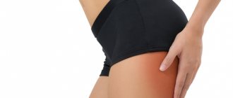
Lumbosacral region
Due to the fact that the lumbar (L-vertebrae) and sacral (S-vertebrae) parts of the spine bear the greatest loads, it is in them that degenerative-dystrophic changes and inflammatory processes most often occur. Therefore, radicular syndrome most often occurs due to damage to the spinal roots in these parts of the spine.
- L1 – pain and sensory disturbances are observed in the groin area and spread to the upper outer quadrant of the buttock;
- L2 – pain is present in the front and inner part of the thigh, weakness is observed when adducting the hip;
- L3 – pain is observed in the greater trochanter, the anterior surface of the thigh and goes down to the lower third of the middle part of the thigh. But sensory disturbances are observed only on its inner surface in the area above the knee.
- L4 – pain spreads along the front of the thigh, extends to the knee and the middle surface of the lower leg up to the ankle. This is associated with a decrease in the quadriceps muscle, as well as a decrease in the strength of the tibialis muscles. That is why, when the spinal roots are damaged at the level of the 4th lumbar vertebra, the foot seems to slam when walking.
- L5 – pain sensations originating in the lower back radiate to the buttock, descend along the lateral surface of the thigh and lower leg and cover the first 2 toes. In these same areas, a decrease in sensitivity is observed, which is combined with a decrease in the tone of the tibialis muscle, as well as only the big toe or the entire foot.
- S1 - damage to the nerve roots at the level of the 1st vertebra of the sacral body is associated with the appearance of pain in the lower back and sacrum, which radiates along the posterolateral surface of the thigh and lower leg to the foot and 3-5 fingers. Sensitivity and muscle tone disturbances are concentrated along the lateral edge of the foot, and the calf muscle also suffers. In this case, there may be a decrease in the ability to turn the foot and extend it.
- S2 – pain and sensory disturbances are present in the sacral area, affecting the back of the thigh and lower leg, the plantar part of the foot, and the big toe. Often there are cramps in the thigh.
- S3-S5 – damage to the spinal roots at this level is called sacral caudopathy, which is usually associated with the appearance of signs of compression of 3 roots simultaneously. This condition is characterized by the presence of pain and lack of sensitivity in the area of the sacrum and perineum, and there is also often a disruption in the functioning of the sphincters of the pelvic organs.
Diagnosis of radiculopathy
For an experienced neurologist, it is not a problem to detect signs of radicular syndrome. In addition to the characteristic symptoms, the results of an examination of the patient (the doctor evaluates the posture that the person involuntarily assumes, muscle tone, the presence of painful sensations on palpation) and specific neurological tests carried out help him make a diagnosis. But in order to accurately determine at what level compression of the spinal root occurs and what causes it, a number of instrumental studies are required. The neurologist will determine which of them are necessary and write a prescription:
- radiography of the spine in 2 projections;
- CT;
- MRI;
- electroneuromyography.
If there is a suspicion of the presence of diseases of the heart, gastrointestinal tract or others, ultrasound, blood tests, urine tests, etc. may be additionally prescribed.
For diagnosing intervertebral disc pathologies, MRI is considered the most informative method. With its help, you can detect even the slightest changes in the structure of the discs, which will not be visible with any other type of examination. But if it is necessary to accurately examine bone elements and detect their deformations, an X-ray, or better yet a CT scan, is required.
Diet for osteochondrosis of the thoracic region
At the first symptoms and treatment of thoracic osteochondrosis, a menu high in collagen, vitamins A, B and C is recommended, which promote the regeneration of intervertebral cartilage and strengthening of ligaments. It is also advisable to “accelerate” metabolism with easily digestible food (small portions every 3 hours).
The diet includes a protein breakfast (eggs, dairy products) and dinner (boiled, stewed, baked in foil fish, poultry, seafood). During the day you should eat legumes, whole grains, vegetables, fruits and berries.
It is worth reducing the consumption of starch, salt, flour products, processed foods and carbonated drinks. Drinking regimen: from 2 liters of clean water per day.
Images designed by Freepik
Treatment of radicular syndrome
Treatment for each patient is selected individually depending on what disease caused the development of radicular syndrome. In the absence of a serious threat to the health and life of the patient, for example, sequestration of an intervertebral hernia or other severe complications, they initially try to relieve the nerves from compression using conservative methods. In such situations, in accordance with the diagnosis, patients may be prescribed:
- drug therapy, which consists in the use of NSAIDs in the form of injections, tablets, ointments or suppositories, chondroprotectants, muscle relaxants, vitamins (if the radicular syndrome is infectious, the use of antibiotics, antiviral and other agents is indicated);
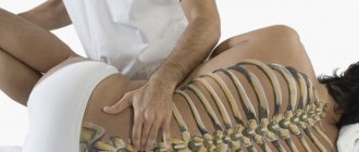
- manual therapy, which helps eliminate functional blocks, normalize muscle tone, and most importantly, restore the normal position of the vertebrae and release compressed spinal roots;
- physiotherapy, which increases the effectiveness of other measures taken to treat radicular syndrome;
- Exercise therapy, which is an obligatory component of treatment and is necessary to improve blood circulation, strengthen muscles and prevent their atrophy.
In case of very severe pain, patients can undergo blockades of the corresponding spinal motion segment. They do not have a therapeutic effect, but are able to eliminate pain of any severity in a matter of minutes.
But conservative therapy does not always bring positive changes in the patient’s well-being. If the patient’s condition does not improve within 3 months after its onset, he will be indicated for appropriate surgery. Also, surgical intervention may be prescribed immediately if:
- large intervertebral hernias;
- sequestered hernias;
- dangerous tumors of the spine or surrounding tissues;
- spondylolisthesis and some other pathologies.
In each case, the method of surgical intervention is selected individually, based not only on what pathology was detected, but also on its severity. For example, for small intervertebral hernias, percutaneous surgery techniques can be used, in particular cold plasma nucleoplasty or hydroplasty. For larger intervertebral disc defects, endoscopic surgery or microdiscectomy is indicated. For large hernias, the only way out of the situation is microdiscectomy or open discectomy with stabilization with a metal structure. If the patient is diagnosed with spondylolisthesis, it will be possible to eliminate the radicular syndrome only by implanting special fixing titanium plates, etc.
Thus, the appearance of symptoms of radicular syndrome or radiculopathy is a reason to immediately contact a neurologist, conduct an appropriate examination and begin treatment. But even if there are indications for surgery, you should not be afraid of it. Modern spinal surgery techniques are highly effective, and they are much safer than non-intervention and the inevitable addition of complications and subsequent disability.
Lifestyle correction
After diagnosing thoracic osteochondrosis, the doctor necessarily recommends making certain changes to your usual lifestyle. If a patient shows signs of excess weight, he is advised to take measures to reduce it. But any diets for weight loss, especially monocomponent ones, are contraindicated. Nutrition must be complete and varied so that the body receives all the substances necessary for proper functioning, and metabolic processes in the intervertebral discs proceed properly. Therefore, it should comply as fully as possible with the principles of rational nutrition.
It is also recommended that all patients increase their level of physical activity, especially those who lead a sedentary lifestyle. This could be daily walking, swimming, yoga, or Pilates. But serious physical activity, in particular intense training on simulators, jumping sports, and weightlifting are contraindicated.
If the patient’s profession involves heavy physical labor, such as heavy lifting, it is recommended to try to change it. This is due to the fact that increased loads on the back in the presence of osteochondrosis can play the role of a trigger for the rapid progression of degenerative changes in the discs.
Absolutely all patients with thoracic osteochondrosis are recommended to change the mattress to an orthopedic one with medium hardness, as well as purchase an orthopedic pillow. This will ensure that the physiological curves of the spine are maintained and will prevent further disc degeneration.