MRI of the pituitary gland with contrast is a magnetic resonance examination of the most important endocrine gland of the human body, due to its small size, the use of contrast agents is required. Contrast agents for MRI contain nanoparticles of gadolinium, a metal that can increase the sensitivity and information content of the method. Once in the blood, gadolinium is evenly distributed throughout all tissues of the human body. Inflammatory processes, tumors and other anomalies, on the contrary, lead to local accumulation of contrast, which is why this area stands out in the images, facilitating diagnosis.
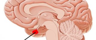
Where is the pituitary gland located?
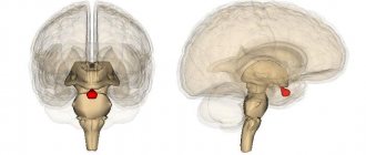
The pituitary gland is the most important organ of the endocrine system (gland), which produces a number of biologically active substances (hormones) that regulate the balance of energy, nutrients and minerals, cell division, sexual functions and reproduction, metabolism and many other aspects of the normal functioning of the human body. This is hard to believe, especially considering the small size of the pituitary gland - only 13 mm in length, 5-6 mm in thickness (the size of a pea or bean). The mass of the pituitary gland does not exceed 0.5 grams.
The pituitary gland is located at the base of the brain, inside the skull, in an area called the sella turcica (due to the shape of the bones, which actually resemble a saddle). The only method for diagnosing pituitary gland diseases that allows obtaining detailed and high-quality images is magnetic resonance imaging. MRI of the pituitary gland is always performed with contrast - this allows to increase the accuracy of diagnosis and the information content of the images, which is important, given the miniature size of the organ.
What to choose - brain tomography or pituitary MRI?
Brain tomography shows abnormalities in the organ in question, but with a small degree of probability. A targeted study allows you to obtain much more information to make the correct diagnosis.
The procedure is prescribed for suspected congenital abnormalities, cysts, inflammation, tumors, or disorders of the hypothalamic-pituitary system. Studies in this area show pathological lesions less than 1 cm and their structure. To increase the information content, an MRI with contrast is performed.
Indications and contraindications for MRI of the pituitary gland with contrast
Indications for the use of contrast-enhanced MRI of the pituitary gland:
- pituitary tumors/cysts of various origins;
- metastases to the pituitary gland;
- empty sella syndrome;
- hypofunction of the pituitary gland (decrease in the levels of pituitary hormones - prolactin, somatotropin, TSH, LH, FSH, etc.);
- hyperfunction of the pituitary gland (increased levels of pituitary hormones);
- apoplexy (hemorrhage) in the pituitary gland, pituitary necrosis (Sheehan syndrome);
- Itsenko-Cushing's disease;
- other endocrine disorders associated with the functional activity of the pituitary gland.
Symptoms in which an MRI of the pituitary gland is recommended:
- headache;
- decreased visual acuity (loss of visual fields, double vision, distortion of visible objects);
- excessively short or high stature inappropriate for age, delayed physical development;
- constant thirst with increased fluid consumption and frequent urination;
- infertility, both male and female;
- impotence in men, irregularities or absence of the menstrual cycle in women;
- obesity.
What does a brain MRI with contrast show?
With the development of diseases in this department, the patient develops various symptoms, for which one should not hesitate to visit a doctor. Medical staff will issue a referral for an MRI of the head with contrast if the following manifestations are present:
- coordination disorders;
- general poor health;
- atypical behavior for a person.
Additionally, a tomographic scan with an additional intensifier is necessary when it is necessary to obtain a medical report when other diagnostic procedures have not been able to show the full picture of the disease. The main indications for undergoing the study are:
- neoplasms of various nature;
- multiple sclerosis;
- damage to the vascular system;
- traumatic injuries, bruises and their consequences.
Screening can identify inflammatory reactions with precise locations. MRI testing is prescribed after surgical procedures. The procedure allows you to evaluate the effectiveness of the operation and the likelihood of relapse. Contrast is introduced for detailed and clear images. This product is safe for humans and is eliminated from the body several hours after the scan.
Contraindications to contrast-enhanced MRI of the pituitary gland
MRI of the pituitary gland with contrast is contraindicated:
- patients who have metal foreign bodies in their bodies (iron filings in the eyes, steel clips on blood vessels, orthopedic structures, etc.) – the metal heats up and can move from its place under the influence of magnetic fields;
- patients with pacemakers, defibrillators, neurostimulators, cochlear implants, insulin pumps - magnetic fields can damage complex electronics;
- pregnant women in the first 13 weeks of pregnancy - MRI is not recommended in the first trimester of pregnancy without good reason;
- children under 5 years of age - MRI in children under 5 years of age is performed in specialized hospitals under anesthesia and medical supervision;
- patients with severe claustrophobia - people with a fear of closed spaces often cannot remain calm and still during an MRI examination.
In addition, MRI diagnostics may be hampered by excess weight (more than 130 kg) and circumference (more than 150 cm in girth) of the body. Patients with such data do not physically fit in the magnetic resonance imaging machine.
Preparing for MRI of the pituitary gland with contrast
Magnetic resonance imaging of the pituitary gland with contrast agents is performed without any preliminary preparation. The study can be carried out at any time convenient for the patient. The main condition for safe MRI is the absence of foreign metal objects on the patient’s body. Magnetic fields generated during the examination can lead to heating of jewelry, piercings, and metal clothing fittings, with the risk of burns. It is strictly prohibited to carry mobile phones, electronic gadgets, credit and other magnetic cards, storage media, external batteries, etc. Otherwise, injury or burns may occur. The patient can leave anything unnecessary in the storage room.
Contrast drugs very rarely cause side effects - the most common of them is a mild allergy in the form of a small rash and itching on the skin. Severe allergic reactions to contrast are rare and also serve as a contraindication for its administration. In such cases, the study is performed without contrast.
What are the advantages of MRI with contrast?
The main advantages of scanning using an additional coloring agent are:
- minimum number of restrictions to the procedure;
- obtaining slices in any projection;
- high level of resolution of the tomograph;
- obtaining images showing all parts of the brain;
- easy portability of the supplement.
In the contrast protocol, several types of drugs are used, which differ from each other in composition and methods of use. The study is available to all patients, except those with individual intolerance to gadolinium salts. Some patients report side effects from using the coloring component:
- skin rashes;
- nausea;
- slight dizziness.
All symptoms go away on their own without medical help. Such complications occur in less than 1% of clinical situations.
How does a contrast-enhanced MRI of the pituitary gland work?
Magnetic resonance imaging is performed on an outpatient basis, using special equipment - an MRI scanner. The research setup is a large cylindrical block with a tunnel equipped with a special bed on which the patient lies during the examination. A lattice structure resembling a helmet is installed around the patient’s head - this is an additional coil that increases the quality and clarity of images of the brain and pituitary gland.
Before the examination, the patient is asked to wear special headphones - they protect the ears from the noise that occurs during the operation of the tomograph. Headphones can transmit music, which helps the patient relieve stress, and also serve to communicate with the operator. To make an emergency call to the operator, the patient can press a pear-shaped button, which is in his hands at all times during the examination.
The duration of an MRI of the pituitary gland is approximately 30 minutes. In the first half, the study is performed without contrast. Then, 15 minutes after the start of the procedure, the nurse administers an intravenous contrast agent, after which the study is repeated with contrast enhancement.
All that is required of the patient during diagnosis is to lie down, remaining calm and still. MRI is an absolutely safe and painless procedure that is not accompanied by any unpleasant sensations. Slight discomfort may occur in people prone to anxiety and panic attacks due to the limited space of the tunnel.
After completing the examination, you must wait for the images to be decrypted (usually this takes a maximum of 15-30 minutes). The images themselves are recorded free of charge on a CD and sent to the patient along with a written report from the radiologist. If desired, you can record pictures on a flash drive or print them on film.
Advantages: why should you do an MRI with us?
The brain coordinates the entire body, and disruptions in its functioning can have detrimental consequences. In diseases of the main organ of the central nervous system, the accuracy of indications and the speed of obtaining results take priority. That is why it is so important that diagnostics are carried out only by competent specialists, using the most modern equipment. Carrying out MRI in our clinic has the following advantages:
- quality and speed of examination (97% accuracy in 30 minutes!)
- affordable prices and experienced specialists
- availability of powerful closed-type tomographs (1.5 Tesla)
All glands without exception are connected to the pituitary gland - it is it that ensures their coordinated work. Delay in examination will inevitably lead to breakdowns in other systems. Malfunctions in its functioning cause mutations, metabolic disorders, atrophy or hyperplasia of internal organs. A timely examination that uses contrast will help bring the situation under control. In this case, it is important not only to carry out the procedure competently, but also to correctly interpret the results obtained. The doctors at our clinic have many years of experience working with tomographs, which ensures that all the nuances of the examination are taken into account.
We invite you to undergo an informative MRI of the pituitary gland at the Kuntsevo multidisciplinary treatment and rehabilitation center. We accept by appointment.
Interpretation of the results of MRI of the pituitary gland with contrast
MRI results of the pituitary gland - a series of thin layer-by-layer images of the sella turcica area. Since the pituitary gland is the size of a pea, each section has a minimum thickness of 1 mm. The radiologist carefully examines each image, looking for deviations from the norm. Normal MRI picture of the pituitary gland: dimensions do not exceed 1-1.3 cm, absence of cysts, tumors and other heterogeneous inclusions, the anterior and posterior lobes of the pituitary gland are clearly distinguishable. When administered, the contrast is evenly distributed throughout the pituitary gland. In addition, the condition of the tissues surrounding the pituitary gland - nerve fibers, blood vessels and other anatomical structures - is described.
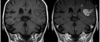
Local accumulation of contrast is the main sign of the presence of a pituitary tumor. Most often, doctors deal with so-called pituitary adenomas - these are hormonally active tumors that secrete pituitary hormones. An excess of certain hormones leads to endocrine diseases with corresponding symptoms.
Adenomas in the photographs look like formations in the pituitary tissue of arbitrary size, intensively accumulating a contrast agent. Moreover, in the absence of contrast, the tumor may not be visible - very small tumors or microadenomas can hardly be detected without contrast. This is why MRI of the pituitary gland is almost always performed with contrast enhancement.