3 January 2021
21580
0
4 out of 5
Spinal injuries today, unfortunately, are far from uncommon. They account for 5.5% to 17.8% of all cases of injuries to the musculoskeletal system. Neither children, nor adults, nor especially the elderly are insured against them. And the consequences of such accidents, especially those leading to spinal cord damage, can be extremely severe, including disability and death, especially if the victim does not receive qualified medical care in the first hours after the incident. Only in the mildest cases or with timely surgery, spinal injuries go away without a trace and are not associated with the risk of complications. Therefore, they always require the fastest possible response and contact a neurosurgeon or spinal surgeon.
Types of spinal injuries
In general, the spine consists of 33 vertebrae, between which are located cartilaginous plates (intervertebral discs). Almost all vertebrae have a bony arch and processes extending from it. A hole is formed between the vertebral body and the arch, the position of which coincides in each spinal motion segment throughout the entire spinal column. The canal formed in this way is called the spinal canal, in which the delicate nervous structure - the spinal cord - is located. A pair of nerve roots depart from it at the level of each vertebra, which together are responsible for the innervation of the entire body.
Each of these anatomical structures can be injured. As a result, they distinguish:
- spinal bruises are the mildest injuries in which mechanical injury to soft tissues is observed, but the integrity of bone structures, intervertebral discs and ligaments is not compromised;
- fractures of the vertebral bodies - can be compression, in which the vertebra is flattened on one side, or comminuted, in which individual bone fragments are formed;
- fractures of the processes and/or arches of the vertebrae;
- vertebral dislocations - a change in the position of the bones that form the joints of the spine (sometimes the dislocation can be self-correcting);
- fracture-dislocations of the vertebrae;
- spondylolisthesis – displacement of the upper vertebral body anteriorly relative to the lower one, which also leads to sprained ligaments;
- partial or complete rupture of the ligaments of the spinal motion segment (distortion) - a minor injury, usually with a favorable outcome;
- intervertebral disc rupture – instantaneous formation of a disc herniation under the influence of a traumatic factor;
- spinal cord concussion – a change in the condition of the spinal cord that is reversible over time;
- contusion of the spinal cord and/or its roots - with rapid restoration of blood flow and elimination of compression, the consequences can be reversible;
- partial or complete rupture of the spinal cord and/or its roots is the main cause of disability.
All these types of damage can be combined with each other in different combinations, so 3 types of spinal injuries can be distinguished:
- uncomplicated spinal injury – accompanied by a violation of the integrity of the spinal structures without affecting the spinal cord;
- spinal cord injury without damage to the spine - consists of damage to the spinal cord;
- spinal cord injury – accompanied by a combination of damage to the spinal column, spinal cord and neurovascular formations of the spinal canal.
In 40-60% of patients with spinal cord injury, damage to internal organs and other tissues is observed.
Like other injuries, spinal injury, depending on the nature of the violation of the integrity of the skin, can be closed, open or penetrating. It is very important to determine exactly how much time has passed since the injury. Depending on this, 5 periods are distinguished:
- acute – up to 8 hours;
- acute – 8-72 hours;
- early – from 3 days to 4 weeks;
- intermediate – 1-3 months;
- late – more than 3 months.
All spinal injuries can be divided into stable and unstable, which largely influences the nature of the necessary treatment.
Accurately determining the type of injury and the period in which the patient is currently in is extremely important for the correct development of treatment tactics. Therefore, after providing emergency care to a patient, a comprehensive diagnosis is always carried out.
Why is a spinal cord rupture dangerous?
Spinal cord rupture is a very serious condition that leads to disability and threatens a person’s life. When the spinal cord is ruptured in areas of the body below the rupture site, communication with the brain is disrupted and they lose their functions.
A particularly severe complication of spinal cord rupture is spinal shock. The duration of spinal shock cannot be predicted. It can be several weeks or months. At this time, it is necessary to support the body and take measures to prevent muscle atrophy.
A rupture of the spinal cord poses a direct threat to human life. If the most difficult period can be survived, the person will need to undergo a long course of rehabilitation to adapt to life with a disability.
Causes
Spinal injury is usually caused by the application of an external force. Most often it occurs as a result of:
- falls from a height, which is called catatrauma – 50%;
- road traffic accidents – 30%;
- diving head down in shallow water – 10%.
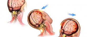
This is often accompanied by damage to the spinal cord, i.e., a spinal cord injury initially occurs. In such situations, the spinal cord suffers due to instant or gradually increasing compression by bone structures, displacement of the vertebrae in an unstable segment of the spine. It can also be stretched and ruptured, which leads to disability.
The spinal cord is a sensitive anatomical structure that can also be damaged, even if initially only the spine was damaged at the time of injury. In such situations, spinal cord injury is secondary and becomes a consequence of impaired microcirculation, severe swelling of the surrounding soft tissues, hypoxia (lack of oxygen), disturbances in electrolyte metabolism and the action of a number of other factors.
However, the situation is extremely serious in both cases, regardless of whether primary or secondary spinal cord injury occurs.
Less commonly, the cause of spinal injury, namely vertebral compression fracture, is osteoporosis. This disease is most common in older people, although it can also occur at a young age without long-term use of corticosteroids and some other drugs. It is characterized by a decrease in bone density, which leads to the fact that the vertebral bodies become more fragile and can flatten, forming a wedge-shaped deformity, even as a result of a sudden movement, coughing, not to mention a fall or blow.
Other causes of spinal injury without significant external influence may be tumors of the spine itself or the formation of metastases in it.
Diagnosis and clinical picture of spinal cord injuries
Diagnosis of spinal cord injury involves collecting complaints from the victim or witness to the incident, examining the patient, neurological examinations, laboratory tests and instrumental methods (lumbar puncture, CT or MRI of the brain, spondylography, vertebral angiography, myelography, CT myelography).
It is extremely important to correctly collect anamnesis, since the timeliness and correctness of further treatment depends on this. Namely, the doctor must find out the time and mechanism of injury, determine the location of pain, sensory and motor disorders, and find out whether the victim made any movements after the injury. If the patient experiences neurological symptoms in the acute period, this indicates a brain contusion. The doctor pays attention to the type of breathing, the presence of weakness in the limbs, and tension in the abdominal wall.
Instrumental methods are used for differential diagnosis of the disease. They help distinguish spinal cord compression from other types of spinal injury that are treated conservatively. Instrumental diagnostics are also indicated for spinal shock and the patient’s inability to empty the bladder independently. To make a diagnosis, the doctor does not need to use the entire range of instrumental methods. The choice of technique depends on the doctor’s suspicions and the results of a neurological examination.
The symptoms of spinal cord injury depend on the period of the disease. In total, there are four main periods of the disease, which reflect the dynamics of restorative and destructive processes:
- The acute period lasts the first two to three days after injury. It is characterized by necrotic and necrobiotic lesions of the spinal cord, circulatory and lymph circulation disorders. During this period of injury, symptoms such as spinal shock and conduction syndrome appear.
- The early period takes 2-3 weeks. This period is characterized by clearing of foci of traumatic necrosis and signs of pathological changes in nerve bundles and nerve fibers.
- The intermediate period lasts about 3-4 months. Patients experience symptoms of fiber regeneration and scar formation. During this period of the disease, all reversible changes and signs of spinal shock disappear.
- The late period starts from the third or fourth month and lasts for a long time. Clinically manifested by the final stage of scarring and cyst formation, pathological processes in the nervous tissue.
Symptoms
The main manifestations of spinal injury are pain and a burning sensation, swelling, and redness at the site of injury. A hematoma or even an open wound may also be observed. But the clinical picture largely depends on the level of damage and its severity. Thus, cervical spine injuries are characterized by:
- impaired sensitivity and mobility of the hands up to complete paralysis (tetraplegia);
- difficulty turning your head;
- respiratory failure, shortness of breath, respiratory arrest.
Injuries to the cervical spine are the most dangerous in terms of disability and death.
Damage at the level of the thoracic and lumbar vertebrae may be accompanied by:
- decreased or loss of sensation in the legs;
- disorders of urination and defecation;
- inability to raise a straight leg while lying on your back;
- back muscle tension.
Thus, a high risk of injury is indicated by the appearance of disturbances in sensitivity and motor functions.
Where does radicular syndrome occur?
As we have already found out, nerve impulses from the spinal nerve diverge throughout the entire zone of innervation of this nerve. Consequently, when a nerve is compressed, pain, muscle weakening, numbness, pins and needles and pins and needles will cover the entire area. It does not happen that radicular syndrome causes pain only in places, and there is no pain in the rest of the area. Remember, radicular syndrome is the simultaneous manifestations of pain, muscle weakness, numbness and other symptoms, covering the entire innervation zone.
In medical terms, radicular syndrome is:
- Sensory disturbance (pins and needles, goosebumps, pain)
- Muscle hypotension (weakness)
- Decreased or complete loss of muscle reflexes
All of these symptoms develop within the zone of innervation of the corresponding spinal nerve.
Diagnostics
Diagnosis of spinal injury is always complex, since the slightest omission can have catastrophic consequences. Therefore, the spine is always carefully examined using various research methods.
Initially, a survey is carried out of the victim himself or the person who witnessed the incident that allegedly led to a spinal injury. The main emphasis is on elucidating the mechanism of its production and timing. If the patient is conscious and able to answer questions, they try to find out where the pain is felt and whether sensory or motor disorders are observed, and if so, when they arose.
The further diagnostic algorithm is as follows:
- examination using palpation methods;
- assessment of neurological condition;
- carrying out instrumental diagnostic procedures.
It is also necessary to immediately draw blood for general and biochemical analysis. In addition, it is necessary to perform a general urine test.
Patient examination
As part of the physical examination, it is possible to establish the approximate location of the injury, detect visible deformations and find out which levels of the spine should be examined using X-rays in order to exclude the presence of combined injuries. Therefore, palpation and examination is carried out throughout the patient’s body, but with great care to avoid causing additional damage to the patient.
Determination of neurological status
The degree of sensitivity and preservation of motor functions must also be assessed. In this case, the international ASIA scale is used, which implies 5 degrees of spinal cord injury (A, B, C, D, E), of which A is the most severe injury, accompanied by a complete absence of motor and sensory functions, and E is the norm.
When determining neurological status, attention is paid to:
- muscle strength;
- sensitivity to pain and touch (tested by touching with cotton wool or a special caliper-like instrument called Frey hairs);
- reflex activity in the anogenital area.
Additional information about the patient’s condition can be provided by a test in which passive movements of the fingers or toes are performed, depending on the level of the lesion.
Instrumental diagnostics
The use of special diagnostic equipment allows you to obtain the most accurate information about the patient’s condition, determine the specific level of damage, the degree of spinal cord injury, assess the nature of changes in the condition of the spine, determine the presence of instability and obtain a lot of other data important for selecting the optimal treatment method. Based on them, a decision is made on the need for surgical intervention, as well as the type of operation required and its volume, or the possibility of conservative treatment.
Thus, when diagnosing spinal injuries, the following are carried out:
- X-ray is an outdated method, which is now being abandoned in favor of CT, since X-rays are not always able to detect all existing bone damage and accurately determine the type of fracture.
- Computed tomography (CT) is the main method for diagnosing fractures and dislocations, providing the most complete and accurate information about the state of the bone structures of the spine and not requiring additional movements of the patient on the couch. With its help, you can detect fractures of vertebral bodies, arches and processes, accurately determine the length of fracture lines, assess the degree of divergence of bone fragments, detect displaced fragments, etc.
- Magnetic resonance imaging (MRI) is the most informative study that allows you to accurately assess the condition of the spinal cord, intervertebral discs, ligaments, the presence of hematomas and soft tissue formations in general.
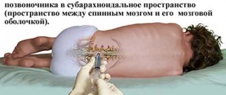
- Myelography is an additional research method used in the presence of neurological disorders, but it is impossible to detect their causes on CT. Using myelography, the presence of a violation of the patency of the subarachnoid space in the spinal cord is determined and it is revealed at what level the deformation occurred, leading to compression of the spinal cord or rupture of its dura mater.
Forecast
About 50% of people with spinal injuries die in the preoperative period, most of them before even reaching medical facilities. After surgery, the mortality rate decreases to 4-5%, but can increase to 75% depending on the complexity of the injuries, the quality of medical care and other related factors.
Complete or partial recovery of patients with SCI occurs in approximately 10% of cases, taking into account that the injury was of a stab-and-cut nature. With gunshot wounds, a favorable outcome is possible in 3% of cases. Complications that arise during the hospital stay cannot be excluded.
High-level diagnostics, operations to stabilize the spine and eliminate compression factors reduce the risks of a negative outcome. Modern implantable systems help to lift the patient faster, eliminating the negative consequences of a long period of immobility.
First aid for spinal injury and choice of treatment tactics
It is important not to move or lift the patient after receiving a blow or other traumatic impact and immediately call an ambulance. Under no circumstances should the victim be allowed to sit or stand, but it is worth giving pain medication and attempting to calm him down. If there is no breathing or heartbeat, resuscitation should be performed. Specialists who arrive at the scene carefully immobilize the patient at the scene by securely fixing him on a rigid shield using a rigid head holder, and then transport him to a medical facility.
Any patient who is suspected of having a spinal injury is immediately treated with a spinal injury protocol until a full investigation has been completed and it is proven that no injury has occurred.
If the presence of an injury is confirmed, its type and characteristics are clarified, it becomes clearly clear what treatment is indicated in a particular case. Conservative therapy is carried out only in the mildest cases in the absence of signs of neurological deficit, when only a violation of the anatomy of bone structures is observed without signs and risks of complications, in particular compression of the spinal cord. It is usually indicated for mild compression fractures, dislocations, fractures of processes, arches, and vertebral bodies without the formation of fragments.
Conservative treatment consists of:
- bed rest or immobilization of the affected part of the spine;
- drug therapy appropriate to the situation;
- traction therapy (spinal traction using a special apparatus);
- physiotherapy;
- manual therapy.
If necessary, vertebral reduction is initially performed.
But in the vast majority of cases, surgery is indicated for spinal injuries. Emergency surgery is required when:
- the presence and especially intensification of signs of neurological deficit;
- severe deformation of the spinal canal due to bone fragments, dislocated vertebrae or significant curvature;
- the presence of a large hematoma formed as a result of injury to a herniated intervertebral disc, injury to the yellow ligament, the presence of a foreign body that threatens compression of the spinal cord;
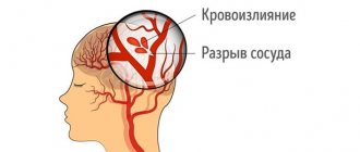
- the presence of isolated hematomyelia, i.e. hemorrhage in the spinal cord;
- compression of a large blood vessel supplying the spinal cord;
- pronounced compression of the spinal roots;
- instability of spinal motion segments when creating a threat of their displacement and compression of the spinal cord;
- the presence of foreign bodies in the spine;
- liquorrhea, i.e. leakage of cerebrospinal fluid through defects at the base of the skull;
- injuries received from gunshot or stab wounds.
But there are also contraindications for performing the operation even if there are compelling indications for it. In such situations, spinal surgery is postponed until the patient's condition is stabilized. This is about:
- traumatic or hemorrhagic shock with unstable hemodynamics;
- the presence of severe damage to internal organs, leading to internal bleeding, associated with the risk of developing peritonitis;
- extremely severe traumatic brain injuries with signs of intracranial hematoma formation.
However, it is also possible to carry out the operation as planned. They are required when conservative therapy is ineffective, but they allow you to carefully prepare the patient for the upcoming surgical intervention and select a treatment tactic that suits him.
Intensive therapy
Intensive therapy is carried out, which is aimed at maintaining the normal functioning of important body systems. The first step is to maintain a normal blood pressure level, since hypotension can aggravate poor circulation in the area of injury. After normalization of blood pressure, doctors begin drug therapy for spinal cord edema, for which they prescribe diuretics and methylprednisolone.
In the first 4 hours after spinal injury, hypothermia of the spinal cord is indicated. To maintain a normal volume of circulating blood during traumatic shock, the patient is shown blood up to 1200 ml, low- and high-molecular dextrans. Giving patients plenty of fluid (at least 2.5 liters) helps prevent hypovolemia, which can worsen circulatory problems. In acute respiratory failure, ventilation is indicated.
Intensive therapy also involves maintaining cardiac activity and electrolyte balance, and correcting metabolic disorders. From the first days, patients are required to be prescribed antibacterial therapy. Also in the acute period, periodic catheterization of the bladder and washing it with a solution of furatsilin are indicated. If the victim has an open wound, its initial treatment is necessary.
Surgery for spinal injury
The goals of surgical treatment are:
- rapid and complete elimination of the factor leading to compression of the spinal cord and other neurovascular formations;
- restoration of the correct axis of the spine;
- fixation and stabilization of the spine, which will allow the patient to be activated as early as possible and avoid deformation in the future.
If there are signs of spinal cord damage, it is recommended to perform surgery as early as possible, since up to 70% of cases of the development of all irreversible changes occur in the first hours after injury. But there are quite a few types of operations that can be indicated for spinal injuries. The choice of a specific one is made by the neurosurgeon in each case individually.
Halotraction
Halotraction is an operation indicated for complicated or unstable injuries of the cervical spine. It involves dynamic reposition and reliable fixation of the vertebrae while maintaining the patient’s physiological activity using a special halo apparatus.
It is a device, part of which reliably connects the damaged vertebrae to the base of the skull, and the other is located outside the body. Its design features make it possible to gradually perform traction, i.e. stretching of damaged spinal motion segments until complete reposition of the vertebrae is achieved.
On average, the halo apparatus is removed after 2.5-3 months. After this, the use of a head holder is prescribed for 3-4 weeks.
Laminectomy
Laminectomy is a decompression operation widely used for various spinal injuries. It allows you to eliminate compression of the spinal cord and its roots by various anatomical structures by removing the vertebral arches or only their processes, or to provide access to the spinal cord to remove foreign bodies or perform other manipulations on it.
Often the situation requires a discectomy, i.e., removal of the intervertebral disc, and in some cases the surgeon is forced to remove the entire damaged vertebra completely, i.e., perform a corpectomy.
Laminectomy requires further installation of stabilizing systems in order to achieve spinal fusion.
Discectomy
Discectomy is a decompression operation indicated for traumatic injuries of the intervertebral disc that have led to compression of the spinal roots or spinal cord. It involves removing the damaged intervertebral disc and can be combined with laminectomy.
The operation is performed through an anterolateral approach (for injuries to the cervical spine) or posterior (for damage to the thoracic or lumbosacral discs). It is performed under general anesthesia, and the removed disc is replaced with a bone graft, synthetic implant, or spinal fusion is achieved.
In cases uncomplicated by serious spinal cord injuries, microdiscectomy may be performed. This operation involves making a much smaller incision and may involve removing only a formed hernia while preserving the disc. Endoscopic surgery can also be an alternative, but spinal injuries are rarely limited to isolated disc damage, so discectomy is often only one of the stages of surgical intervention.
Spinal fusion
Spondylodesis is an operation whose main goal is the reliable fusion of several spinal motion segments with each other, which leads to their complete immobilization, and therefore eliminating the risk of displacement and subsequent injury to the spinal cord. For this purpose, a fragment of the patient’s own bone (autograft) or artificial bone can be used. But more often special titanium structures are used, i.e., transpedicular fixation of the vertebrae is performed, since this technique is associated with fewer risks and is more reliable.
Transpedicular fixation involves a strong connection of adjacent vertebrae using special screws and rods passed through their heads. Titanium screws are screwed into the intersection of the transverse process of the vertebra with the superior articular process. In this case, it is necessary to fix at least 3 vertebrae, even if only one of them is affected, since otherwise the proper degree of stabilization of the spine will not be achieved.
The transpedicular fixation method is the main way to achieve stabilization of the spinal column in various spinal injuries.
Spinal fusion can be performed in isolation or as one of the stages of surgical treatment of spinal injury, for example, after decompression of the spinal cord or its roots. The operation may also involve removal of the intervertebral disc, which is especially important in case of traumatic damage. In this case, a modern, precisely sized cage is installed in place of the removed disc.
If initial decompression of spinal structures is necessary, this task can be solved through an anterior or posterior approach. Most often, the operation is performed through a posterior approach, since this is associated with lower intraoperative risks and is less likely to lead to postoperative complications.
Vertebroplasty and kyphoplasty
Vertebroplasty and kyphoplasty are two similar techniques that can be used for vertebral compression fractures. Their essence is to restore the strength of a broken vertebral body using specially developed, quickly hardening bone cement.
Both operations are minimally invasive and involve the introduction of a thin cannula into the body of a broken vertebra, through which its integrity will be restored by injecting freshly mixed bone cement into it. It will fill all natural bone pores and provide high strength to the vertebra within 10 minutes, since this is exactly the time it takes for it to completely harden.
But when choosing kyphoplasty, a special balloon is first inserted into the vertebral body through the same cannula and filled with liquid. As a result, it swells and helps restore the anatomy of the fractured vertebral body. After this, the balloon is deflated and removed, and the resulting space and the entire vertebral body are filled with bone cement. Once it has hardened, the cannula is removed and the remaining puncture is covered with a sterile bandage.
The features of these two percutaneous surgery techniques determine the specifics of their application. Thus, vertebroplasty is indicated for mild compression fractures, when the height of the vertebral body decreases by less than 70%. At the same time, kyphoplasty has wider possibilities, so it can be used for severe compression fractures with a decrease in vertebral height by more than 70%.
Thus, no one is immune from spinal injury. But if it happens, it is important not to try to cope with the situation on your own, but to immediately call an ambulance, ensuring the victim is completely immobile. The prognosis of a spinal injury largely depends on both its type and the speed of receiving qualified medical care. In mild, uncomplicated cases, complete recovery usually occurs, but if the spinal cord is damaged, there is a high risk of complications, including loss of control over urination and defecation, and disability.
Treatment of radicular syndrome
Treatment of radicular syndrome depends on the reasons that caused it.
- If radicular syndrome is caused by compression of the root by a tumor, cyst or fracture, then these problems can only be eliminated surgically. And then a neurosurgeon deals with these issues.
- If the picture of the disease is dominated by radicular-vascular syndrome, then drug treatment comes to the fore. And then your doctor is a neurologist.
- If you have been diagnosed with “osteochondrosis - radicular syndrome”, and also in cases where the radicular syndrome is associated with a disc herniation or protrusion, the leading doctor should be a chiropractor.
And, of course, it is unnecessary to talk about how important it is, if pain occurs and radicular syndrome is suspected, to promptly and promptly consult a doctor and take all possible measures to treat and prevent the critical development of the disease. It remains only to add that this should be an experienced and knowledgeable doctor.
When choosing a clinic, the main thing is to get to an experienced and knowledgeable doctor.
We can understand the symptoms of your disease, establish an accurate diagnosis and eliminate not only the disease, but also its causes.
Advantages of treatment at the Spina Zdorova clinic
- Guarantee of complete and qualified treatment. The word “full” is key in our work.
- High qualifications and extensive practical experience - 30 years.
- We consider each case individually and comprehensively - no formalism.
- Synergy effect.
- Guaranteed fair treatment and fair price.
- The location is a stone's throw from the metro in the very center of Moscow.
