Tuberous sclerosis: about the disease and its treatment
Tuberous sclerosis (Bourneville disease) is a rare genetic disorder that can be inherited from a parent or occur as a result of a spontaneous genetic mutation.
One in 6,000 to 10,000 newborns has a mutation in one of the genes that causes this disease. 1 in 6–10 thousand people has a gene mutation that causes tuberous sclerosis.
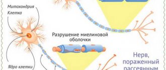
The disease causes the occurrence of multiple benign tumors (hamartomas), which lead to disruption of the vital systems of the body.
The multisystem nature of tuberous sclerosis is expressed in damage to almost all organs - the brain, central nervous system, kidneys, heart, skin, retina, digestive tract, lungs, etc.
The manifestations of tuberous sclerosis that most affect quality of life are most often associated with its effects on the brain, and include epileptic seizures, developmental delays, including mental retardation, and autism. However, many patients are able to live full independent lives and be professionally in demand, working, for example, as doctors, lawyers, teachers and engineers.
Advantages of treatment in Israel
- Highly qualified doctors.
- Excellent material and technical base of the clinics.
- High quality of low-traumatic neurosurgical operations.
- Use of modern medications.
- Loyal prices.
Effective treatment of tuberous sclerosis requires high professionalism of doctors and high-tech medical equipment, presented in Israeli clinics. Contact your chosen clinic without delay and undergo therapy under the guidance of world-class specialists.
- 5
- 4
- 3
- 2
- 1
(1 vote, average: 5 out of 5)
Symptoms
This disease is quite rare and therefore not fully studied. Its signs are varied, many organs and systems are affected, and therefore sometimes an accurate diagnosis can be made only after a comprehensive examination.
The signs of tuberous sclerosis are varied, many organs and systems are affected, and therefore sometimes an accurate diagnosis can be made only after a comprehensive examination.
Usually the first symptoms of the disease are polymorphic formations on the skin. Nodular tubercles with a diameter of 0.5 to 1 cm appear on different parts of the body (usually on the face - nose, cheeks, on the scalp in the scalp), as well as spots in the shape of “leaves”. Such depigmented areas of coffee-milky or whitish color on the skin can appear anywhere on the body. The probability of their appearance in newborns up to one year is 80%, in the second year – up to 100%. The number of areas with uneven depigmentation increases as the child grows older. In 15% of cases, both types of spots can occur - whitish and coffee-milky.
In addition to skin symptoms, signs of this type of sclerosis include: • myoclonic seizures, • tonic-clonic generalized seizures, • hemianopsia.
During myoclonic convulsions, the patient does not lose consciousness; individual muscle groups contract involuntarily and randomly. This pathological shock muscle twitching quickly passes. During generalized tonic-clonic seizures, a person suddenly loses consciousness. With massive spasm, involuntary separation of urine and trauma to the tongue may occur (the patient may bite it).
Hemianopsia is the partial loss of a person’s field of vision with limited space around.
Skin changes
Hypopigment spots, facial angiofibromas, areas of “shagreen skin”, periungual fibromas, fibrous plaques, white strands of hair, eyelashes and eyebrows.
Hypopigment spots (oval, polygonal, or in the form of scattered “confetti”) are often detected from birth; their number may increase with age. They are located asymmetrically, mainly on the torso and buttocks.
Hypopigmented spot on the skin of the leg
Angiofibromas of the face (Pringle's adenoma) are pink or red nodes with a smooth, shiny surface, located symmetrically on both sides of the face on the cheeks and nose (like “butterfly wings”) and chin. They usually develop after 4 years of age.
Angiofibromas of the face
Areas of “shagreen leather” (peau chagrine in French - “untanned, rough, hard leather”), yellowish-brown or pink in color, protrude moderately above the surface of the surrounding skin and resemble the skin of an orange. Their size ranges from a few millimeters to 10 or more centimeters. More often they appear in the second decade of life. They are located predominantly in the lumbosacral region.
Shagreen skin area
Fibrous plaques are beige-colored formations, rough to the touch, and protrude somewhat above the surrounding skin. They often appear already in the first year of life. The favorite location for fibrous plaques is the forehead. Their size and number vary.
Periungual fibromas are dull, red or meat-colored nodules that grow from the nail bed or around the nail plate. In most cases, periungual fibromas appear in the second decade of life. More common on the legs than on the arms. The size varies from 1 millimeter to 1 centimeter in diameter.
Soft fibromas are located on the trunk and neck, less often on the limbs. There are two types:
- multiple or single, soft saccular formations on the legs
- multiple formations, slightly raised above the surface of the skin, smaller than a pinhead (reminiscent of “goose bumps”)
Periungual fibromas
Forms and causes of the disease. Prevention of the disease
There are two forms of tuberous sclerosis: - hereditary - spontaneous (de novo mutations)
Tuberous sclerosis is a hereditary disease. But even if the parents have a mutation in the genes, the birth of a healthy baby is quite likely. To do this, when planning a pregnancy, it is important to undergo a complete examination by a geneticist.
Bourneville disease is an autosomal dominant type. This is a type of inheritance in which a genetic disease manifests itself if a person has at least one mutated gene corresponding to it. The disease can be inherited from either parent with a 0-50% chance. Boys and girls get sick with the same frequency. The disease is not diagnosed immediately - either in infancy (up to 1 year) or in adolescence. Approximately one third of cases of tuberous sclerosis are due to heredity, the remaining two thirds are spontaneous, unpredictable genetic mutations.
Genes on chromosomes 9 and 16 are responsible for controlling the synthesis of hamartin and tuberin, proteins that suppress tumor processes. But when these genes are mutated, the barrier to the pathological growth of abnormal tissue disappears - and tumors form.
If the gene on chromosome 9 is damaged, tuberous sclerosis type I (TSC1) develops. With a mutation on chromosome 16, a diagnosis is made of TSC2 - type II of the disease.
But even if the parents have a mutation in the genes, the birth of a healthy baby is quite likely. Therefore, if there are hereditary diseases in the family, before planning a child, it is necessary to visit a geneticist for consultation and examination in order to exclude the possibility of the birth of a sick baby.
If tuberous sclerosis is suspected, it is necessary to consult a specialist (neurologist, dermatologist, geneticist, ophthalmologist, nephrologist, cardiologist, gastroenterologist, etc.) as soon as possible and undergo a comprehensive examination to reduce the risk of complications.
Epilepsy: there will be more operations to remove the source of the disease
In Belarus today, about 4,500 children with epilepsy are under the supervision of doctors; for the first time, more than 600 cases are being registered per year. Medicines do not help everyone with this disease. For many, surgery is the only effective treatment to avoid disability, achieve seizure control, and even avoid drug therapy entirely.
Our country has already mastered neurosurgical methods for treating epilepsy in children. Over the past 4 years, dozens of operations have been performed to remove the focus of the disease (the focus of epileptiform activity) with good results.
However, for most such operations were inaccessible due to the lack of ability to diagnose epileptic foci. Therefore, some people had to travel abroad for expensive examination and treatment.
What has changed: This year, for the Republican Scientific and Practical Center of Neurology and Neurosurgery, philanthropists will purchase medical diagnostic equipment that will allow long-term EEG monitoring. Patients will no longer have to travel abroad. According to forecasts, this will increase the number of operations to remove the source of the disease to 40-50 per year. Also, to develop this area of neurosurgical care, the center’s doctors will undergo the necessary internships. For its part, the Ministry of Health guarantees the timely introduction of this medical equipment.
Diagnostics and types of examination
When you first contact a doctor, you should tell about how long ago the first signs of the disease appeared, what kind of complaints are bothering you (convulsions, nodules on the tongue, skin, its depigmentation, partial blindness with the inability to see some part of the space), as well as whether there were any such illnesses in someone's family.
Mandatory examinations:
- General blood analysis
- General urine analysis
- Radiography
- Consultations with neurologists, ophthalmologists, dermatologists and geneticists
- MRI and CT
- EEG and ECG
- Echocardiography
- Ultrasound
General clinical tests - blood and urine - are required. If there is pathology in the blood test, the level of urea and creatinine may increase, since they are products of protein metabolism and indicate kidney function. If the kidneys are affected by the disease, blood may appear in the urine (hematuria).
Radiography is also prescribed to detect tumor growths in tissues.
At an appointment with a neurologist, you need to draw the doctor's attention to disturbing attacks of seizures, headaches, vomiting, changes in behavior and intellectual disorders.
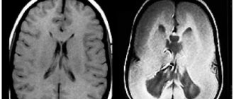
Today it is impossible to undergo a comprehensive examination without tomography - magnetic resonance imaging (MRI) and computer computed tomography (CT). Thanks to their use, specialists can: • engage closely in a layer-by-layer study of the structure of the brain; • identify tumors in it (they are more often found at the border of gray matter with white matter); • detect signs of abnormal development of brain matter and areas with reduced density of its nervous tissue; • identify accumulations of cerebrospinal fluid (cerebrospinal fluid that supplies the brain with nutrition and metabolic processes) in the cavities of the brain.
Using electroencephalography (EEG), specialists assess brain activity in different areas. This method of examination makes it possible to search for foci with increased impulses that provoke convulsive attacks in tuberous sclerosis.
To determine whether there are growths in the heart muscle (rhabdomyomas), the patient is sent for echocardiography.
Electrocardiography (ECG) is recommended for all patients because Cardiac arrhythmia, although rare, can lead to sudden death. Ultrasound (ultrasound) will help check for the presence of kidney tumors and cysts in their tissue.
The ophthalmologist will conduct a series of examinations: • using a slit lamp (biomicroscopy) to examine the fundus of the eye; • using tables to check visual acuity (visometry); • perimetry to determine the edge of the visual field to identify defects in it; • assessment of the condition of the retina, the fundus of the eye and its vessels, the disc (nipple) of the optic nerve (ophthalmoscopy) to determine the presence of pathological changes (swelling of the nipple, retinal hamartomas).
At your appointment, the dermatologist will definitely pay attention to the presence of formations on the skin in the form of reddish nodules around the nails, tubercles, growths, fibrous plaques and areas with insufficient pigmentation.
A comprehensive examination cannot be done without the help of a geneticist, who will prescribe a special test to identify mutational changes in the genes of the 9th and 16th chromosomes (TSC1, TSC2). If necessary, a consultation with a neurosurgeon or psychiatrist is prescribed.
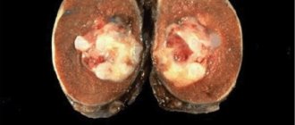
Possibilities of pathogenetic therapy of kidney AML associated with TS
AML of the kidneys occurs in more than half of patients with TS. AML is a benign mesenchymal tumor, the composition of which in various proportions is represented by adipose tissue, spindle-shaped and epithelioid smooth muscle cells, and abnormal thin-walled blood vessels. In women, progesterone receptors are often detected in tumors. AMLs account for about 1% of all surgically removed renal tumors; Moreover, 33% of all cases of renal AML are determined among patients with TS. In TS, tumors are often multiple and can be combined with lung LAM [9].
Sirolimus (another mTOR inhibitor) was studied in 25 adult patients with renal AML associated with TC or sporadic pulmonary LAM (NCT00457808 ClinicalTrial.gov). According to the study design, patients received treatment with sirolimus for 12 months. Patients who completed treatment were followed up for another 12 months. It was shown that the volume of AML kidneys after 12 months of treatment averaged 53.2 ± 26.6 cm³ compared with the volume before therapy (p < 0.001). For 5 patients, volume reduction of more than 30% was maintained at 24 months. Additionally, external respiratory function was assessed.
After 12 months of therapy, an improvement was noted in all assessed indicators (forced expiratory volume per second, forced vital capacity, residual lung volume). Five patients experienced the development of serious adverse events (diarrhea, pyelonephritis, stomatitis, respiratory infections) [10].
The results of the phase II clinical trial NCT00490798 of sirolimus therapy for renal AML associated with TS and sporadic pulmonary LAM have also been published. The study included 16 patients, 10 of them were diagnosed with TS (including 3 with LAM), 6 patients were diagnosed with only sporadic LAM. Patients received sirolimus at an initial dose of 0.5 mg/m² daily, followed by adjustment to achieve plasma drug concentrations in the range of 3–6 ng/ml; the duration of therapy in the study was 2 years. The overall response rate to treatment for renal AML according to RECIST (Response Evaluation Criteria in Solid Tumors) criteria was 50% (8 out of 16) in the overall group and 80% (8 out of 10) in the group of patients with TC. A partial response in the group of patients with renal AML and TS after 2 years of therapy was observed in 40% of cases (4 out of 10). During therapy, some improvement in lung function was noted in patients with both sporadic pulmonary LAM and LAM in TS. However, during serial computed tomographic studies of the chest organs, no significant changes in the lung tissue were recorded [11].
Currently, two phase 3 clinical multicenter studies of everolimus are ongoing: EXIST-1 (NCT00789828 ClinicalTrial.gov) and EXIST-2 (NCT00790400 ClinicalTrial.gov). 10 countries are participating, including Russia.
EXIST-1 everolimus for SELA associated with TS: study design – randomization into everolimus and placebo groups in a 2:1 ratio with the possibility of switching to open administration of everolimus in case of tumor progression. Treatment regimen: everolimus 4.5 mg/m² per day orally continuously. Parameters assessed: change in the frequency of epileptic attacks, tumor response to treatment, time to registration of response, duration of response, response from skin lesions, angiogenesis markers, safety. EXIST-2 everolimus for renal AML: study design – randomization into everolimus and placebo groups in a 2: 1 ratio with the possibility of switching to open administration of everolimus in case of tumor progression. Treatment regimen: everolimus 10 mg/day orally continuously. Parameters assessed: tumor response to treatment, time to response registration, duration of response, response from skin lesions, angiogenesis markers, safety.
Data from preclinical studies of mTOR inhibitors and encouraging first results of early clinical studies give hope that in the near future doctors will have the opportunity to carry out pathogenetic therapy for TS, which will improve the results of treatment of patients with various manifestations of this disease.
Literature
1. Verdecchia M, Bombardieri R, Curatolo P. Diagnostic Criteria and evolution of patients with tuberous sclerosis complex. In: Neurocutaneous syndromes in children. Ed. by Curatolo P, Riva D. John Libbey Eurotext, 2006:81–90. 2. Whittemore VH. The history of Tuberous Sclerosis Complex. In: Tuberous sclerosis complex. Genes, Clinical features and therapeutics. Ed. by Kwiatkovski DJ, Whittemore VH, Thiele EA. Wiley-Blackwell 2010:3–9. 3. Gomez MR. History of Tuberous Sclerosis Complex. In: Tuberous Sclerosis Copmplex. Ed. by. Gomez MR, Sampson JR, Whittemore VH. Oxford University Press, 1999:3–9. 4. Curatolo P. Historical background. In: Tuberous Sclerosis complex: From Basic Science to Clinical Phenotypes. Mac Keith Press, 2003:1–10. 5. Napolioni V, Moavero R, Curatolo P. Recent advances in neurobiology of Tuberous Sclerosis Complex. Brain and Dev 2009;31:104–13. 6. Krueger D, Care M, et al. Everolimus for Subependymal Giant-Cell Astrocytomas in Tuberous Sclerosis Complex. New Eng J Med 2010;363:1801–11. 7. Franz D, Leonard J, Tudor C, et al. Rapamycin causes regression of astrocytomas in tuberous sclerosis complex. Anals of Neurology 2006;59(3):490–98. 8. Jozwiak S, Perek-Polnik M, Kotulska K, et al. Eur Journal of Neurology 2011;70:09.6–870. 9. 19. Mantignoni G, Amin M. Angiomyolipoma. In: Pathology and Genetics of Tumors of the Urinary System and Male Genital Organs Ed. By Eble J, et al. IARC Press, 2004:65–9. 10. Bissler J, McCormack F, et al. Sirolimus for angiomyolipoma in Tuberous Sclerosis Complex or Lymphangioleiomyomatosis. New Eng J Med 2008;358:140–51. 11. Davies M, de Vries P, Johnson P, et al., Sirolimus Therapy for Angiomyolipoma in Tuberous Sclerosis and Sporadic Lymphangioleiomyomatosis: a Phase 2 Trial. Clin Cancer Res 2011;17(12);4071–81.
About the authors / For correspondence
Dorofeeva M.Yu. – Candidate of Medical Sciences, Senior Researcher of the Department of Psychoneurology and Epileptology of the Federal State Budgetary Institution Moscow Research Institute of Pediatrics and Pediatric Surgery of the Ministry of Health and Social Development of the Russian Federation Belousova E.D. – Doctor of Medical Sciences, Professor, Head of the Department of Psychoneurology and Epileptology, Federal State Budgetary Institution Moscow Research Institute of Pediatrics and Pediatric Surgery, Ministry of Health and Social Development of Russia, Moscow