Problems with using or understanding language (aphasia) appear, according to statistics from studies conducted by NABI (National Stroke Association), in 25 patients out of 100. Every fourth patient experiences difficulties associated with verbal communication - the ability to speak, write or understand spoken language. and written language. This indicates that acute cerebrovascular accident affected one or more language control centers.
Aphasia is a symptom of brain damage, not a disease. As individual as the picture of damage is, the manifestation of aphasia in each patient is as individual. This condition determines the individual nature of the selection of speech restoration techniques after a stroke for each patient.
There are four main types of post-stroke speech disorders
Damage to the language center in the dominant hemisphere - Broca's area - leads to the fact that patients lose the ability to convey their thoughts using coherent and grammatically correct language structures. Conversely, pathological changes in Wernicke's area (sensory center) lead to problems in receptivity to language. Such patients have difficulty understanding spoken or written language. They often use correct grammatical structures, but their statements may not make sense. The most severe form of aphasia (global or total) occurs in patients with extensive damage to several areas of the brain responsible for understanding and language. The mildest form of aphasia—amnestic—leads to difficulty working with vocabulary.
How do these types of aphasia manifest themselves in practice?
Damage to the language center in the dominant hemisphere - Broca's area - leads to the fact that patients lose the ability to convey their thoughts using coherent and grammatically correct language structures. Conversely, pathological changes in Wernicke's area (sensory center) lead to problems in receptivity to language. These patients have difficulty understanding spoken or written language. They often use correct grammatical structures, but their statements may not make sense. The most severe form of aphasia (global or total) occurs in patients with extensive damage to several areas of the brain responsible for understanding and language. The mildest form of aphasia—amnestic—leads to difficulty working with vocabulary.
How do these types of aphasia manifest themselves in practice?
- Motor aphasia. You know what you want to say, but you can’t find (remember, use and pronounce) the right words.
- Sensory aphasia. You hear someone speak or see printed text, but cannot understand the meaning of the words.
- Amnestic aphasia. You find it difficult to select and use the correct words to refer to certain people, objects, places or events.
- Global aphasia. You cannot speak, write, read and do not understand what is being said to you.
Post-stroke aphasia: clinical picture, differential diagnosis, treatment
Speech disturbances are a common symptom of stroke (15–38%). They often lead to permanent disability, significantly complicate rehabilitation during the recovery period, reduce the quality of life of both the patients themselves and those around them, cause negative psycho-emotional reactions, and increase the economic costs of treatment. Compared to patients with stroke but without speech disorders, patients with post-stroke aphasia have a higher mortality rate and stay in hospital longer. Predictors of good recovery of speech function are mild to moderate severity of speech disorders in the acute period of stroke, the amount of brain damage (the smaller the ischemic focus, the higher the chances of recovery), young age, high Barthel index, and high level of education. In addition to the treatment of the underlying vascular disease, patients with speech disorders need systematic speech therapy sessions and medications in order to optimize cerebral neuroreparative processes.
Table. Types of aphasia
Aphasia is a disorder of a person’s higher mental functions, which consists of a loss or decrease in the ability to verbally communicate, including constructing one’s own speech utterance and/or understanding spoken speech. As a rule, patients with aphasia have pathology in both oral and written speech (reading, writing), and also have difficulties in using sign language and Braille (a dotted font for writing and reading for the blind).
Neuroanatomy of aphasia
Speech areas are a complex neurocognitive network located in the dominant hemisphere. In approximately 95% of people, the left hemisphere is dominant in speech, and in 5% of cases, either both hemispheres are involved in the innervation of speech, or the right hemisphere becomes dominant. Already at birth, in more than half of newborns, the cortex in Wernicke’s area and the angular gyrus in the left hemisphere is approximately 50% larger in volume than in the right [1]. If for some reason in very early childhood the left speech areas suffer, then the right hemisphere of the brain acquires signs of dominance [1].
Speech centers include the posterior parts of the left frontal lobe (Broca's area) and the left superior temporal gyrus (Wernicke's area), as well as connections between these areas. The motor program for speech utterance is formed in Broca's area. Broca's area projects directly to the neurons of the precentral gyrus, which innervate the muscles of the larynx and oral cavity. Wernicke's area is responsible for comparing auditory information with visual and kinesthetic images, which is necessary for understanding spoken speech. Comparison of information is provided by connections between Wernicke's area and the occipital and parietal cortex. Another cerebral region important for speech is the angular gyrus in the inferior parietal lobule, which is responsible for the perception of written speech and sign language.
In addition to the classical speech centers, other areas of the brain also play an important role in the formation of speech. These include the insula (important for articulation), zones of the frontal and temporal lobe (processing sentences), as well as zones of the occipital and parietal cortex of the brain (responsible for memory for the meanings of words) [2–5].
The subcortical nuclei of the brain also make a significant contribution to maintaining normal speech function. Diffusion-weighted and perfusion-weighted magnetic resonance imaging (MRI) of the brain in patients with ischemic damage to the basal ganglia and aphasia showed a secondary decrease in perfusion in the subcortical gray ganglia. However, the prognosis for subcortical aphasia is more favorable than for cortical damage [6]. In recent years, the role of cerebellar damage has also been actively discussed, since it can also lead to dysphasic disorders in the form of disturbances in the grammatical structure of speech [7].
Etiology of aphasia
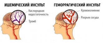
The most common cause of aphasia is ischemic stroke. Less commonly, speech disorders are observed with hemorrhagic strokes, space-occupying brain lesions, infectious lesions (abscess, encephalitis), and traumatic brain injury. Rare cases of the development of aphasia in demyelinating diseases have been described [8, 9].
Transient aphasia can be observed with transient ischemic attacks, epilepsy, migraine. The presence of aphasia during a transient ischemic attack is one of the high risk factors for developing stroke in the coming days and weeks [10].
Gradually progressive aphasia may also be a manifestation of a neurodegenerative disease. Most often, gradually progressive aphasia is associated with frontotemporal degeneration, less often with Alzheimer's disease or other dementing diseases. Moreover, in some cases, the clinical picture does not show any other cognitive and/or behavioral disorders for many years (the so-called primary progressive aphasia) [11–13].
Speech status study
To diagnose and analyze the characteristics of dysphasia, it is necessary to carefully listen to the patient’s speech, examine the understanding of spoken speech, reading and writing. You should pay attention to the number of words spoken per minute (speech fluency), unmotivated repetitions of individual words and phrases (perseverations), shortened phrases (less than five words), errors in the grammatical construction of statements (case endings, prepositions, conjunctions, word order in a sentence etc.) and/or difficulties in understanding grammatical structures. In addition, it is necessary to assess the ability to control the movements of articulatory muscles (speech praxis). To do this, you can ask the patient to repeat the phrase “artillery brigade” several times.
An important part of speech research is the assessment of its nominative function.
. The patient is shown certain objects and asked to name them, starting with familiar ones (for example, a spoon, pen, mug) and moving on to unusual ones (for example, a phonendoscope). Insufficiency of the nominative function of speech is noted in many aphasias, sometimes becoming one of the main manifestations of dysphasia.
Assessing oral language comprehension
is carried out by checking the execution of verbal commands, first simple and then complex (“close your eyes”, “show me two fingers”, “touch your left ear with your right hand”). We can then move on to explore understanding of more complex grammatical structures (“Are my brother’s father and my father’s brother the same person?” or “Is my aunt’s uncle a man or a woman?”). Final trials may reveal a lack of understanding, including in those who have followed simple verbal commands.
When checking the reading function
the patient is asked to read aloud a paragraph from a newspaper or magazine, assessing the correct pronunciation of the words. Comprehension of written language can be tested using written commands (eg, “take this piece of paper, fold it in half and place it on the floor” or “close your eyes”).
Letter evaluation
– the patient is asked to write any sentence. You can also dictate any text to the patient or offer to write the names of objects drawn in the pictures.
The above methods make it possible to diagnose dysphasia without leaving the patient’s bedside, which is of great importance in the acute period of stroke. When diagnosing speech disorders in a patient, it is necessary to examine them in more detail, analyze quantitative and qualitative features, and also evaluate other higher brain functions: attention, memory, praxis, visual-spatial orientation, etc.
Epidemiology and types of aphasia
According to the literature, aphasia is a common symptom of stroke (15–38%). Typically, the clinical picture also includes other symptoms of damage to the dominant hemisphere (right-sided hemiparesis, hemihypesthesia, hemianopsia) [14]. Let's look at the main types of aphasia (table).
Broca's aphasia (motor aphasia)
characterized by a violation of the construction of one’s own speech utterance, as well as the repetition of phrases. The patient's speech is laconic, poorly articulated, and characterized by auditory and verbal perseverations. The letter is broken. Comprehension of addressed speech may be incomplete in the first few days after acute cerebrovascular accident, but then quickly recovers. Motor aphasia develops as a result of acute ischemic stroke in the anterior branches of the left middle cerebral artery and is often combined with hemiparesis and hemihypesthesia.
Wernicke–Kozhevnikov aphasia (sensory aphasia)
characterized primarily by impaired understanding of oral and written speech. The patient’s own speech, as a rule, maintains normal tempo and intonation, but is meaningless, as it contains numerous replacements of syllables and words with similar ones in sound, but meaningless in meaning (literal and verbal paraphasias), as well as new unusual words (neologisms). With significant severity of these disorders, speech production takes on the character of so-called verbal okroshka. However, many patients are not aware of their defect. Sensory aphasia develops when the upper parts of the temporal lobe and the lower parts of the parietal lobe are damaged as a result of a stroke in the territory of the left middle cerebral artery. It is often combined with right upper quadrant hemianopia.
Total sensorimotor aphasia
– a set of symptoms of motor and sensory aphasia. Develops as a result of extensive strokes in the left middle cerebral artery, usually combined with hemiparesis, hemihypesthesia and hemianopsia. Rarely, encephalitis and late manifestations of neurodegenerative processes can be the cause of total aphasia.
Dynamic aphasia (transcortical motor aphasia)
is largely reminiscent of Broca's motor aphasia. There is a violation of the initiation of speech activity, perseveration and grammatical errors occur, while the understanding of addressed speech does not suffer. The main difference between dynamic aphasia and motor aphasia is preserved repeated speech: the patient can repeat words and phrases after the doctor. Typically, dynamic aphasia develops with a heart attack in the territory of the left anterior cerebral artery.
Transcortical sensory aphasia
is characterized by a violation of the understanding of addressed speech, which resembles Wernicke–Kozhevnikov aphasia, but the severity of these disorders is somewhat lesser. The patients’ own speech is fluent, but lacks content, and verbal paraphasia may be observed. However, unlike Wernicke's aphasia, repeated speech is preserved in transcortical sensory aphasia. Patients may also read aloud, but without understanding the meaning of what they read. Transcortical sensory aphasia develops with damage to the temporo-occipital or temporo-parietal areas adjacent to Wernicke's area as a result of a stroke, and can be combined with hemianopsia.
Transcortical mixed aphasia
– patients have signs of transcortical motor and sensory aphasia, but the ability to repeat words and phrases after the doctor remains intact. Understanding of written and spoken language also deteriorates significantly. Occurs when there is damage in the anterior and posterior cerebral arteries during repeated cerebral embolisms, infarctions in areas of adjacent blood supply associated with systemic circulatory disorders, such as acute heart failure.
Amnestic aphasia
– patients with amnestic aphasia cannot name a word or object, but they can describe the meaning and functions of the object. Spontaneous speech is characterized by pauses, word substitutions, and paraphasias are possible. Repetition of words and understanding of oral speech are not impaired. Amnestic aphasia has been described with damage to various anatomical areas, including the basal temporal lobe, anterior temporal lobe, temporo-parieto-occipital junction, and inferior parietal lobe.
Alexia without agraphia
– patients can write, but not read. Understanding of oral speech is intact, spontaneous speech is not affected. It develops when the left occipital lobe and splenium of the corpus callosum are damaged during an ischemic stroke in the territory of the left posterior cerebral artery [15].
Differential diagnosis
In most cases, in patients with risk factors for stroke (in old age, in the presence of arterial hypertension, diabetes mellitus, hypercholesterolemia, concomitant cardiac pathology, atrial fibrillation, etc.) with the acute development of neurovascular syndrome, characteristic of damage to the middle and/or posterior cerebral artery dominant hemisphere, the diagnosis of ischemic stroke is not in doubt. Neuroimaging methods are used to verify the diagnosis.
Impaired understanding of speech and speaking, reminiscent of dysphasia, can develop with acute dysmetabolic encephalopathy (delirium)
. In this case, difficulties often arise in understanding the spoken speech; the patient does not follow commands. Grammatical errors and paraphasia appear in the patient’s own speech. Signs of a confused state of consciousness, the presence of hallucinations, tremors, psychomotor agitation and delirium help in diagnosis.
Akinetic mutism
develops when the medial frontal region is damaged. In such patients, there is an absence or extreme paucity of spontaneous speech, poor execution of commands, a decrease in motor reactions, and signs of catatonia (waxy flexibility).
Patients suffering from depression
, may avoid communicating with others. They do not look the doctor in the eyes, they lie with their faces turned away from the people around them. In this case, anamnesis collected from the patient’s relatives and/or loved ones plays an important role in the differential diagnosis.
If the patient has episodes of transient aphasia
The differential diagnosis is made between
transient ischemic attack and epilepsy
. 24-hour electroencephalographic monitoring, angiography of cerebral vessels, and duplex scanning of the brachiocephalic arteries are performed.
Aphasias with gradual onset and slow progression, especially in middle-aged and elderly people, require a differential diagnosis between neurodegenerative diseases and brain space-occupying lesions
. In this case, an MRI of the brain is indicated.
Treatment and prognosis of post-stroke aphasia
Speech disorders significantly disable patients, reduce the quality of life of both themselves and those around them, interfere with full neurorehabilitation, and increase the economic costs of treatment. In addition, patients with post-stroke aphasia, compared with patients with stroke but without speech disorders, have higher mortality rates and stay in hospital longer [16].
The degree of speech restoration depends primarily on the location and extent of damage to the brain. For example, sensorimotor aphasia as a result of ischemic stroke of the cardioembolic type, usually in combination with right-sided hemiparesis and hemihypesthesia, has a less favorable prognosis than motor aphasia as a result of a stroke in the anterior cortical branches of the middle cerebral artery [17].
Predictors of good recovery of speech function are young age, high Barthel index, high level of education and hemorrhagic nature of the stroke [17].
Management of a patient with aphasia involves, in addition to treating the underlying vascular disease, systematic speech therapy exercises [17–19]. They are carried out by patients independently under the supervision of relatives. The clinical effectiveness of speech rehabilitation has been assessed in a number of studies [20, 21]. It has been shown that the specific method of speech therapy correction is not of fundamental importance, while the frequency and intensity of classes significantly influence the prognosis [22, 23]. Speech therapy exercises must be started already in the acute phase of a stroke and continued throughout the entire recovery period, as long as the patient continues to have speech disorders. Currently, computer programs and applications for phones and tablets have been developed that allow the patient to communicate with others and independently perform speech exercises.
Restoration of speech in the first months after a stroke is associated with reperfusion of the corresponding cortical centers, activation of adjacent cortical areas and homologous brain areas in the contralateral hemisphere. This is evidenced by studies using diffusion-weighted and perfusion MRI methods [24, 25].
Drug therapy in patients with aphasia should be aimed at metabolic support of neuroreparative processes and improvement of cerebral blood flow.
To date, several randomized, double-blind studies of medications for post-stroke aphasia have been conducted. The effectiveness of bromocriptine [26] and amphetamine [27] has not been proven, and a meta-analysis of clinical studies of piracetam indicated a rather modest effect of this drug [28]. There is little clinical experience with the use of anticholinesterase inhibitors such as donepezil and galantamine, but their clinical effectiveness requires further study [29, 30].
Currently, the use of citicoline in patients with post-stroke aphasia seems promising. Citicoline (Ceraxon) is an endogenous mononucleotide containing ribose, cytosine, pyrophosphate and choline in its chemical structure. Being a necessary intermediate substance in the synthesis of structural phospholipids of the cytoplasmic and mitochondrial membranes of neurons, citicoline restores their integrity during ischemic damage. A number of experimental studies have shown that citicoline inhibits the enzyme phospholipase A2, normalizes energy processes in mitochondria, restoring the functioning of membrane sodium-potassium and mitochondrial adenosine triphosphatases. In addition, it enhances the activity of antioxidant systems, thereby providing a neuroprotective effect and preventing the processes of oxidative stress and apoptosis. Another mechanism of action of Ceraxon is to replenish cerebral acetylcholinergic deficiency, which is of great importance for metabolic support of cognitive activity in general. Finally, this drug affects dopamine and glutamatergic neurotransmission. Citicoline has a pleiotropic effect, also due to its influence on neurorepair processes, which play a key role in restoring lost functions [31]. A number of experiments demonstrated the effect of the drug on the processes of neuroglial activation, enhancement of post-ischemic neurogenesis, angiogenesis and neuroplasticity [32, 33]. It has been established that citicoline reduces the volume of brain damage during experimental ischemia and hypoxia, increases learning ability and has a beneficial effect on memory in experimental animals with age-related changes in the brain.
In clinical studies, Ceraxon improved functional recovery and accelerated rehabilitation of patients with ischemic stroke [34]. Currently, the effectiveness of citicoline in post-stroke cognitive and motor disorders has been shown [35, 36]. Moreover, the drug is characterized by a satisfactory safety and tolerability profile. The positive effect of the drug on speech disorders in patients after stroke has not been separately assessed. However, it can be assumed that the neuroregenerative properties of Ceraxon and its effects in the cognitive sphere will contribute to the improvement of speech functions due to the close relationship of these disorders.
Conclusion
When treating patients with aphasia, regular speech therapy sessions and long-term drug therapy are required, aimed at metabolic support of neuroreparative processes and improvement of cerebral blood flow.
Stroke: restoration of speech. Exercises
Patients diagnosed with aphasia and speech disorders are concerned with the following questions: is it possible to fully recover from a stroke and how long will it take?
First of all, it is necessary to take into account the irreversibility of the process of destruction of brain cells. To restore the patient’s ability to speak, specialists will need to select means of recovery after a stroke that will help switch speech functions to the closest areas of the brain not affected by the disease. And the timing depends, among other things, on the high inertia of brain cells - after all, it is necessary to switch them to functions unusual for them
Many methods have been developed for restoring speech after a stroke that can speed up this process - speech, melodic, visual, artistic, etc. These speech therapy methods are used along with drug therapy - the patient is prescribed drugs that can stimulate the formation of new neural connections.
The method of intonation (melodic) therapy is based on “singing”. The patient is asked to sing words that cannot be pronounced clearly. Visual therapy uses the ability to form associations between words denoting an object and their visual image.
The most common method of treating aphasia is speech therapy. Classes on the method are conducted with a speech therapist-aphasiologist and are included in the course of intensive rehabilitation of patients after a stroke to the extent necessary for a particular patient.
Exercises to restore speech after a stroke begin with a complex that is designed to gradually restore the patient’s articulatory skills. This could be pronouncing individual sounds, syllables or words, or pronouncing tongue twisters. The exercises are performed with a speech therapist, and the results are consolidated independently in your free time. The more intense the exercise, the greater the likelihood of recovery.
Knew, but forgot
Amnestic aphasia is characterized by the inability to use long-known words. The person has preserved hearing and intelligence, he understands what is said well and speaks clearly himself, but there are frequent pauses in his speech. He replaces words with inappropriate meanings, and sometimes with just invented ones. Sometimes it is possible to remember a word only with the help of loved ones or a speech therapist. The patient says: “This is what they eat”, he is upset that he cannot name the object, but after the first syllable “lo” he remembers: “Spoon!” The signs of amnestic aphasia resemble the stage of speech formation, when the child still has a poor active vocabulary. But the causes of the disease are completely different.
Who destroyed the bridge in the head?
Typically, a particular type of speech disorder is associated with problems in the functioning of a specific area of the cerebral cortex, and this helps doctors make a diagnosis. But not in this case. In both left-handers and right-handers, amnestic aphasia develops most often with damage to:
1) basal or anterior parts of the temporal lobe,
2) temporo-parietal-occipital junctions,
3) inferior parietal lobule.
Foci are also found in other areas of the cerebral cortex. The causes of violations are various. In children, in most cases, amnestic aphasia occurs when the brain is damaged as a result of trauma, but the disease also occurs in those who have experienced severe poisoning with brain damage, neuroinfection, or suffer from epilepsy and tumor diseases of the brain. But the essence is always the same - the transferred condition led to the death of neurons in a certain area of the cortex. Speech is the result of complex brain work. Therefore, amnestic aphasia is observed in a wide variety of lesions.
Like a child!
Speech is a complex process in which different areas of the cerebral cortex actively interact. Thus, Wernicke's area is responsible for understanding; signals are formed in Broca's area, which ultimately turn into words and phrases. The role of auditory and visual analyzers is important. When a person talks, the whole brain is actively working. It establishes connections, for example, between the image of a spoon and the word that denotes this object. It is these connections that are destroyed in patients with amnestic aphasia. This is the difference between patients of the NeuroSpectrum Center for Pediatric Speech Neurology and Rehabilitation, who are being treated for amnestic aphasia, and healthy children. The normal process of forming connections goes by itself, and if connections are destroyed, their restoration requires outside help. Fortunately, children's brains are plastic, and it is easier to help our patients than adults and especially older people.
Sensory aphasia
Wernicke's sensory aphasia
is a violation of the understanding of speech, both someone else’s and one’s own, while maintaining the ability to speak.
Occurs when the left temporal lobe of the brain is damaged. As a rule, the patient speaks quite willingly and a lot, but due to the loss of understanding of what he hears, he does not understand either himself or those around him. The form of sensory aphasia
is characterized by two main features:
- the patient’s speech is incomprehensible to others due to the lack of meaning in it (misunderstanding of one’s own speech leads to loss of control over it, the patient invents new words, replaces letters or syllables in existing ones, swaps words, pronounces incoherent strings of words and sentences)
- the speech of others and his own is incomprehensible to the patient (due to impaired perception of sound, loss of distinction in the sound composition of words, the patient does not understand the meaning of what he hears)
What is amnestic aphasia?
Aphasia is a neurological disease of organic nature, which is formed as a result of the death of neurons located in the cerebral cortex area, responsible for the synthesis and formation of speech function, as well as the perception of speech and non-verbal signals of surrounding people. Aphasia is always an acquired pathology, i.e. it is observed in people with initially normally functioning speech function, so it is important not to confuse aphasia with alalia if the disease is observed in childhood.
A wide range of reasons can lead to the development of pathology, but the final pathogenetic mechanism of this disease is the same and consists in extensive death of nervous tissue localized in specific areas of the brain responsible for speech.
Why look for a reason?
Amnestic aphasia is only a symptom of severe brain damage. To successfully fight it, you need to make sure that the patient does not get worse. Therefore, neurologists are actively involved in the treatment of the disease. If the cause is not removed, the localization of damage will expand, and amnestic aphasia will be combined with other disorders. Often this is facial agnosia, in which the patient cannot even recognize relatives, not only in photos, but also in life.
Therefore, patients undergo various studies. Computed tomography or magnetic resonance imaging will help determine if there are any abnormal changes inside the skull. In this way, it is not difficult to detect tumors, bone fragments after traumatic brain injuries, areas of tissue that are left without blood supply, and much more. Electroencephalography - recording the electrical signals that the brain sends - will help determine whether there are areas of hidden excitation of the cortex, that is, whether forgetting words is associated with signs of epilepsy. This insidious disease does not necessarily manifest itself in large convulsive seizures; there are also forms with subtle manifestations.
Examination by an experienced neurologist is extremely important. Our center’s specialists work a lot with speech problems in children. They know how to find an approach to any child, help him feel at ease, and sometimes notice signs that usually remain hidden from the eyes of the doctor, not only in the clinic, but also in the hospital.
DYNAMIC APHASAIA
Stage of severe disorders
1. Increasing the level of general activity of the patient, overcoming speech inactivity, organizing voluntary attention:
- performing various types of non-verbal activities (drawing, modeling, etc.);
— assessment of distorted images, words, phrases, etc.;
- situational, emotionally significant dialogue for the patient;
- listening to plot texts and answering questions about them in the form of affirmative-negative gestures or words “yes”, “no”.
2. Stimulating simple types of communicative speech:
— automation in dialogical speech of communicatively significant words: “yes”, “no”, “can”, “want”, “will”, “must”, etc.;
— automation of individual cliches of communicative, incentive and interrogative speech: “give”, “come here”, “who’s there?”, “hush!” etc.
3. Overcoming speech programming disorders:
- stimulation of answers to questions with a gradual decrease in the answer of words borrowed from the question;
- constructing phrases of the simplest syntactic models based on chips and a simple plot picture;
- performing simple grammatical transformations to change words that make up a phrase, but are presented in nominative forms;
- laying out a series of sequential pictures according to the plot contained in them.
4. Overcoming grammatical structuring disorders
5. Stimulating written speech:
— laying out captions under pictures;
- reading ideogram words and phrases.
Moderate stage of disorders
1. Restoration of communicative phrasal speech:
— construction of a simple phrase;
- composing phrases based on a plot picture using the chip method and gradually “collapsing” the number of external supports;
- compiling a story based on a series of sequential pictures;
— detailed answers to questions in the dialogue;
- compiling simple dialogues like speech sketches: “In a store” - a dialogue between a buyer and a seller, “In a savings bank”, “In an atelier”, etc.
2. Overcoming perseverations in independent oral and written statements:
- display of objects in pictures and in the room, parts of the body (in random order, by individual names and by series of names);
- ending phrases with different words;
- selection of words of given categories and in given quantities, for example, two words related to the topic “Clothing” and one word related to the topic “Tableware”, etc.;
- writing numbers and letters broken down (from dictation);
- writing from dictation of words and phrases that contribute to the development of semantic and motor switching;
— elements of sound-letter analysis of the composition of a word: folding simple words from letters of a split alphabet;
- filling in gaps in words;
- writing simple words from memory and dictation.
Stage of mild disorders
1. Restoration of spontaneous communicative phrasal speech:
- extensive dialogue on various topics;
- constructing phrases based on a plot picture with a gradual decrease in the number of external supports;
— automation of phrases of certain syntactic models in spontaneous speech;
- accumulation of a verbal dictionary and “revitalization” of the semantic connections behind the predicate (with the help of questions posed to it);
- reading and retelling texts;
- “role-playing conversations”, playing out a certain situation;
- “speech improvisations” on a given topic;
- detailed presentations of texts, essays;
- drafting greeting cards, letters, etc.
Remember all!
Disturbances in amnestic aphasia require coordinated work of a team of doctors and defectologists. The baby’s family also needs to get involved. The main thing is to do it right. After all, the task is to build new connections to replace the lost ones, which means that you won’t achieve much by simple repetition. The image of an object is made up of a set of factors, and the task of adults is to connect as many of the child’s sensations as possible. Is the baby unable to remember the word “spoon” because the connection between the visual image and the name of the object is broken? It doesn’t matter, because you can use a spoon:
- touch,
- twirl in your hands
- lick
- listen to her fall to the floor
- smell.
One of the familiar sensations may well turn out to be the key to a forgotten word and will tell specialists possible ways of rehabilitation. The child’s brain is plastic, and when the cause of forgetting words is eliminated, it builds up new connections quite quickly. Especially if you give it support in the form of a dosed load, which will stimulate the activity of various zones.
Methods of correctional work for aphasia
The same teaching methods are used for left- and right-handed people. The main principle of speech restoration is to use the compensatory capabilities of the undamaged area of the brain. The duration of speech therapy sessions for all forms of aphasia is two to three years (in the hospital, then at home), but the patient should not be told about this. After examining the patient, the neurologist determines the form of aphasia. Corrective and rehabilitation work with a speech therapist begins with permission and under the supervision of the attending physician from the first weeks after a stroke or injury. In the early stages, the duration of classes should not exceed 15 minutes twice a week. In the later stages it lasts 30-40 minutes three times a day. The first stage is the same for all types of aphasia: speech disinhibition. They talk to the patient, observe his auditory perception, answers to questions, and understanding of speech. Further work is carried out depending on the form of the disease on all aspects of speech.
Motor aphasia
Broca's motor aphasia
is a violation of the ability to speak while maintaining the ability to understand speech. Occurs when the frontal lobe of the brain (Broca's motor speech center) is damaged. The patient has difficulty pronouncing or does not pronounce words, mainly pronouncing simple words or syllables. The main features of the form of motor aphasia are:
- the patient’s speech is poorly distinguishable, but is often understandable and meaningful, accompanied by eloquent gestures (the patient has difficulty pronouncing and moving from one word to another)
- the speech of others is well understood by the patient